. 2022 Jun 14;9(1):333.
doi: 10.1038/s41597-022-01409-z.

The two decades brainclinics research archive for insights in neurophysiology (TDBRAIN) database

Affiliations

The two decades brainclinics research archive for insights in neurophysiology (TDBRAIN) database

Hanneke van Dijk et al. Sci Data. .

Abstract

In neuroscience, electroencephalography (EEG) data is often used to extract features (biomarkers) to identify neurological or psychiatric dysfunction or to predict treatment response. At the same time neuroscience is becoming more data-driven, made possible by computational advances. In support of biomarker development and methodologies such as training Artificial Intelligent (AI) networks we present the extensive Two Decades-Brainclinics Research Archive for Insights in Neurophysiology (TDBRAIN) EEG database. This clinical lifespan database (5-89 years) contains resting-state, raw EEG-data complemented with relevant clinical and demographic data of a heterogenous collection of 1274 psychiatric patients collected between 2001 to 2021. Main indications included are Major Depressive Disorder (MDD; N = 426), attention deficit hyperactivity disorder (ADHD; N = 271), Subjective Memory Complaints (SMC: N = 119) and obsessive-compulsive disorder (OCD; N = 75). Demographic-, personality- and day of measurement data are included in the database. Thirty percent of clinical and treatment outcome data will remain blinded for prospective validation and replication purposes. The TDBRAIN database and code are available on the Brainclinics Foundation website at www.brainclinics.com/resources and on Synapse at www.synapse.org/TDBRAIN .


Conflict of interest statement

MA is unpaid chairman of the Brainclinics Foundation, a minority shareholder in neuroCare Group (Munich, Germany), and a co-inventor on 4 patent applications related to EEG, neuromodulation and psychophysiology, but receives no royalties related to these patents; Research Institute Brainclinics received research funding from Brain Resource (Sydney, Australia), Urgotech (France) and neuroCare Group (Munich, Germany), and equipment support from Deymed, neuroConn and Magventure. SO is Co-Founder of DeepPsy, a deep learning based application for prediction of treatment response in depression.

Figures

Fig. 1
Fig. 1
Electrode positions (blue dots) shown from different perspectives: Top, back, front, left and right views. For exact position coordinates (x,y,z) see Table 3.
Fig. 2
Fig. 2
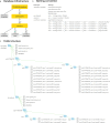
Database design and naming convention. (a) shows the infrastructure, the TDBRAIN consists of a file containing the participants metadata and multiple participants folders, these in turn may include multiple session folders. In the session folders, session specific information is stored in the session metadata, the condition files (EEG data) measured within this session are stored as.csv files and their specific information in condition metadata. (b) the naming convention: participants always have 8 digit IDcodes, sessions are described with the participants IDcode and then ‘-’ + <sessionnumber>. Each EEG measurement additionally acquires a condition, such as ‘.EO’ or ‘.EC’ in the current database. These measurements possibly will be complimented with several additional conditions, with condition having a maximum of 4 characters. (c) shows an example of one participants’ folder and file structure.
Fig. 3
Fig. 3
Age distribution for female (green) and male (blue) participants, for the whole heterogenous database.
Fig. 4
Fig. 4
The frequency response of the two amplifiers used in this dataset for the two EEG channels, (a) Fz and (b) Pz.
Fig. 5
Fig. 5
Averaged logPower measured at Pz, for Eyes Open (green) and Eyes closed (blue). The difference is significant between 7 and 13 Hz (p < 0.001, d’ = 0.9).
Fig. 6
Fig. 6
The iAPF (at Pz) related with age, and iAPF predicted from age. (a) the iAPF of all participants sorted by age (green) and the logGaussian function modeling the iAPF from age (blue). The model explains 4% of the variance and shows an initial steep increase of iAPF up till an age of approximately 18 years after which a slight decrease sets in. b) the distribution of the residuals that shows to be normal with a mean of 0.003 +/− 1.06 (Shapiro test for normality; stat = 0.99, p < 0.001).

References

    1. Berger H. Über das Elektrenkephalogramm des Menschen. Archiv Für Psychiatrie Und Nervenkrankheiten. 1929;87:527–570. doi: 10.1007/BF01797193. - DOI
    1. Widge AS, et al. Electroencephalographic biomarkers for treatment response prediction in major depressive illness: a meta-analysis. Am J Psychiatry. 2019;176:44–56. doi: 10.1176/appi.ajp.2018.17121358. - DOI - PMC - PubMed
    1. Ioannidis JPA. Why Most Published Research Findings Are False. Plos Med. 2005;2:e124. doi: 10.1371/journal.pmed.0020124. - DOI - PMC - PubMed
    1. van der Vinne N, Vollebregt MA, van Putten MJAM, Arns M. Frontal alpha asymmetry as a diagnostic marker in depression: Fact or fiction? A meta-analysis. Neuroimage Clin. 2017;16:79–87. doi: 10.1016/j.nicl.2017.07.006. - DOI - PMC - PubMed
    1. Arns M, Conners CK, Kraemer HC. A Decade of EEG Theta/Beta Ratio Research in ADHD. J Atten Disord. 2013;17:374–383. doi: 10.1177/1087054712460087. - DOI - PubMed