

A longitudinal multi-scanner multimodal human neuroimaging dataset
- PMID: 35701471
- PMCID: PMC9198098
- DOI: 10.1038/s41597-022-01386-3
A longitudinal multi-scanner multimodal human neuroimaging dataset
Abstract
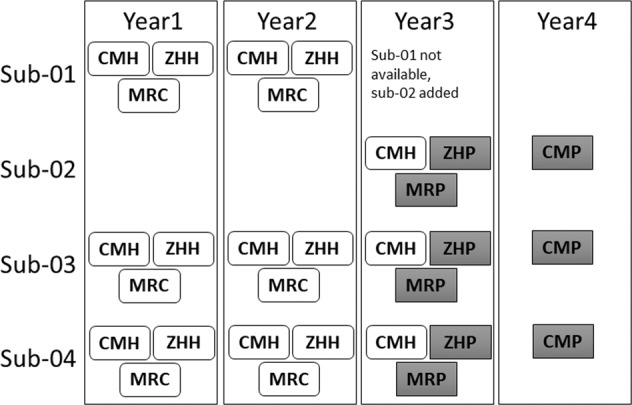
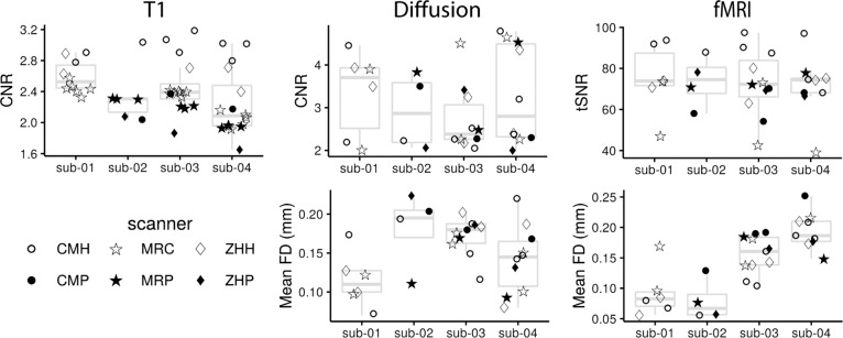
Human neuroimaging has led to an overwhelming amount of research into brain function in healthy and clinical populations. However, a better appreciation of the limitations of small sample studies has led to an increased number of multi-site, multi-scanner protocols to understand human brain function. As part of a multi-site project examining social cognition in schizophrenia, a group of "travelling human phantoms" had structural T1, diffusion, and resting-state functional MRIs obtained annually at each of three sites. Scan protocols were carefully harmonized across sites prior to the study. Due to scanner upgrades at each site (all sites acquired PRISMA MRIs during the study) and one participant being replaced, the end result was 30 MRI scans across 4 people, 6 MRIs, and 4 years. This dataset includes multiple neuroimaging modalities and repeated scans across six MRIs. It can be used to evaluate differences across scanners, consistency of pipeline outputs, or test multi-scanner harmonization approaches.
© 2022. The Author(s).
Conflict of interest statement
Dr. Buchanon is on data safety monitoring boards for Roche, Newron, Merck, as well as advisory boards for Acadia, Boehringer-Ingelheim, and GW Pharma Ltd.
Figures
References
-
- Plitman, E. et al. The Impact of the Siemens Tim Trio to Prisma Upgrade and the Addition of Volumetric Navigators on Cortical Thickness, Structure Volume, and 1H-MRS Indices: An MRI Reliability Study with Implications for Longitudinal Study Designs. Neuroimage 118172 (2021). - PubMed
Publication types
MeSH terms
Grants and funding
- R01MH102313/U.S. Department of Health & Human Services | NIH | National Institute of Mental Health (NIMH)
- R01MH102318/U.S. Department of Health & Human Services | NIH | National Institute of Mental Health (NIMH)
- R01MH102324/U.S. Department of Health & Human Services | NIH | National Institute of Mental Health (NIMH)
LinkOut - more resources
Full Text Sources
Medical
Research Materials
