Review
. 2022 Jun 14;15(1):23.
doi: 10.1186/s13039-022-00600-6.

Mechanisms of structural chromosomal rearrangement formation

Affiliations
Review

Mechanisms of structural chromosomal rearrangement formation

Bruna Burssed et al. Mol Cytogenet. .

Abstract

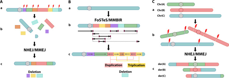
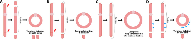
Structural chromosomal rearrangements result from different mechanisms of formation, usually related to certain genomic architectural features that may lead to genetic instability. Most of these rearrangements arise from recombination, repair, or replication mechanisms that occur after a double-strand break or the stalling/breakage of a replication fork. Here, we review the mechanisms of formation of structural rearrangements, highlighting their main features and differences. The most important mechanisms of constitutional chromosomal alterations are discussed, including Non-Allelic Homologous Recombination (NAHR), Non-Homologous End-Joining (NHEJ), Fork Stalling and Template Switching (FoSTeS), and Microhomology-Mediated Break-Induced Replication (MMBIR). Their involvement in chromoanagenesis and in the formation of complex chromosomal rearrangements, inverted duplications associated with terminal deletions, and ring chromosomes is also outlined. We reinforce the importance of high-resolution analysis to determine the DNA sequence at, and near, their breakpoints in order to infer the mechanisms of formation of structural rearrangements and to reveal how cells respond to DNA damage and repair broken ends.

Keywords: Chromoanagenesis; Fork stalling and template switching (FoSTeS); Inv dup del; Mechanisms of formation; Microhomology-mediated break-induced replication (MMBIR); Non-allelic homologous recombination (NAHR); Non-homologous end-joining (NHEJ); Ring chromosome; Structural chromosomal rearrangements.


Conflict of interest statement

The authors declare that they have no competing interests.

Figures

Fig. 1
Fig. 1
Recurrent and Non-recurrent rearrangements. A Representation of a genomic region with two Low Copy Repeats (LCRs). B Recurrent rearrangements show similar breakpoints clustered in regions of Low Copy Repeats. C Non-recurrent rearrangements show diverse breakpoints and sizes. Each grey bar represents deletions, duplications, or inversions in unrelated individuals
Fig. 2
Fig. 2
Repetitive sequences and associated non-B DNA structures that can cause replication fork stalling or breakage. A Inverted Repeats (equidistant DNA bases are Watson–Crick complements) form cruciform in double-stranded DNA and hairpin in single-stranded DNA, B Mirror Repeats (equidistant DNA bases are identical) form H-DNA (triple-helical DNA), C Direct Tandem Repeats (simple, noninterrupted repeats) form S-DNA (slipped-stranded DNA), and D Direct Tandem Repeats with G-runs form G quartet or quadruplex. Based on [32]
Fig. 3
Fig. 3
Non-Allelic Homologous Recombination (NAHR) mechanism. A NAHR leading to the formation of duplication and deletion: (a) Normal chromosome pairing and alignment of Low Copy Repeats (LCRs) in the same orientation. (b) A misalignment between LCRs due to their high level of sequence identity leads to an unequal crossing over event that can generate (c) a duplication and (d) a deletion. The scheme represents the LCRs of region 22q11.2. NAHR between LCR-A and LCR-B leads to a deletion seen in 8% of the cases of 22q11.2 deletion syndrome. B NAHR leading to the formation of inversions: (a) Normal chromosome pairing and alignment of Low Copy Repeats (LCRs). LCR-X and LCR-Z present similar DNA sequences but in opposite orientations. (b) A misalignment between LCRs due to their high level of sequence identity leads to an unequal crossing over event that can generate (c) an inversion
Fig. 4
Fig. 4
Mechanisms of double-strand break (DSB) repair. A Canonical Non-Homologous End-Joining (c-NHEJ) mechanism: The repair of (a) the DSB is done by molecularly bridging and rejoining the broken ends. This can happen (b) without nucleotide edition or through the editing of the broken ends with (c) addition or (d) loss of nucleotides. B Microhomology-Mediated End-Joining (MMEJ) mechanism: After (a) the DSB, (b) a 5′ to 3′ resection results in two 3′ single-stranded overhangs with exposed nucleotides of microhomology (purple), which (c) anneal. (d) After the trimming of the 3′ strands, the gaps are filled (light orange) and ligated, resulting in the repair with a deletion. C Homologous Recombination (HR) mechanism: after (a) a DSB in one of the sister chromatids, (b) a 5′ to 3′ resection creates a 3′ overhang with an exposed region of homology (yellow), (c) which helps the broken end to invade its sister chromatid and anneal, allowing for the start of DNA synthesis. (d) The DSB can be solved using homologous chromosomes without errors, therefore without the creation of rearrangements. It is important to note that HR can lead to rearrangements when it uses repetitive elements and not the homology located at the sister chromatid
Fig. 5
Fig. 5
Replication mechanisms. A Fork Stalling and Template Switching (FoSTeS) mechanism: (a) When a replication fork stalls, (b) the lagging strand disengages from its original template and, due to the presence of microhomology (purple), invades and switches to another template (dashed line) at another active replication fork and restarts DNA synthesis. (c) The nascent lagging strand can disengage again and invade other replication forks. Eventually, the strand can return to its original template and (a,d) restart synthesis. (e) The final product contains segments from different parts of the genome that were brought together due to microhomology. B Microhomology-Mediated Break-Induced Replication (MMBIR) mechanism: (a) A replication fork collapses when it encounters a DNA lesion, forming (b) a single-ended double-strand break. (c) A 5′–3′ resection creates a 3′ overhang with an exposed region of microhomology (purple), which serves as a template for (d) the invasion of a different region of the genome, where DNA synthesis is restarted. (e, f) The process can be repeated, and other regions of the genome can be invaded due to the presence of microhomology. (g) The final product presents a complex rearrangement with distinct parts of the genome united due to microhomology. In FoSTeS and MMBIR, the low processivity of the DNA polymerase leads to constant strand switching, especially at the beginning of the process, which may lead to short insertions in the junction points. As the invasion goes on, the DNA polymerase is switched and becomes more processive, allowing the replication to proceed until the end of the chromosome. The Break-Induced Replication (BIR) mechanism process is similar to MMBIR but uses larger homologous sequences instead of microhomologies
Fig. 6
Fig. 6
Chromoanagenesis. A Chromothripsis: (a) A missegregated chromosome is encapsulated inside a micronucleus and suffers multiple double-strand breaks, leading to (b) the shattering of the chromosome. (c) The chromosome is reassembled by c-NHEJ or MMEJ and reincorporated in the nucleus. Deletions due to loss of DNA fragments can occur. B Chromoanasynthesis: (a) A chromosome undergoes (b) DNA segment re-synthesis in a process that involves FoSTeS or MMBIR, forming (c) a new chromosome, which can present inversions, deletions, duplications, and triplications. C Chromoplexy: (a) Different chromosomes suffer (b) double-strand breaks and, after recombination by NHEJ or MMEJ, (c) form chromosomes with translocations. Based on (59)
Fig. 7
Fig. 7
Mechanisms of formation of inverted duplications associated with terminal deletions (inv dup del rearrangements). A U-type Exchange Mechanism: (a) a double-strand break in sister chromatids leads to a terminal deletion. (b) Fusion of the broken ends forms a symmetric U-type structure and produces a dicentric chromosome, which undergoes another double-strand break forming the (c) inv dup del chromosome without the spacer between the duplicated region (shown as blue arrows). B Low Copy Repeat-dependant Mechanism: (a) LCRs in inverted orientation located in sister chromatids lead to (b) a partial folding of a chromatid onto itself so that the LCRs can pair and align with the other chromatid. A crossing over event can happen between them and form (c) a dicentric chromosome, which undergoes a double-strand break and forms the (d) inv dup del chromosome, which presents a spacer flanked by the LCRs between the duplicated region (shown as blue arrows). C Paracentric Inversion-dependant Mechanism: (a) Two homologous chromosomes with one (bottom) presenting an inversion (e′–d′). (b) Formation of an inversion loop to allow for proper chromosome pairing with the normal homolog during meiosis. (c) A crossing over event happens within the loop and leads to the formation of (d) a dicentric chromosome, which undergoes a double-strand break and forms the (d) inv dup del chromosome, which presents a spacer between the duplications (shown as blue arrows). D Fold-back Mechanism: (a) Chromosome with microhomologies (shown in purple). (b) A double-strand break forms the terminal deletion. (c) A 5′ to 3′ resection creates a 3′ overhang with exposed microhomologies. (d) Due to the microhomologies, the 3′ overhang folds back onto itself and allows for intrastrand pairing. (e) DNA synthesis fills the resected gap forming a monocentric fold back chromosome, and (f) DNA replication forms a dicentric chromosome, which suffers a (g) double strand break forming an (h) inv dup del chromosome, which presents a spacer between the duplicated region (shown as blue arrows)
Fig. 8
Fig. 8
Ring Chromosome Formation. A Two terminal double-strand breaks in each chromosome arm and subsequent fusion of the broken ends lead to the formation of a ring chromosome with terminal deletions in both arms. B A terminal double-strand break in one arm and subsequent fusion of the broken end with the opposite arm's telomeric or subtelomeric region leads to the formation of a ring chromosome with terminal deletions in one arm. C Fusion of the telomeric or subtelomeric regions of a chromosome without terminal deletions leads to the formation of a complete ring chromosome. D An inv dup del chromosome can be stabilized via circularization after fusion of the end of the inverted duplication with the opposite arm's telomeric or subtelomeric region, leading to the formation of a ring chromosome with inverted duplication (blue arrows) and terminal deletion
Fig. 9
Fig. 9
Junction point sequencing highlights different mechanisms signatures. A Junction point from a translocation between chromosome 3 and chromosome X with insertion of nucleotides from Non-Homologous End-Joining (NHEJ) mechanism. At the top, the chromatogram displays chromosome 3 (underlined in pink) and chromosome X (underlined in blue). At the bottom, the alignment of the derivative (der) chromosome 3 (middle) with the reference sequence chromosomes 3 (pink) and X (blue). In red, four nucleotides were added for the ligation of the chromosomes by NHEJ. B Junction point from an inv dup del(13q) with microhomologies from the Fold-back mechanism. At the top, the chromatogram displays the spacer (underlined in pink) and the inverted duplication (underlined in blue). At the bottom, the alignment of the derivative chromosome 13 with the reference sequence of the spacer (pink) and the inverted duplication (blue). In purple, three nucleotides of the microhomology that prompted the Fold-back mechanism's occurrence. C Junction point from a complex rearrangement involving both arms of chromosome 18 with an insertion from a replication (FoSTeS/MMBIR) mechanism. At the top, the chromatogram displays the normal region of the long arm (underlined in pink), the inserted region of the long arm (underlined in green), and the inverted duplication of the short arm (underlined in blue). At the bottom, the alignment of the altered derivative 18 with the reference sequence of the long arm (pink), the insertion of the long arm (green), and the short arm (blue). In purple, four nucleotides of microhomology between the two regions of the long arm, and the two nucleotides of microhomology between the insertion and the short arm

References

    1. Lupski JR, Stankiewicz P. Genomic disorders: molecular mechanisms for rearrangements and conveyed phenotypes. PLoS Genet. 2005;1(6):0627–633. doi: 10.1371/journal.pgen.0010049. - DOI - PMC - PubMed
    1. Weckselblatt B, Rudd MK. Human structural variation: mechanisms of chromosome rearrangements. Trends Genet. 2015;31(10):587–599. doi: 10.1016/j.tig.2015.05.010. - DOI - PMC - PubMed
    1. Shaw CJ, Lupski JR. Implications of human genome architecture for rearrangement-based disorders: the genomic basis of disease. Hum Mol Genet. 2004;13(REV. ISS. 1):57–64. doi: 10.1093/hmg/ddh073. - DOI - PubMed
    1. Lupski JR. Genomic disorders: structural features of the genome can lead to DNA rearrangements and human disease traits. Trends Genet. 1998;14(10):417–422. doi: 10.1016/S0168-9525(98)01555-8. - DOI - PubMed
    1. Shaffer LG, Lupski JR. C Hromosomal R earrangements in H umans. Annu Rev Genet. 2000;34:297–329. doi: 10.1146/annurev.genet.34.1.297. - DOI - PubMed

LinkOut - more resources