. 2022 Jun 14;20(1):88.
doi: 10.1186/s12958-022-00959-6.

High androgen concentrations in follicular fluid of polycystic ovary syndrome women

Affiliations

High androgen concentrations in follicular fluid of polycystic ovary syndrome women

Alice Bongrani et al. Reprod Biol Endocrinol. .

Abstract

Background: According to current definitions of Polycystic Ovary Syndrome (PCOS), hyperandrogenism is considered as a key element in the pathogenesis of this common endocrinopathy. However, until now, studies about ovarian androgen profile in women are very rare. Our aim was then to characterise the expression profile of the androgens in follicular fluid of 30 PCOS patients, and compare it to those of 47 Control women and 29 women with only polycystic ovary morphology on ultrasounds (ECHO group).

Methods: A retrospective, single-centre cohort study was performed. The intrafollicular concentrations of the key androgens were assessed and correlated with the intrafollicular levels of some adipokines of interest. Androgens were quantified by mass spectrophotometry combined with ultra-high-performance liquid chromatography, while adipokine concentrations were measured by ELISA assays.

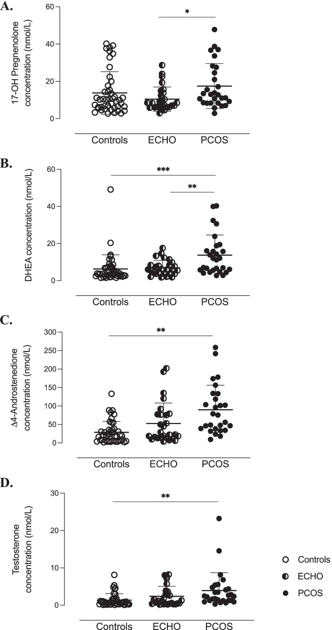
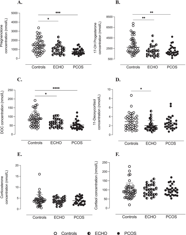
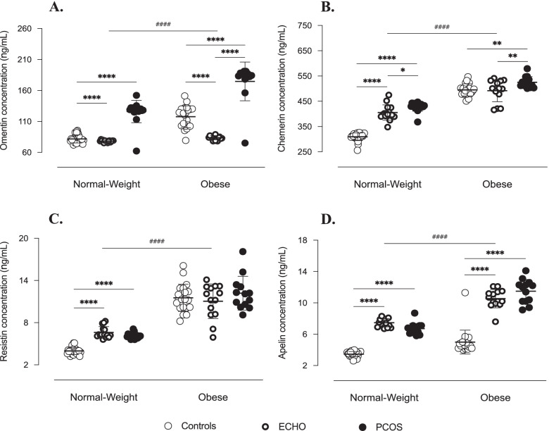
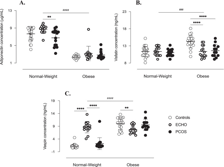
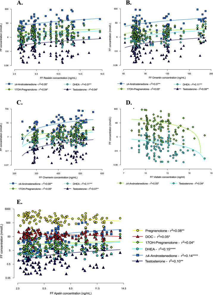
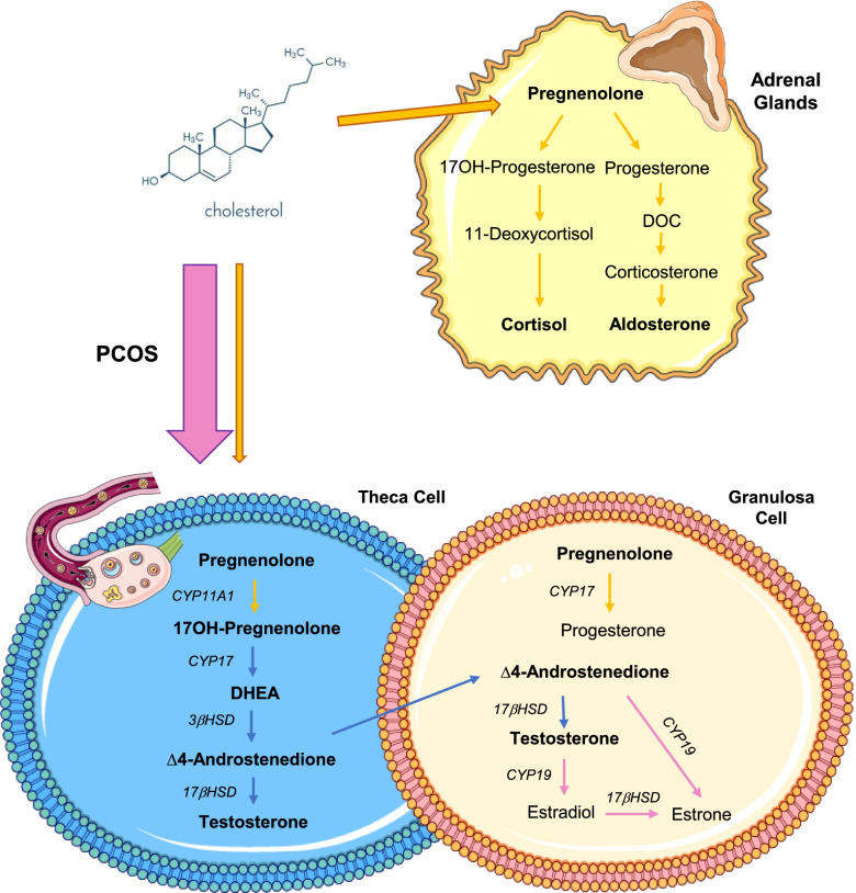
Results: In PCOS patients, the intrafollicular concentrations of the androgens synthesised by ovarian theca cells, i.e., 17OH-pregnenolone, dehydroepiandrosterone, Δ4-androstenedione and testosterone, were significantly higher than those of the androgens of adrenal origin, and positively correlated with the main PCOS clinical and biological features, as well as with the adipokines mostly expressed in the follicular fluid of PCOS patients, i.e. resistin, omentin, chemerin and apelin. Conversely, Control women showed the highest levels of 17OH-progesterone, deoxycorticosterone and 11-deoxycortisol. Confirming these results, apelin levels were negatively associated with pregnenolone and deoxycorticosterone concentrations, while visfatin levels, which were higher in the Control group, negatively correlated with the Δ4-androstenedione and testosterone ones.

Conclusions: PCOS is characterised by a selective increase in the intrafollicular levels of the androgens synthesised by theca cells, strengthening the hypothesis that ovarian hyperandrogenism plays a central role in its pathogenesis. Further, the significant correlation between the intrafollicular concentrations of the androgens and most of the adipokines of interest, including apelin, chemerin, resistin and omentin, confirms the existence of a close relationship between these two hormonal systems, which appear deeply involved in ovarian physiology and PCOS physiopathology.

Keywords: Adipokines; Androgens; Follicular fluid; Polycystic ovary syndrome (PCOS); Theca cells.


Conflict of interest statement

The authors declare that there is no conflict of interest that could be perceived as prejudicing the impartiality of the research reported.

Figures

Fig. 1
Fig. 1
Ovary-synthetised androgens in follicular fluid of study women. Follicular concentrations of 17OH-pregnenolone (A), dehydroepiandrosterone (DHEA) (B), ∆4-androstenedione (C) and testosterone (D) quantified by mass spectrophotometry combined with ultra-high performance liquid chromatography (LC-MS/MS). ***p < 0.001, **p < 0.01, *p < 0.05 (two-way ANOVA followed by Bonferroni post-hoc tests)
Fig. 2
Fig. 2
Adrenal-originated androgens in follicular fluid of study women. Follicular concentrations of pregnenolone (A), 17OH-progesterone (B), deoxycorticosterone (DOC) (C), 11-deoxycortisol (D), corticosterone (E) and cortisol (F) quantified by mass spectrophotometry combined with ultra-high performance liquid chromatography (LC-MS/MS). ****p < 0.0001, *** p < 0.001, ** p < 0.01, *p < 0.05 (two-way ANOVA followed by Bonferroni post-hoc tests)
Fig. 3
Fig. 3
Correlations between androgen concentrations in follicular fluid and anthropometric features of study women. Correlations of androgen levels in follicular fluid (FF) with BMI (A) and age (B) (simple linear regression). *p ≤ 0.05 and **p < 0.01
Fig. 4
Fig. 4
Correlations between androgen follicular concentrations and clinical-biological parameters of study women. Correlations of androgen levels in follicular fluid (FF) with cycles duration (A), follicles count (B), plasma AMH (C) and plasma LH (D) concentrations (simple linear regression). *p ≤ 0.05, **p < 0.01, ***p < 0.001 and ****p < 0.0001
Fig. 5
Fig. 5
Adipokines in follicular fluid of study women. Omentin (A), chemerin (B), resistin (C) and apelin (D) concentrations measured by ELISA assay in follicular fluid. Data are shown as individual values with means as horizontal bars. Groups were compared by two-way ANOVA followed by Bonferroni post-hoc tests. As interactions between BMI and pathological condition were statistically significant (p < 0.001), normal-weight and obese groups were analysed separately. ****/####p < 0.0001, **p < 0.01, *p < 0.05
Fig. 6
Fig. 6
Adipokines in follicular fluid of study women. Adiponectin (A), visfatin (B) and vaspin (C) concentrations measured by ELISA assay in follicular fluid. Data are shown as individual values with means as horizontal bars. Groups were compared by two-way ANOVA followed by Bonferroni post-hoc tests. As interactions between BMI and pathological condition were statistically significant (p < 0.001), normal-weight and obese groups were analysed separately. ****/####p < 0.0001, ###p < 0.001, **p < 0.01
Fig. 7
Fig. 7
Correlations between androgen and adipocytokine concentrations in follicular fluid of study women. Correlations between androgen levels in follicular fluid and resistin (A), omentin (B), chemerin (C), visfatin (D) and apelin (E) follicular concentrations (simple linear regression). *p ≤ 0.05, **p < 0.01, ***p < 0.001 and ****p < 0.0001. DHEA = dehydroepiandrosterone; DOC = deoxycorticosterone
Fig. 8
Fig. 8
Overview of androgen synthesis pathways in women. Metabolic pathways of androgen synthesis in the adrenal glands and ovarian theca and granulosa cells. In PCOS (pink arrow), steroidogenesis seems to be diverted from adrenal to ovarian metabolic pathways due to CYP11A1, CYP17 and 3𝛽HSD hyperactivity in theca cells, resulting in increased intrafollicular levels of 17OH-pregnenolone, DHEA, ∆4-androstenedione and testosterone. PCOS = Polycystic Ovary Syndrome; DHEA = dehydroepiandrosterone; DOC = deoxycorticosterone

Similar articles

Cited by

References

    1. Teede HJ, Misso ML, Costello MF, Dokras A, Laven J, Moran L, et al. Recommendations from the international evidence-based guideline for the assessment and management of polycystic ovary syndrome†‡. Hum Reprod. 2018;33(9):1602–1618. doi: 10.1093/humrep/dey256. - DOI - PMC - PubMed
    1. Rotterdam ESHRE/ASRM-Sponsored PCOS consensus workshop group Revised 2003 consensus on diagnostic criteria and long-term health risks related to polycystic ovary syndrome (PCOS) Hum Reprod. 2004;19(1):41–47. doi: 10.1093/humrep/deh098. - DOI - PubMed
    1. Azziz R, Carmina E, Dewailly D, Diamanti-Kandarakis E, Escobar-Morreale HF, Futterweit W, et al. The androgen excess and PCOS society criteria for the polycystic ovary syndrome: the complete task force report. Fertil Steril. 2009;91(2):456–488. doi: 10.1016/j.fertnstert.2008.06.035. - DOI - PubMed
    1. Gilling-Smith C, Story H, Rogers V, Franks S. Evidence for a primary abnormality of thecal cell steroidogenesis in the polycystic ovary syndrome. Clin Endocrinol. 1997;47(1):93–99. doi: 10.1046/j.1365-2265.1997.2321049.x. - DOI - PubMed
    1. Rosenfield RL, Ehrmann DA. The pathogenesis of polycystic ovary syndrome (PCOS): the hypothesis of PCOS as functional ovarian Hyperandrogenism revisited. Endocr Rev. 2016;37(5):467–520. doi: 10.1210/er.2015-1104. - DOI - PMC - PubMed