. 2022 Jul 8;131(2):e2-e21.
doi: 10.1161/CIRCRESAHA.121.320565. Epub 2022 Jun 14.

Piezo1-Regulated Mechanotransduction Controls Flow-Activated Lymphatic Expansion

Affiliations

Piezo1-Regulated Mechanotransduction Controls Flow-Activated Lymphatic Expansion

Dongwon Choi et al. Circ Res. .

Abstract

Background: Mutations in PIEZO1 (Piezo type mechanosensitive ion channel component 1) cause human lymphatic malformations. We have previously uncovered an ORAI1 (ORAI calcium release-activated calcium modulator 1)-mediated mechanotransduction pathway that triggers lymphatic sprouting through Notch downregulation in response to fluid flow. However, the identity of its upstream mechanosensor remains unknown. This study aimed to identify and characterize the molecular sensor that translates the flow-mediated external signal to the Orai1-regulated lymphatic expansion.

Methods: Various mutant mouse models, cellular, biochemical, and molecular biology tools, and a mouse tail lymphedema model were employed to elucidate the role of Piezo1 in flow-induced lymphatic growth and regeneration.

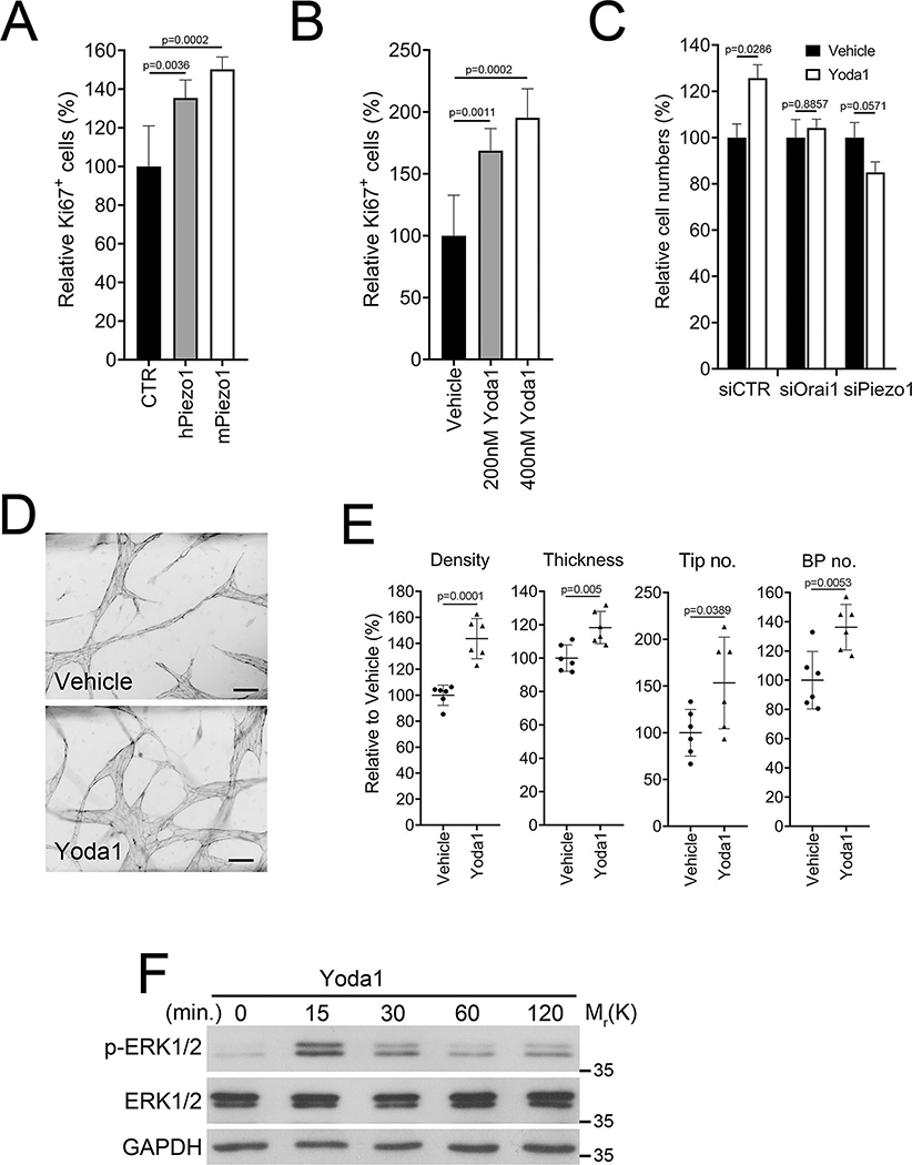
Results: Piezo1 was found to be abundantly expressed in lymphatic endothelial cells. Piezo1 knockdown in cultured lymphatic endothelial cells inhibited the laminar flow-induced calcium influx and abrogated the flow-mediated regulation of the Orai1 downstream genes, such as KLF2 (Krüppel-like factor 2), DTX1 (Deltex E3 ubiquitin ligase 1), DTX3L (Deltex E3 ubiquitin ligase 3L,) and NOTCH1 (Notch receptor 1), which are involved in lymphatic sprouting. Conversely, stimulation of Piezo1 activated the Orai1-regulated mechanotransduction in the absence of fluid flow. Piezo1-mediated mechanotransduction was significantly blocked by Orai1 inhibition, establishing the epistatic relationship between Piezo1 and Orai1. Lymphatic-specific conditional Piezo1 knockout largely phenocopied sprouting defects shown in Orai1- or Klf2- knockout lymphatics during embryo development. Postnatal deletion of Piezo1 induced lymphatic regression in adults. Ectopic Dtx3L expression rescued the lymphatic defects caused by Piezo1 knockout, affirming that the Piezo1 promotes lymphatic sprouting through Notch downregulation. Consistently, transgenic Piezo1 expression or pharmacological Piezo1 activation enhanced lymphatic sprouting. Finally, we assessed a potential therapeutic value of Piezo1 activation in lymphatic regeneration and found that a Piezo1 agonist, Yoda1, effectively suppressed postsurgical lymphedema development.

Conclusions: Piezo1 is an upstream mechanosensor for the lymphatic mechanotransduction pathway and regulates lymphatic growth in response to external physical stimuli. Piezo1 activation presents a novel therapeutic opportunity for preventing postsurgical lymphedema. The Piezo1-regulated lymphangiogenesis mechanism offers a molecular basis for Piezo1-associated lymphatic malformation in humans.

Keywords: calmodulin; endothelial cell; lymphedema; mechanotransduction, cellular; mutation.


Conflict of interest statement

Disclosures

Alex S. Huang discloses his relationships with Santen Pharmaceutical (Consultant), Allergan (Consultant), Aerie Pharmaceuticals (Consultant), Heidelberg Engineering (Research Support), Glaukos Corporation (Research Support), and Diagnosys (Research Support). All other authors disclose no conflict of interest with this study.

Figures

Figure 1.
Figure 1.. Piezo1 Is Required for Flow-Activated Notch Downregulation
(A) Western blot analyses showing the expression of Piezo1, Orai1, Prox1, and β-actin in three sets of the same donor-derived BECs and LECs. (B) Detection of endogenous Piezo1 expression in developing lymphatics using a novel Piezo1 reporter mouse with Piezo1-CreERT2-BFP (blue fluorescence protein) driver allele and Ai47 (CAG-LSL-3XGFP) reporter allele. The Piezo1-CreERT2-BFP driver was created by CRISPR/Cas9-mediated insertion of a DNA cassette (CreERT2-P2A-BFP-WPRE-polyA) immediately following the ATG initiation codon in the endogenous Piezo1, allowing CreERT2 and BFP to mirror the endogenous Piezo1expression pattern. Tamoxifen (6 mg) was injected into pregnant females bearing Piezo1 reporter embryos at E12.5 or E13.5. After two days, embryos were harvested at E14.5 or E15.5, respectively. Embryonic back skins were stained with an anti-Prox1 antibody to detect lymphatic vessels. White arrows indicate Prox1-negative blood vessels. Scale bars: 100 μm. (C) Flow-activated calcium influx is blocked by knockdown of Piezo1 or Orai1 in LECs. Cells were transfected with Piezo1 siRNA (siPiezo1), Orai1 siRNA (siOrai1), or scrambled siRNA (siCTR) for 24 hours and then subjected to laminar flow (2 dyne/cm2) . N=10 per group. Statistics: two-way repeated-measures ANOVA. (D, E) Orai1 and Piezo1 knockdown similarly abrogated the flow-induced downregulation of NICD in LECs. Cells were transfected with the corresponding siRNA for 24-hours and subjected to laminar flow (LF, 2 dyne/cm2) for 12- or 24-hours, or alternatively cultured under the static condition for 24 hours. (F) Piezo1 knockdown in LECs abrogated the flow-induced regulation of Orai1 downstream genes such as Dtx1, Dtx3L, Klf2, and Notch1. Experiments were repeated at least three times.
Figure 2.
Figure 2.. Piezo1 is a Mechanosensor for the Orai1-Regulated Notch Pathway.
(A) Piezo1 overexpression (O.E.) mimicked the flow-mediated regulation of NICD1, Dtx1, Dtx3L, and Klf2 in the absence of flow. LECs were transfected with a control vector or a mouse Piezo1-expressing vector for 48 hours before cells were harvested. (B) Yoda1 regulates the Orai1-downstream genes. LECs were treated with Yoda1 at the indicated concentrations for 24 hours under the static condition. (C) Regulation of Orai1 downstream genes by Yoda1 was inhibited by Piezo1 knockdown in LECs for 24 hours, followed by Yoda1 treatment (250 nM, 24 hours). (D) Orai1 inhibition abrogated the NICD-downregulation caused by Piezo1 overexpression (O.E.). LECs were transfected with a control or a Piezo1 vector for 24 hours and then treated with an Orai1 inhibitor SKF-96365 (SKF, 10 nM) for 24 hours. (E) Orai1 inhibition suppressed Yoda1-activated calcium influx. LECs were loaded with a calcium dye (Fluo-4) and treated with SKF-96365 (SKF, 5 or 10 μM) for 10 minutes, followed by Yoda1 treatment (2 μM). N=10 per group. Statistics: two-way repeated-measures ANOVA. (F) An Orai1 agonist IA65 recapitulates the flow-mediated mechanotransduction phenotypes: NICD downregulation and Dtx1/Dtx3L upregulation. LECs were treated with IA65 at the indicated concentrations for 24 hours under the static condition. (G) NICD downregulation caused by IA65 treatment was suppressed by Orai1 knockdown but not by Piezo1 knockdown.
Figure 3.
Figure 3.. Piezo1 KO Phenocopies Lymphatic Defects of Orai1 KO, Klf2 KO, and NICD TG.
(A) Diagram showing lymphatic-specific conditional knockout or transgenic strategy for Piezo1, Orai1, Klf2, or NICD1. Defective lymphatic sprouting in embryonic back skins caused by lymphatic-specific deletion of Piezo1 (B-E), Orai1 (F-I), and Klf2 (J-M) or by ectopic NICD1 expression (N-Q), compared to their wild-type control littermates. Lymphatics were visualized by anti-Lyve1 whole-mount staining. Scale bars: 500 μm in B, D, F, H, J, L, N, and P; 100 μm in C, E, G, I, K, M, O, and Q. Graphs showing the relative numbers of lymphatic vessel tip (R), branches (S), and vessel thickness (T) in embryos of wild-type (WT), lymphatic-specific Piezo1 (P1ΔL), Orai1 (O1ΔL), and Klf2 (K2ΔL) deletions, or NICD transgenic expression (NICD TG). Successful Cre-mediated genomic recombination was confirmed using PCR-based genotyping analyses (Figure S2). Embryos from multiple litters per group were analyzed (WT n=5 mice vs. P1ΔL n=5 mice, WT n=4 mice vs. O1ΔL n=8 mice, WT n=6 mice vs. K2ΔL n=4 mice, and WT n=4 mice vs. NICD TG n=4 mice). Statistics: Mann-Whitney U test.
Figure 4.
Figure 4.. Piezo1 Is Required for Postnatal Lymphatic Development and Maintenance
(A) Diagram illustrating postnatal induction of lymphatic-specific Piezo1 KO. Tamoxifen was injected into the 3-week-old juvenile control or Piezo1ΔLEC mice, followed by lymphatic analyses at 7 weeks. Lymphatics were visualized using Prox1-tdTomato reporter allele . Lymphatic Piezo1 KO was induced in young adults (3 weeks old) by Tamoxifen injection at Days 21, 23, and 25. The colon mesentery (B-E) and hindlimb skins (F-I) were collected for lymphatic analyses at 7 weeks. Branch points per lymphatic vessel length (BP/Length) and distance between two branch points (BP-BP) were compared in the mesentery (J,K) and hindlimb (L,M) between wild type and Piezo1ΔLEC mice. Each dot represents one animal (WT n=6 mice vs. Piezo1ΔLEC n=7 mice) (J,K), or one limb (n=10 mice per group) (L,M). Scale bars: 100 μm. Statistics: Mann-Whitney U test (J and K), and two-tailed t-test (L and M).
Figure 5.
Figure 5.. Dtx3L Overexpression Rescues Lymphatic Defects Caused by Piezo1 KO
(A) Genetic components of the mice for this experiment: Prox1-tdTomato, Prox1-CreERT2, Piezo1 fl/fl, and/or Dtx3L transgenic (TG) allele. pA, poly-A sequences. (B-I) Lymphatic vessels in control back skin (B,C), lymphatic-specific Dtx3L transgenic back skin (D,E), lymphatic-specific Piezo1 KO back skin (F,G), and lymphatic-specific Dtx3L transgenic/Piezo1 KO back skin (H,I). Pregnant mice were injected with Tamoxifen at E11.5 and 13.5, and embryos were harvested at E15.5. Scale bars: 1 mm (B, D, F, H), 100 μm (C, E, G, I). (J-M) Graphs showing the percent changes in lymphatic tip number (J), branch point number (BP No.) (K), and the distance between two branch points (BP-BP dis.) (M). Six different litters were analyzed (CTR n=14, Dtx3lTG n=8, Piezo1ΔLEC, n=12, and Piezo1ΔLEC; Dtx3lTG n=12 mice). Images in panel B-I were obtained from one representative litter. Lymphatic vessels were visualized by the Prox1-tdTomato reporter. Statistics: two-tailed t-test.
Figure 6.
Figure 6.. Transgenic Piezo1 Expression Promotes Lymphatic Sprouting and Network Formation
(A) Genetic components of the mice for this experiment: Prox1-tdTomato, Prox1-CreERT2, and/or mouse Piezo1 cDNA transgenic (TG) allele. (B, C) Pregnant females were injected with Tamoxifen at E11.5 and 13.5, and their embryos were harvested at 15.5 for lymphatic analyses. Lymphatic vessel tip number (Tip no.), branch point (BP no.), and lymphatic density of Piezo1 TG embryos (n=6 mice) were compared to those of their control litter embryos (CTR, n=4 mice). (D, E) Alternatively, newborns (P0) were injected with Tamoxifen, and their mesenteries were harvested at P4. Mesenteric lymphatics in the colon are shown (D). The number of branch points per lymphatic vessel length and distance between two branch points in the colon and intestine mesentery lymphatics of the control vs. Piezo1 TG pups (n=6 mice per group) (E). Each dot represents one bunch of mesentery lymphatic pre-collectors. Scale bars: 1 mm. Lymphatic vessels were visualized by the Prox1-tdTomato reporter. Statistics: Mann-Whitney U test (C), and two-tailed t-test (E).
Figure 7.
Figure 7.. Piezo1 Activation Stimulates Lymphangiogenesis in Adults
(A) Primary LECs were transiently transfected with a control (CTR), human Piezo1-, or mouse Piezo1- vector for 48 hours and then subjected to Ki67 immunostaining (n=6 per group). (B) LECs were treated with vehicle or Yoda1 at 200 or 400 nM for 24 hours and subjected to Ki67 immunostaining (n=6 per group). (C) LECs were transiently transfected with siRNA for control (siCTR), Orai1 (siOrai1), or Piezo1 (siPiezo1). After 24 hours, cells were treated with vehicle or Yoda1 (300 nM) for 24 hours, followed by total cell number counting (n=4 per group). (D) The effect of Yoda1 on 3-D lymphatic network formation was assessed. LECs are allowed to form 3-D lymphatic networks in fibrin gels containing vehicle or Yoda1 (500 nM) in a vessel-on-a-chip device . After 3 days, lymphatic networks were fixed and stained against VE-Cadherin. Scale bars: 100 μm. (E) Morphometric analyses of the 3-D lymphatic vessels. BP, branch point (n=6 per group). (F) Western blots showing ERK1/2 phosphorylation in LECs treated with Yoda1 (400 nM) for the indicated time. The vehicle-treated sample is marked 0 minutes, and Glyceraldehyde-3-Phosphate Dehydrogenase (GAPDH) levels are also shown as loading controls. (G) Yoda1 activated dermal lymphangiogenesis in the mouse ears. These ears were harvested from mice subjected to the tail lymphedema model (Figure 8). Adult Prox1-EGFP lymphatic reporter mice were i.p. injected daily with vehicle or Yoda1 (71 μg/kg/day) over 35 days. (H) The numbers of lymphatic tips and valves were compared between the two groups (Vehicle n=8 and Yoda1 n=9 mice). (I) Yoda1 activated postnatal growth of the ocular lymphatics. Vehicle (10 μL, DMSO) or Yoda1 (10 μL, 50 μM in DMSO) was injected into the subconjunctival area of adult Prox-1-tdTomato mice every other day. The eyes were harvested 6 days after the first injection. (J) Numbers of lymphatic tip and valve were compared between the vehicle (n=7 mice) vs. Yoda1-injected groups (n=8 mice). (K) Matrigel matrix pre-mixed with vehicle or Yoda1 (500 nM) was intradermally injected into the flank of adult Prox1-EGFP mice. After two weeks, Matrigel plugs were harvested, and lymphatic ingrowth was visualized. Mouse VEGFC (50 ng/mL) was added to both groups to promote lymphatic expansion. 7 mice were used in each group. (L) Lymphatic vessels numbers and vessel length were compared between vehicle- vs. Yoda1-containing Matrigel plugs (n=14 Matrigel plugs per group). Scale bars: 200 μm in G and I; 500 μm in K. Statistics: Mann-Whitney U test (C and L), and two-tailed t-test (A, B, E, H, and J).
Figure 8.
Figure 8.. Yoda1 Prevents Lymphedema Formation in Mouse: Potential Therapeutic Efficacy
(A) Experimental tail lymphedema images demonstrating a therapeutic efficacy of Yoda1. The tail lymphedema model was introduced to adult Prox1-EGFP mice. Vehicle control (n=18) or Yoda1 at low (n=15, 71 μg/kg/day) or high (n=13, 213 μg/kg/day) was i.p. injected daily from post-operation day (POD) 0 for 35 days. (B) Graph showing the degree of lymphedema by charting percent changes in the tail volume of each group over 35 days. Tail volume was measured weekly. (C) ICG-based lymphangiography assessing lymphatic function. At POD 35, ICG was injected at the tip of the tail, and fluorescent images were captured over 96 hours. Representative images are shown after 3 min., 24 hours, and 48 hours post-ICG-injection. (D) Graph showing the rate of ICG clearance reflecting lymphatic function. ICG fluorescence intensity was measured from captured images. N = 10/group. p < 0.001 from 4 hours to 96 hours post-ICG-injection. Scale bars: 5 mm in panels A and C (F) H&E staining of paraffin sections prepared from an immediately distal area of the surgery site. (G) Thickness of the subcutaneous area was measured, and the relative values are charted. (H) Anti-Lyve1 immunohistochemistry visualizing lymphatic vessels (n=4 mice per group). (I) Graphs showing relative lymphatic vessel number (LV No), vessel size, and size/number (n=4 mice per group). Vessel size is determined by measuring their circumferences. Scale bars: 100 μm in F and H. Statistics: ANCOVA (B and D), Mann-Whitney U test (G and I).

Comment in

References

    1. Escobedo N and Oliver G. Lymphangiogenesis: Origin, Specification, and Cell Fate Determination. Annu Rev Cell Dev Biol. 2016;32:677–691. - PubMed
    1. Breslin JW, Yang Y, Scallan JP, Sweat RS, Adderley SP and Murfee WL. Lymphatic Vessel Network Structure and Physiology. Compr Physiol. 2018;9:207–299. - PMC - PubMed
    1. Geng X, Ho YC and Srinivasan RS. Biochemical and mechanical signals in the lymphatic vasculature. Cell Mol Life Sci. 2021. - PMC - PubMed
    1. Choi D, Park E, Jung E, Seong YJ, Hong M, Lee S, Burford J, Gyarmati G, Peti-Peterdi J, Srikanth S, Gwack Y, Koh CJ, Boriushkin E, Hamik A, Wong AK and Hong YK. ORAI1 Activates Proliferation of Lymphatic Endothelial Cells in Response to Laminar Flow Through Kruppel-Like Factors 2 and 4. Circ Res. 2017;120:1426–1439. - PMC - PubMed
    1. Choi D, Park E, Jung E, Seong YJ, Yoo J, Lee E, Hong M, Lee S, Ishida H, Burford J, Peti-Peterdi J, Adams RH, Srikanth S, Gwack Y, Chen CS, Vogel HJ, Koh CJ, Wong AK and Hong YK. Laminar flow downregulates Notch activity to promote lymphatic sprouting. J Clin Invest. 2017;127:1225–1240. - PMC - PubMed

Publication types