. 2022 Jul 22;131(3):222-235.
doi: 10.1161/CIRCRESAHA.121.319817. Epub 2022 Jun 14.

Mitochondrial H2S Regulates BCAA Catabolism in Heart Failure

Affiliations

Mitochondrial H2S Regulates BCAA Catabolism in Heart Failure

Zhen Li et al. Circ Res. .

Erratum in

Abstract

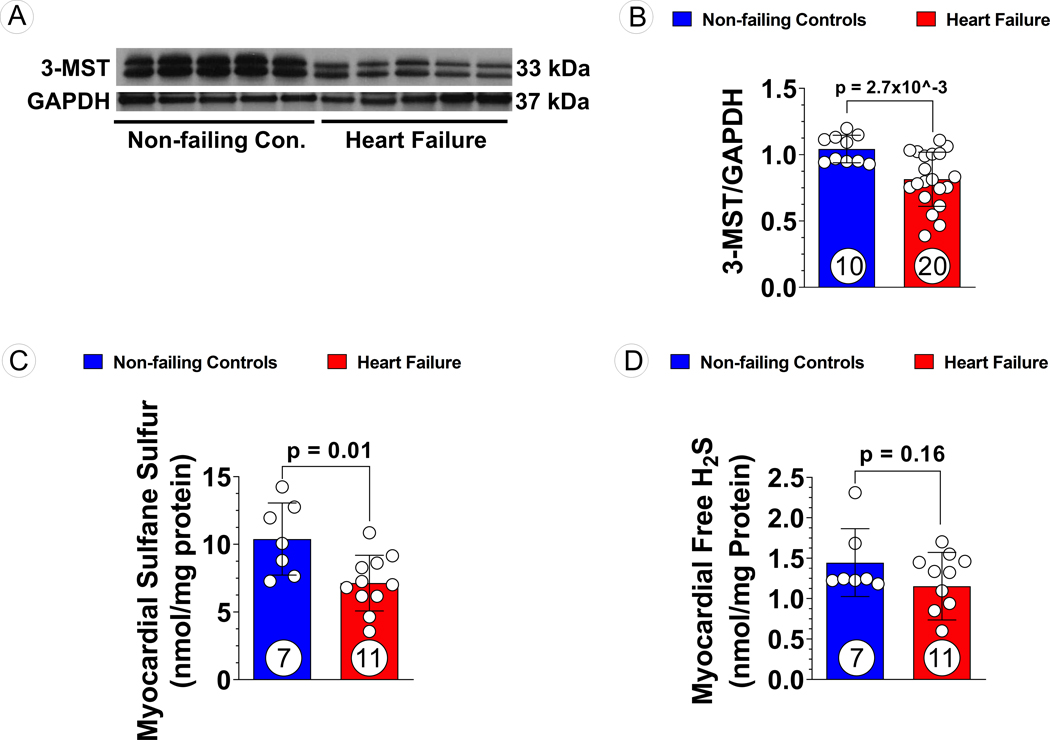
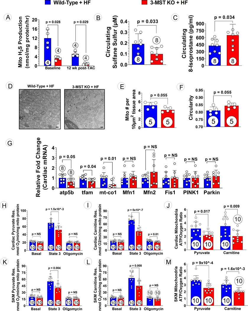
Background: Hydrogen sulfide (H2S) exerts mitochondria-specific actions that include the preservation of oxidative phosphorylation, biogenesis, and ATP synthesis, while inhibiting cell death. 3-MST (3-mercaptopyruvate sulfurtransferase) is a mitochondrial H2S-producing enzyme whose functions in the cardiovascular disease are not fully understood. In the current study, we investigated the effects of global 3-MST deficiency in the setting of pressure overload-induced heart failure.

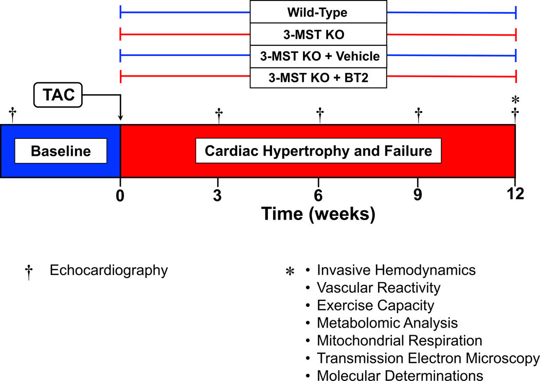
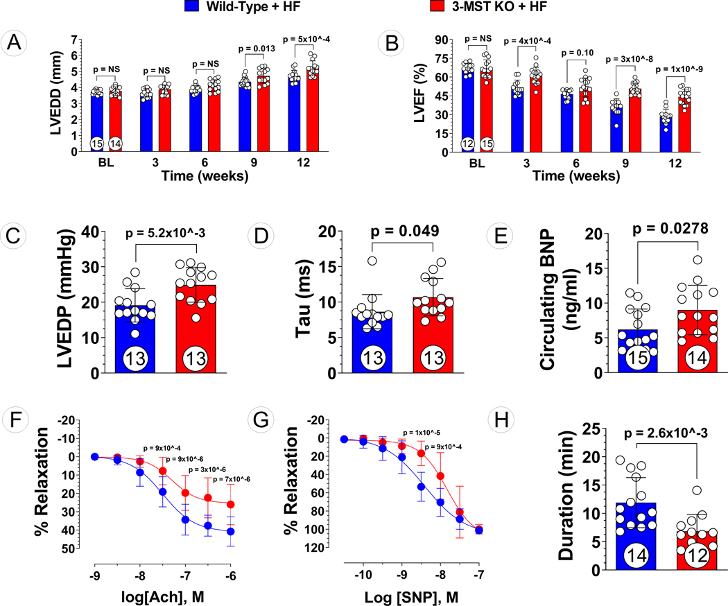
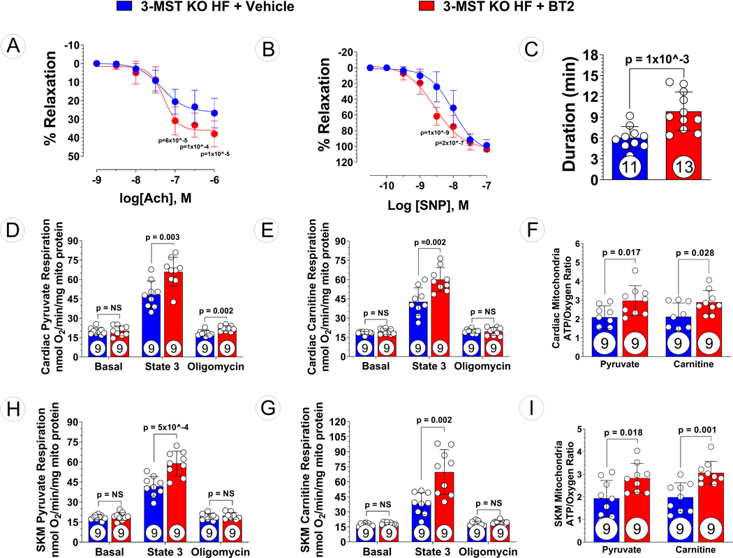
Methods: Human myocardial samples obtained from patients with heart failure undergoing cardiac surgeries were probed for 3-MST protein expression. 3-MST knockout mice and C57BL/6J wild-type mice were subjected to transverse aortic constriction to induce pressure overload heart failure with reduced ejection fraction. Cardiac structure and function, vascular reactivity, exercise performance, mitochondrial respiration, and ATP synthesis efficiency were assessed. In addition, untargeted metabolomics were utilized to identify key pathways altered by 3-MST deficiency.

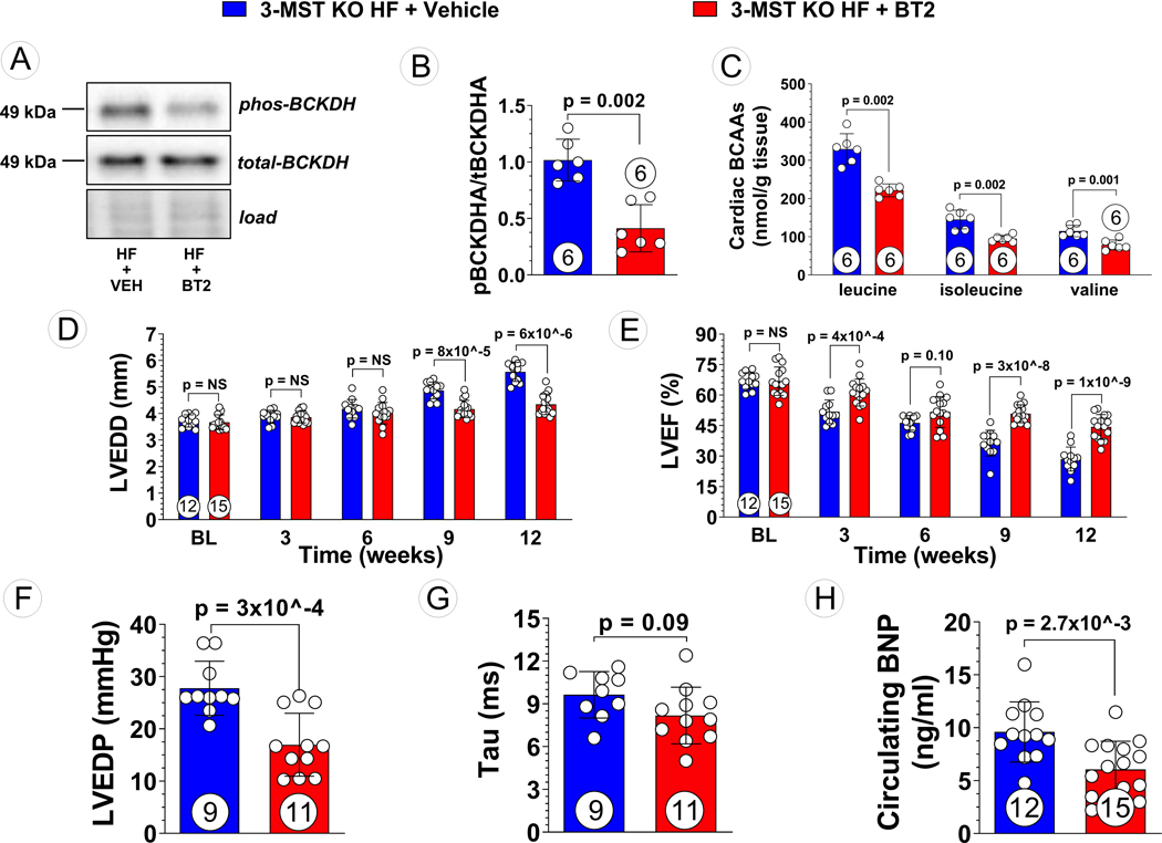
Results: Myocardial 3-MST was significantly reduced in patients with heart failure compared with nonfailing controls. 3-MST KO mice exhibited increased accumulation of branched-chain amino acids in the myocardium, which was associated with reduced mitochondrial respiration and ATP synthesis, exacerbated cardiac and vascular dysfunction, and worsened exercise performance following transverse aortic constriction. Restoring myocardial branched-chain amino acid catabolism with 3,6-dichlorobenzo1[b]thiophene-2-carboxylic acid (BT2) and administration of a potent H2S donor JK-1 ameliorates the detrimental effects of 3-MST deficiency in heart failure with reduced ejection fraction.

Conclusions: Our data suggest that 3-MST derived mitochondrial H2S may play a regulatory role in branched-chain amino acid catabolism and mediate critical cardiovascular protection in heart failure.

Keywords: amino acids, branched-chain; cell death; heart failure; hydrogen sulfide; mitochondrial respiration.


Conflict of interest statement

CONFLICT OF INTEREST DISCLOSURES

Dr. David Lefer serves as scientific consultant for Sulfagenix Inc, a company focusing on developing H2S-based therapy for clinical use. Dr. Lefer also has stock in both NovoMedix and SAJE Pharma, biotech companies that are developing novel therapeutics for cardiovascular diseases. Other authors declare no conflict of interests.

Figures

Figure 1:
Figure 1:. 3-MST and H2S/Sulfane Sulfur Levels in Human Non-Failing Control Hearts and Failing Hearts
(A) Representative Western blots of human 3-MST, (B) Quantification of the Western blots images, (C) sulfane sulfur, and (D) free H2S levels in human myocardial samples obtained from non-failing vs. failing hearts. Circles inside bars indicate samples size. Data were analyzed with student unpaired 2-tailed t test and presented as mean ± SD.
Figure 2:
Figure 2:. Study Timeline of Murine Model of Pressure Overload Heart Failure
Echocardiography was performed prior to the induction of HF by TAC procedure then once every 3 weeks for a duration of 12 weeks. Invasive hemodynamic, vascular reactivity, exercise capacity, mitochondrial function assessments, metabolomic analysis, and other molecular determination were performed at 12 weeks post TAC.
Figure 3:
Figure 3:. Exacerbated Pressure Overload HF in 3-MST KO Mice.
(A) LV end-diastolic diameter (LVEDD) and (B) LV ejection fraction (LVEF) throughout the 12 weeks study for 3-MST KO and wildtype (WT) control mice. (C) LV end-diastolic pressure (LVEDP), (D) LV relaxation constant Tau, (E) circulating B-type natriuretic peptide (BNP), (F) aortic vascular reactivity to acetylcholine (Ach), (G) aortic vascular reactivity to sodium nitroprusside (SNP), and (H) treadmill running duration in 3-MST KO and control mice at 12 weeks post TAC. Circles inside bars indicates samples size. Data in (A), (B), (F) and (G) were analyzed with ordinary 2-way ANOVA; data in other panels were analyzed with student unpaired 2-tailed t test. Data are presented as mean ± SD.
Figure 4:
Figure 4:. Mitochondrial Morphology, Respiration and ATP Synthesis Efficiency in 3-MST KO Mice after TAC
(A) Mitochondrial H2S generation capacity using 3-mercaptopyruvate as substrate in Wild-type and 3-MST KO mice at baseline and 12 weeks post TAC. Circulating (B) Sulfane Sulfur and (C) 8-isoprostane in wildtype or 3-MST KO mice at 12 weeks post TAC. (D) Representative transmission electron microscopy microphotography of cardiac mitochondria from 3-MST KO + HF vs. WT + HF mice. Scale bar at the bottom right corner represents 1 μm. (E) Number of mitochondria per area and (F) circularity of the mitochondria quantified. (G) Relative fold changes in gene expression of atp5b, tfam, mt-co1, Mfn1, Mfn2, Fis1, PINK1, and Parkin. Respiration using (H) pyruvate or (I) carnitine as substrate and (J) ATP synthesis efficiency in mitochondria isolated from wildtype or 3-MST KO hearts. Respiration using (K) pyruvate or (L) carnitine as substrate and (M) ATP synthesis efficiency in mitochondria isolated from wildtype or 3-MST KO skeletal muscle. Circles inside bars indicates samples size. Data in (A), (E), and (F) were analyzed with Mann-Whitney test; data in other panels were analyzed with student unpaired 2-tailed t test. Data are presented as mean ± SD.
Figure 5:
Figure 5:. Metabolomic Analysis of Wildtype and 3-MST KO Hearts
(A) Z-score plot analysis of metabolic changes unique to WT hearts following TAC. (B) Z-score plot analysis of metabolic changes unique to 3-MST KO hearts following TAC. Red arrows represent the significantly elevated levels of BCAA while blue arrows indicate the significant reduction in the downstream metabolites of BCAA. (C) Z-score plot analysis of metabolic changes common to WT and 3-MST KO hearts following TAC. In each plot, the data are shown as standard deviation from the mean of the respective shams. Each dot represents a single metabolite in 1 sample. n=5 per group. (D) Dotplot showing the top 25 enriched metabolite sets for WT hearts following TAC. (E) Dotplot showing the top 25 enriched metabolite sets for 3-MST KO hearts following TAC. For these analyses, metabolites showing significant changes were analyzed using MetaboAnalyst metabolite set enrichment analysis.
Figure 6:
Figure 6:. Reduced BCAA Accumulation and Attenuated Severity of Pressure Overload HF in BT2 Treated 3-MST KO Mice
(A) Representative Western blots of branched-chain ketoacid dehydrogenase kinase (BCKDK) and phosphorylated BCKDK, (B) quantified pBCKDK/BCKDK ratio, and (C) targeted metabolomic analysis of BCAA in vehicle treated vs. ,6-dichlorobenzo1[b]thiophene-2-carboxylaic acid (BT2) treated 3-MST KO mice. (D) LV end-diastolic diameter (LVEDD) and (E) LV ejection fraction (LVEF) throughout the 12 weeks study for vehicle or BT2 treated mice. (F) LV end-diastolic pressure (LVEDP), (G) LV relaxation constant Tau, (H) circulating B-type natriuretic peptide (BNP), in vehicle vs. BT2 treated 3-MST KO mice at 12 weeks post TAC. Circles inside bars indicates samples size. Data in (B) and (C) were analyzed with Mann-Whitney test; data in (D) and (E) were analyzed with ordinary 2-way ANOVA; data in (F) to (H) were analyzed with student unpaired 2-tailed t test. Data are presented as mean ± SD.
Figure 7:
Figure 7:. Ameliorated Vascular Dysfunction, Exercise Intolerance, and Mitochondrial Dysfunction in BT2 Treated 3-MST KO Mice after TAC
(A) Aortic vascular reactivity to acetylcholine (Ach), (B) aortic vascular reactivity to sodium nitroprusside (SNP), (C) treadmill running duration in 3-MST KO mice treated with Vehicle or BT2. Respiration using (D) pyruvate or (E) carnitine as substrate and (F) ATP synthesis efficiency in mitochondria isolated from Vehicle or BT2 treated 3-MST KO hearts. Respiration using (G) pyruvate or (H) carnitine as substrate and (I) ATP synthesis efficiency in mitochondria isolated from from Vehicle or BT2 treated 3-MST KO skeletal muscle. Circles inside bars indicates samples size. Data in (A) and (B) were analyzed with ordinary 2-way ANOVA; data in other panels were analyzed with student unpaired 2-tailed t test. Data are presented as mean ± SD.

Comment in

References

    1. Bozkurt B, Hershberger RE, Butler J, Grady KL, Heidenreich PA, Isler ML, Kirklin JK and Weintraub WS. 2021 ACC/AHA Key Data Elements and Definitions for Heart Failure: A Report of the American College of Cardiology/American Heart Association Task Force on Clinical Data Standards (Writing Committee to Develop Clinical Data Standards for Heart Failure). Circ Cardiovasc Qual Outcomes. 2021;14:e000102. - PMC - PubMed
    1. Heidenreich PA, Albert NM, Allen LA, Bluemke DA, Butler J, Fonarow GC, Ikonomidis JS, Khavjou O, Konstam MA, Maddox TM, Nichol G, Pham M, Pina IL, Trogdon JG, American Heart Association Advocacy Coordinating C, Council on Arteriosclerosis T, Vascular B, Council on Cardiovascular R, Intervention, Council on Clinical C, Council on E, Prevention and Stroke C. Forecasting the impact of heart failure in the United States: a policy statement from the American Heart Association. Circ Heart Fail. 2013;6:606–19. - PMC - PubMed
    1. Nie C, He T, Zhang W, Zhang G and Ma X. Branched Chain Amino Acids: Beyond Nutrition Metabolism. Int J Mol Sci. 2018;19. - PMC - PubMed
    1. Sun H and Wang Y. Branched chain amino acid metabolic reprogramming in heart failure. Biochim Biophys Acta. 2016;1862:2270–2275. - PubMed
    1. Jang C, Oh SF, Wada S, Rowe GC, Liu L, Chan MC, Rhee J, Hoshino A, Kim B, Ibrahim A, Baca LG, Kim E, Ghosh CC, Parikh SM, Jiang A, Chu Q, Forman DE, Lecker SH, Krishnaiah S, Rabinowitz JD, Weljie AM, Baur JA, Kasper DL and Arany Z. A branched-chain amino acid metabolite drives vascular fatty acid transport and causes insulin resistance. Nat Med. 2016;22:421–6. - PMC - PubMed

Publication types