. 2022 May 9;14(5):e24864.
doi: 10.7759/cureus.24864. eCollection 2022 May.

Correlation of Chest X-Ray Scores in SARS-CoV-2 Patients With the Clinical Severity Classification and the Quick COVID-19 Severity Index

Affiliations

Correlation of Chest X-Ray Scores in SARS-CoV-2 Patients With the Clinical Severity Classification and the Quick COVID-19 Severity Index

Vo Tan Duc et al. Cureus. .

Abstract

Objectives This study aimed to assess the role of chest X-ray (CXR) scoring methods and their correlations with the clinical severity categories and the Quick COVID-19 Severity Index (qCSI). Methods We conducted a retrospective study of 159 COVID-19 patients who were diagnosed and treated at the University Medical Center between July and September 2021. Chest X-ray findings were evaluated, and severity scores were calculated using the modified CXR (mCXR), Radiographic Assessment of Lung Edema (RALE), and Brixia scoring systems. The three scores were then compared to the clinical severity categories and the qCSI using Spearman's correlation coefficient. Results Overall, 159 patients (63 males and 96 females) (mean age: 58.3 ± 15.7 years) were included. The correlation coefficients between the mCXR score and the Brixia and RALE scores were 0.9438 and 0.9450, respectively. The correlation coefficient between the RALE and Brixia scores was marginally higher, at 0.9625. The correlation coefficients between the qCSI and the Brixia, RALE, and mCXR scores were 0.7298, 0.7408, and 0.7156, respectively. The significant difference in the mean values of the three CXR scores between asymptomatic, mild, moderate, severe, and critical groups was also noted. Conclusions There were strong correlations between the three CXR scores and the clinical severity classification and the qCSI.

Keywords: brixia score; chest x-ray; chest x-ray (cxr); covid-19; covid-19 severity; rale score.


Conflict of interest statement

The authors have declared that no competing interests exist.

Figures

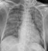
Figure 1
Figure 1. Typical radiographic pattern in a moderate SARS-CoV-2 pneumonia case
This CXR showed reticular, ground-glass opacities and consolidations in bilateral lower and mid-zones. In the bilateral mid-zones, the lesion had a peripheral predominant distribution. The Brixia score was 9, the RALE score was 5, and the mCXR score was 7. This case was classified as moderate pneumonia based on clinical severity. The qCSI was 4.
Figure 2
Figure 2. Atypical radiographic pattern in a moderate SARS-CoV-2 pneumonia case
This CXR showed ground-glass opacity in the right peripheral lower zone and consolidations in the right upper lobe and left basal zone. The Brixia score was 12, the RALE score was 4, and the mCXR score was 8. This case was classified as moderate pneumonia based on clinical severity. The qCSI was 2.
Figure 3
Figure 3. Typical radiographic pattern in a critical case of SARS-CoV-2 pneumonia
This CXR showed ground-glass opacities and consolidations in bilateral, peripheral lower, and mid-zones. The Brixia score was 12, the RALE score was 7, and the mCXR score was 9. This case was classified as a critical situation based on clinical severity. The qCSI was 9.
Figure 4
Figure 4. Atypical radiographic pattern in a critical SARS-CoV-2 pneumonia case
The CXR showed diffuse consolidations in bilateral lung fields with the air bronchogram sign. The Brixia score was 18, the RALE score was 8, and the mCXR score was 12. This case was classified as a critical situation based on clinical severity. The qCSI was 10.

References

    1. Radiographic severity index in COVID-19 pneumonia: relationship to age and sex in 783 Italian patients. Borghesi A, Zigliani A, Masciullo R, Golemi S, Maculotti P, Farina D, Maroldi R. Radiol Med. 2020;125:461–464. - PMC - PubMed
    1. Clinical and laboratory factors associated with coronavirus disease 2019 (Covid-19): a systematic review and meta-analysis. Minh LH, Abozaid AA, Ha NX, et al. Rev Med Virol. 2021;31:0. - PMC - PubMed
    1. Chest radiography in COVID-19: no role in asymptomatic and oligosymptomatic disease. Schaefer-Prokop C, Prokop M. Radiology. 2021;298:0–7. - PMC - PubMed
    1. Chest X-ray for predicting mortality and the need for ventilatory support in COVID-19 patients presenting to the emergency department. Balbi M, Caroli A, Corsi A, et al. Eur Radiol. 2021;31:1999–2012. - PMC - PubMed
    1. Development and validation of the quick COVID-19 severity index: a prognostic tool for early clinical decompensation. Haimovich AD, Ravindra NG, Stoytchev S, et al. Ann Emerg Med. 2020;76:442–453. - PMC - PubMed

LinkOut - more resources