Review
. 2022;6(7):451-469.
doi: 10.1038/s41570-022-00393-7. Epub 2022 Jun 10.

The chemistry of snake venom and its medicinal potential

Affiliations
Review

The chemistry of snake venom and its medicinal potential

Ana L Oliveira et al. Nat Rev Chem. 2022.

Abstract

The fascination and fear of snakes dates back to time immemorial, with the first scientific treatise on snakebite envenoming, the Brooklyn Medical Papyrus, dating from ancient Egypt. Owing to their lethality, snakes have often been associated with images of perfidy, treachery and death. However, snakes did not always have such negative connotations. The curative capacity of venom has been known since antiquity, also making the snake a symbol of pharmacy and medicine. Today, there is renewed interest in pursuing snake-venom-based therapies. This Review focuses on the chemistry of snake venom and the potential for venom to be exploited for medicinal purposes in the development of drugs. The mixture of toxins that constitute snake venom is examined, focusing on the molecular structure, chemical reactivity and target recognition of the most bioactive toxins, from which bioactive drugs might be developed. The design and working mechanisms of snake-venom-derived drugs are illustrated, and the strategies by which toxins are transformed into therapeutics are analysed. Finally, the challenges in realizing the immense curative potential of snake venom are discussed, and chemical strategies by which a plethora of new drugs could be derived from snake venom are proposed.

Keywords: Biochemistry; Drug discovery.


Conflict of interest statement

Competing interestsThe authors declare no competing interests.

Figures
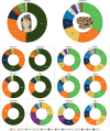
Fig. 1
Fig. 1. Composition of the venom of snakes from the Elapidae and Viperidae families.
The large charts show the averaged composition of the venom of snake species from the Elapidae (elapids) or Viperidae (viperids) families. Each entry in the charts corresponds to a protein family, in which we group tens to hundreds of isoforms. Only protein families with an average abundance of >1% of the total venom proteome are represented, except for the SVSPs in elapids, which are included for comparison with the viperids, and defensins, which although seldom present, can be abundant in the venom of certain species. The distribution of the proportion of the most abundant protein families is shown in Supplementary Fig. 1. Data are from the proteomic studies of the past 15 years; 143 entries for 2007–2017 are from Isbister and Tasoulis’s database of snake venom proteomes; we assembled the additional entries for 2017–2021 from the literature. The Atractaspididae and Colubridae snake families are not included in the study because most are non-venomous or their venoms are weak, not medically important and poorly studied (for venomics studies on colubrids see ref.). Each species contributes with the same weight to the average; subspecies or species from different locations were averaged within the entry for the species. The entry ‘Other’ corresponds to unidentified components or components with an average abundance of <1%. The smaller charts decompose the snake venom composition at the genus level, which reveals the compositional diversity. Only the most well-studied genera are included, which comprise almost all the medically relevant snakes. Supplementary Table 1 details the composition of the venom of each species included in the study together with the relevant reference. 3FTx, three-finger toxin; CRiSP, cysteine-rich secretory protein; CTL/SNACLEC, C-type lectin and C-type lectin-like protein; DEF, defensin; DIS, disintegrin; KSPI, Kunitz-type serine protease inhibitor; LAAO, l-amino acid oxidase; NP, natriuretic peptide; PLA2, phospholipase A2; SVMP, snake venom metalloproteinase; SVSP, snake venom serine protease.
Fig. 2
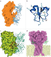
Fig. 2. The three main types of PLA2 bound to their targets.
a | Myotoxin I (MT-I), a strongly myotoxic phospholipase A2 (PLA2) from the venom of a terciopelo viper (Bothrops asper), attached to the sarcolemma. MT-I (PDB ID: 5TFV) is shown with a phospholipid substrate bound to the active centre. In the phospholipid, oxygen is red, phosphorus is orange, nitrogen is blue, carbon is grey and hydrogen is white; the enzyme is shown in light red, and the Ca2+ ion is shown in light green. The residues that form the protein–membrane interface and the PLA2–membrane binding geometry were identified through mutagenesis, fluorescence and X-ray crystallography studies,. b | The PLA2 homologue myotoxin II (MT-II), also from terciopelo venom (PDB ID: 1Y4L), bound to the sarcolemma. The C-terminal region destabilizes and permeabilizes the membrane. The protein is shown in light green, and the C-terminal KKYRYYLKPLCKK sequence is shown in pink. c | β-Bungarotoxin (PDB ID: 1 BUN) from the Taiwan banded krait (Bungarus multicinctus) bound to a neuronal membrane. The toxin travels silently through the victim’s body until its Kunitz (KUN) domain (green) recognizes and binds a presynaptic voltage-gated K+ channel (violet, PDB ID:6PBX) with high specificity, trapping the PLA2 domain (light blue) at the neuronal membrane, where its active site, otherwise occluded, opens and starts degrading the adjacent phospholipids (bound phospholipid coloured by element).
Fig. 3
Fig. 3. Structures of SVMPs and their substrates.
a | The structure of the factor X activating enzyme RVV-X (PDB ID: 2E3X) from the eastern Russell’s viper (Daboia siamensis). RVV-X is a P-III snake venom metalloproteinase (SVMP) isoform that is ubiquitous in species from the Indian subcontinent. RVV-X activates blood coagulation factor X by hydrolysing the Arg194–Ile195 position with such high specificity that it is used as a diagnostic tool for haematologic disorders,,,. The catalytic domain (MET) is coloured yellow, the disintegrin (DIS) domain is coloured green and the cysteine-rich domain (CR) is coloured pink. RVV-X has an additional C-type lectin and C-type lectin-like protein (CLT/SNACLEC) domain, which is shown in blue. The inset shows the Zn2+ cofactor with its coordination shell and the peptidomimetic inhibitor GM6001, whose two coordinated oxygen atoms mimic the positions of the water molecule and the carbonyl of the substrate (superimposed on top of GM6001 with a translucent ball and stick representation). b | Illustrative scheme of daborhagin, a highly haemorrhagic SVMP from Russell’s viper venom, bound to collagen IV at the basement membrane of capillaries. The colour scheme of the enzyme domains is the same as that of RVV-X in part a. A collagen IV fibre is shown in light green, with a tropocollagen unit emphasized in dark green and drawn in a cartoon and tube representation. The hydrolysis of collagen IV weakens the mechanical stability of the capillary wall, which breaks down under regular haemodynamic forces, leading to massive haemorrhage. Daborhagin was modelled with the active site facing collagen IV.
Fig. 4
Fig. 4. Structure of SVSPs and their substrates.
a | The factor V activating enzyme from Russell’s viper venom (RVV-V; PDB ID: 3S9C), which is a snake venom serine protease (SVSP) with specificity for blood coagulation factor V. RVV-V is depicted in a complex with the 14-residue terminal fragment of factor Va (residues 1533–1546), called FV14. RVV-V releases the last 61 residues of factor V by hydrolysing its Arg1545–Ser1546 bond, generating procoagulant factor Va and mimicking one of the physiological roles of thrombin. The inset shows the active site and factor V hydrolysis product. RVV-V recognizes factor V through a selective induced-fit mechanism that opens an otherwise closed subpocket. The strict specificity of RVV-V for factor V makes it a useful diagnostic tool for measuring factor V levels, lupus anticoagulant levels and resistance to activated protein C,. b | Illustrative representation of the thrombin-like Brazilian lancehead pit viper (Bothrops moojeni) SVSP batroxobin (Defibrase),,,, bound to fibrinogen. Thrombin cleaves the Aα and the Bβ chains of fibrinogen and converts factor XIII into factor XIIIa, which generates crosslinked fibrin, whereas most SVSPs cleave either the Aα or the Bβ chain only. Batroxobin cleaves only the Aα chain,. SVSPs therefore form abnormal, easily degradable fibrin clots that lead to fibrinogen depletion and hypofibrinogenaemia. The clotting time in the presence of batroxobin (reptilase time) is used in the clinic to diagnose several diseases,. Batroxobin was modelled from the homologue saxthrombin (PDB ID: 3S69). c | Collinein-1 from the neotropical rattlesnake (Crotalus durissus collilineatus) is the first example of an SVSP with specific K+-channel blocking activity. Through a mechanism that is independent of its enzyme activity, collinein-1 selectively inhibits the oncogenic hEAG1 channel (PDB ID:6PBX) among 12 tested voltage-gated K+-channels, with obvious antitumour implications. As K+ channels are known targets for many animal neurotoxins, the discovery of collinein-1 makes it tempting to speculate that some yet unknown SVSP isoforms might have found a neurotoxic role. Collinein-1 was modelled from the homologue thrombin-like enzyme AhV_TL-I (PDB ID: 4E7N) and is illustratively bound to the oncogenic hEAG1 channel.
Fig. 5
Fig. 5. Approved drugs derived from snake venoms.
Several drugs derived from snake venoms have been approved by the US Food and Drug Administration (FDA) and the European Medicines Agency (EMA). The chemical structures of these drugs are shown, with the region that mimics the snake toxin highlighted in grey. a | Nine hypotensive bradykinin potentiating peptides (BPPs) were isolated from the venom of the jararaca viper; they inspired the design of the antihypertensive drugs captopril and enalapril. These drugs mimic the Trp–Ala–Pro (WAP) motif by which BPP5a (top right) recognizes its target: the angiotensin-converting enzyme (ACE). ACE is shown on the left in a complex with BPP5b, another BPP (PDB ID: 6QS1). The ACE Zn2+ cofactor is shown in orange. b | The drug tirofiban was inspired by a disintegrin called echistatin found in the venom of the saw-scaled viper. Echistatin, shown on the left (PDB ID: 6LSQ), binds specifically to the αIIBβ3 integrin through its Arg–Gly–Asp (RGD) motif (coloured spheres and top right), which prevents platelet aggregation. In tirofiban, the piperidine moiety replicates arginine, the aliphatic linker replicates glycine, and the tyrosine carboxyl group replicates the aspartic acid carboxylate. The (S)-NHSO2nC4H9 group increases the affinity of tirofiban for its αIIBβ3 target. c | Eptifibatide is an antiplatelet drug inspired by the disintegrin babourin purified from the venom of Barbour’s pygmy rattlesnake. A homology model of babourin (template PDB ID: 1J2L) is shown on the left. Most disintegrins recognize the αIIBβ3 integrin through the RGD motif, but babourin uses a Lys–Gly–Asp (KGD) motif (coloured spheres and top right). Eptifibatide achieves maximum selectivity owing to the fusion of the two motifs into the unnatural homoRGD motif. Additional peripheral residues and cyclization endow further molecular recognition capabilities and resistance to proteolysis.
Fig. 6
Fig. 6. Drugs derived from snake venoms in clinical or preclinical trials.
a | Anfibatide (blue cartoon) is a snake C-type lectin-like protein that is predicted to bind to platelet glycoprotein Ib α-chain GPIbα (orange surface) at a site that partially overlaps with the GPIbα–von Willebrand factor binding surface (PDB ID: 1SQ0), thus inhibiting the association of von Willebrand factor and consequently platelet aggregation. Anfibatide is a promising anticoagulation candidate that has passed phase I clinical trials. b | Crotamine is an amphipathic and highly basic defensin that penetrates cells and is resistant to proteolysis. Crotamine exhibits antiproliferative, antinociceptive and analgesic activity in vivo upon oral administration. Cationic residues are shown as sticks and the disulfide bonds are shown in yellow. c | Dendroaspis natriuretic peptide (DNP) from the eastern green mamba (ochre tube with the disulfide bond in yellow) bound to the dimeric particulate guanylyl cyclase A receptor (shown as a lime surface and a green transparent cartoon) (PDB ID: 7BRI). Cenderitide is a natriuretic peptide chimaera resulting from the fusion of human C-type natriuretic peptide (CNP) to DNP and co-activates both DNP and CNP transmembrane receptors. d | The three-finger toxins mambalgin-1 and mambalgin-2 bind to the acid-sensing ion channels 1a and 1b, locking the channels in the closed state and impairing their function, with an analgesic effect as potent as that of morphine but with much lower toxicity in rodents. The complex of mambalgin-1 (green) with the transmembrane (light yellow) acid-sensing ion channel 1a (violet) is shown (PDB ID: 7CFT). The mambalgins are promising scaffolds for the development of a new generation of analgesics.

References

    1. Holford M, Daly M, King GF, Norton RS. Venoms to the rescue. Science. 2018;361:842–844. doi: 10.1126/science.aau7761. - DOI - PubMed
    1. Casewell NR, Wüster W, Vonk FJ, Harrison RA, Fry BG. Complex cocktails: the evolutionary novelty of venoms. Trends Ecol. Evol. 2013;28:219–229. doi: 10.1016/j.tree.2012.10.020. - DOI - PubMed
    1. King GF. Venoms as a platform for human drugs: translating toxins into therapeutics. Expert Opin. Biol. Ther. 2011;11:1469–1484. doi: 10.1517/14712598.2011.621940. - DOI - PubMed
    1. Herzig V, et al. Animal toxins — nature’s evolutionary-refined toolkit for basic research and drug discovery. Biochem. Pharmacol. 2020;181:114096. doi: 10.1016/j.bcp.2020.114096. - DOI - PMC - PubMed
    1. Pineda SS, et al. Structural venomics reveals evolution of a complex venom by duplication and diversification of an ancient peptide-encoding gene. Proc. Natl Acad. Sci. USA. 2020;117:11399–11408. doi: 10.1073/pnas.1914536117. - DOI - PMC - PubMed