Review
. 2022 Jun;12(6):210395.
doi: 10.1098/rsob.210395. Epub 2022 Jun 15.

MicroRNAs: master regulators in host-parasitic protist interactions

Affiliations
Review

MicroRNAs: master regulators in host-parasitic protist interactions

Maura Rojas-Pirela et al. Open Biol. 2022 Jun.

Abstract

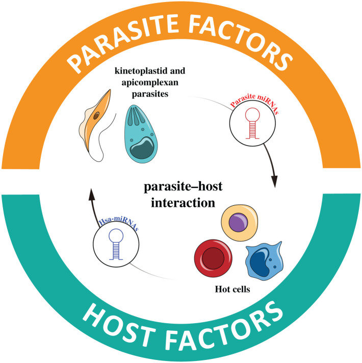
MicroRNAs (miRNAs) are a group of small non-coding RNAs present in a wide diversity of organisms. MiRNAs regulate gene expression at a post-transcriptional level through their interaction with the 3' untranslated regions of target mRNAs, inducing translational inhibition or mRNA destabilization and degradation. Thus, miRNAs regulate key biological processes, such as cell death, signal transduction, development, cellular proliferation and differentiation. The dysregulation of miRNAs biogenesis and function is related to the pathogenesis of diseases, including parasite infection. Moreover, during host-parasite interactions, parasites and host miRNAs determine the probability of infection and progression of the disease. The present review is focused on the possible role of miRNAs in the pathogenesis of diseases of clinical interest caused by parasitic protists. In addition, the potential role of miRNAs as targets for the design of drugs and diagnostic and prognostic markers of parasitic diseases is also discussed.

Keywords: apicomplexan; diagnostic and therapeutic tools; host–parasite interactions; kinetoplastids; microrna.


Figures

Figure 1.
Figure 1.
MicroRNA biogenesis pathways. (a) Canonical pathway: miRNAs are synthesized by RNApolII and processed in the nucleus by the microprocessor complex (Drosha-DGCR8). Pre-miRNA is exported to the cytosol through Exportin-5 (Exp-5)-RanGTPase. In the cytoplasm, DICER processes miRNAs to bind to the RISC-AGO. (b) Non-canonical pathways: miRNAs are processed through microprocessor-independent, TUT-dependent or DICER-independent pathways. In the cytoplasm, all of them are modified by DICER to bind to the RISC-AGO complex, except for DICER-independent processing that binds directly to AGO2 in the cytosol.
Figure 2.
Figure 2.
MicroRNAs’ location and functions in the cell. (a) Cytoplasm: post-transcriptional silencing in the cytoplasm is the classic function mediated by miRNA via RISC. (b) Nucleus: regulatory mechanisms of nuclear miRNAs. (c) Mitochondria: mitochondrial miRNA (MitomiR)-targeting mitochondrial mRNAs. MiRNAs are synthesized in the nucleus (mitomiRNAs) and are imported into the mitochondria. (d) Endoplasmic reticulum: repression of translation occurs at MBP. (e) P-bodies: miRNAs are involved in P-bodies formation. (f) Golgi apparatus: MiRNAs are involved in resistance to and trans-Golgi or the RT. (g) Extracellular vesicles: MiRNA in cell-to-cell communication. EVs released by donor cells can fuse directly with the plasma membrane of recipient cells to discharge their content.
Figure 3.
Figure 3.
MicroRNAs in T. cruzi–host interaction. Regulatory mechanisms of miRNA genes in different organs during T. cruzi pathogenesis. Cell diagram: cardiac cells (Red portion). T. cruzi infection induces in cardiomyocytes the dysregulation of some miRNAs involved in regulating fatty acid metabolism and some pathways related to the immune response during pathogenesis. The downregulation of some miRNAs, such as hsa-miR-322, positively regulates the expression of FGF2 and FGFR1 mRNA. The products of these genes, FGF2 and FGR1 proteins, are incorporated into the host cell's plasma membrane and could be a possible target for parasite–host interaction. Additionally, infection by T. cruzi can induce tolerance. Thymus cell (Blue portion). Parasite infection causes upregulation of some miRNAs, including mmu-let-7a, mmu-let-7 g, mmu-miR-101a, mmu-miR-148b and mmu-miR-193 in.TEC. Several of these miRNAs repress the expression of TGF-β and its receptor TGFBR1, impairing the normal function of the thymus and thus affecting some processes such as ontogeny, negative selection and the accumulation of autoreactive lymphocytes involved in autoimmune diseases. Placenta cells (brown portion). T. cruzi infection of cytotrophoblasts induces the dysregulation of C19MC miRNAs and immunomiRs. The upregulation of hsa-miR-512-3p is responsible for activating the caspase 8 pathway shown to be involved in trophoblast differentiation and apoptosis mechanisms. The upregulation of this miRNA is possibly associated with the autophagy induction in the infected cell. Also, hsa-miR-515-5p downregulation allows the expression of some genes crucial for the differentiation of human trophoblasts during T. cruzi infection. Under an inflammatory environment, the upregulation of immunomiRs (hsa-miR-21, hsa-miR-146a and hsa-miR-210) occur, influenced by the NF-kB pathway.
Figure 4.
Figure 4.
MicroRNAs in leishmania–host interaction. (a) miRNAs during Leishmania donovani infection. miR-30 family miRNAs govern several processes during L. donovani infection of macrophages, including the repression of autophagy-related proteins BECN1, ATG3 and ATG9. Post-transcriptional regulation of inflammatory products by hsa-miR-210 also occurs. Additionally, the accumulation and availability of iron (orange dots) and the overexpression of ABC transporters responsible for drug efflux are also modulated by miRNAs. Alternatively, L. donovani infection influences miRNA expression in T cells that compromise cell polarization. (b) miRNA dysregulation and target genes during infection of macrophages with different species of leishmania. (c) Targets of miRNA-like molecules identified through in silico studies in Leishmania major.
Figure 5.
Figure 5.
(a) MicroRNAs during plasmodium pathogenesis. (a) CM. Plasmodium-infected RBCs accumulate in the brain blood vessels and cross the BBB. It causes the alteration of brain cells' microRNAs and their function regarding proliferation and adhesion of cells, signalling pathways, apoptosis and carbohydrate metabolism. On the other hand, the release of EVs by host cells is involved in inflammation and BBB dysfunction, mainly because of hsa-miR-146a, hsa-miR-193b and hsa-miR-155. (b). MicroRNAs in NCB. Several events are caused by miRNA dysregulation in the parasite, the erythrocyte and endothelial cells. Inside the parasite, miRNAs affect directly ribosomal loading and translation, invasion process, survival and gametogenesis (hsa-miR-451), but also the microtubules stability (hsa-miR-157-5p) and the parasite replication due to the action of imported human AGO (hAGO2) and the regulation of Rad54 and the Lipid/sterol: H symporter by hsa-let-7a and hsa-miR-15a. In iRBC, the release of EVs containing hAG2-hsa-miR-451a/hsa-let-7b (upper right box) fuse with the recipient EC to induce the expression of V-CAM molecules that are required to interact with iRBC through pfEMP1 on their surface. Additionally, ECs release IL-1 and IL-6, which promote vascular dysfunction. (c) MiRNAs during plasmodium pathogenesis in the placenta and liver. (i) Liver. Parasite infection downregulates various miRNAs associated with lipid metabolism. Alteration of the expression of some miRNAs such as hsa-miR-192 and hsa-miR-98, promotes overexpression of sterol-regulatory element-binding proteins (SRBPE1/2) and the subsequent lipid accumulation in liver tissue, which causes the downregulation of hepatocyte nuclear factor 4 alpha (HNF4-α) and hsa-miR-101 and hsa-miR-192. In addition, hsa-miR-101 downregulation induces the expression of a histone-lysine methyltransferase (EZH2) involved in the epigenetic regulation of immunological genes and the maintenance of negative feedback that keeps hsa-miR-101 downregulated. Dysregulation of miRNA during P. chabaudi in mice and miRNA altered during vaccination are shown at the right of (ii). (ii) Placenta. Infected cytotrophoblasts release EVs containing hsa-miR-517c (C19MC) involved in immune pathology, specifically pre-eclampsia and spontaneous abort. Genetic constitution is also relevant since homozygosity can increase parasite infection.
Figure 5.
Figure 5.
(a) MicroRNAs during plasmodium pathogenesis. (a) CM. Plasmodium-infected RBCs accumulate in the brain blood vessels and cross the BBB. It causes the alteration of brain cells' microRNAs and their function regarding proliferation and adhesion of cells, signalling pathways, apoptosis and carbohydrate metabolism. On the other hand, the release of EVs by host cells is involved in inflammation and BBB dysfunction, mainly because of hsa-miR-146a, hsa-miR-193b and hsa-miR-155. (b). MicroRNAs in NCB. Several events are caused by miRNA dysregulation in the parasite, the erythrocyte and endothelial cells. Inside the parasite, miRNAs affect directly ribosomal loading and translation, invasion process, survival and gametogenesis (hsa-miR-451), but also the microtubules stability (hsa-miR-157-5p) and the parasite replication due to the action of imported human AGO (hAGO2) and the regulation of Rad54 and the Lipid/sterol: H symporter by hsa-let-7a and hsa-miR-15a. In iRBC, the release of EVs containing hAG2-hsa-miR-451a/hsa-let-7b (upper right box) fuse with the recipient EC to induce the expression of V-CAM molecules that are required to interact with iRBC through pfEMP1 on their surface. Additionally, ECs release IL-1 and IL-6, which promote vascular dysfunction. (c) MiRNAs during plasmodium pathogenesis in the placenta and liver. (i) Liver. Parasite infection downregulates various miRNAs associated with lipid metabolism. Alteration of the expression of some miRNAs such as hsa-miR-192 and hsa-miR-98, promotes overexpression of sterol-regulatory element-binding proteins (SRBPE1/2) and the subsequent lipid accumulation in liver tissue, which causes the downregulation of hepatocyte nuclear factor 4 alpha (HNF4-α) and hsa-miR-101 and hsa-miR-192. In addition, hsa-miR-101 downregulation induces the expression of a histone-lysine methyltransferase (EZH2) involved in the epigenetic regulation of immunological genes and the maintenance of negative feedback that keeps hsa-miR-101 downregulated. Dysregulation of miRNA during P. chabaudi in mice and miRNA altered during vaccination are shown at the right of (ii). (ii) Placenta. Infected cytotrophoblasts release EVs containing hsa-miR-517c (C19MC) involved in immune pathology, specifically pre-eclampsia and spontaneous abort. Genetic constitution is also relevant since homozygosity can increase parasite infection.
Figure 6.
Figure 6.
(a) MicroRNAs during toxoplasma pathogenesis. (a) Role of T. gondii-infected human and porcine macrophage miRNAs. During infection of porcine macrophages by toxoplasma RH/ME49, various miRNAs regulate the expression of NO synthesis and FCγR signalling pathway by directly targeting mRNAs of NOS1/NOS3, Rac2 and Vav3. In human macrophages, the parasites in the PV release ROP16, a protein kinase that phosphorylates and activates the transcription factor STAT3. The activated factor travels to the nucleus and stimulates the miR-17-92 cluster; this miR cluster is responsible for post-transcriptional regulation of the mRNA of the pro-apoptotic protein ‘Bim’ through the binding to the 3′-UTR region of the mRNA molecule. This process prevents the infected macrophage from activating the apoptosis pathway during toxoplasma infection, thus allowing the survival of the parasites. (b)(i) Role of miRNAs in the pathogenesis of T. gondii in the placenta. (ii) In toxoplasma-infected placenta (cytotrophoblasts), dysregulation of immunomiRs, and the C14MC and C19MC clusters inhibit the NF-kB response and alter the STAT-dependent IL-4 response and pDC differentiation, respectively. (iii) In pDCs, the downregulation of hsa-miR-127 during toxoplasma infection elicit an altered migration by these cells, causing them to function as Trojan horses to cross the placental barrier and reach the fetus environment.
Figure 7.
Figure 7.
MiRNAs and their role in parasite–host interaction: MiRNAs modulate parasite and host responses during parasite infection, determining disease probability. In the parasite, miRNAs modulate cellular proliferation, differentiation, metabolism and drug resistance. In the host, miRNAs play essential roles in host defence mechanisms.

Similar articles

Cited by

References

    1. Wu J, Xiao J, Zhang Z, Wang X, Hu S, Yu J. 2014. Ribogenomics: the science and knowledge of RNA. Genomics Proteomics Bioinform. 12, 57-63. (10.1016/j.gpb.2014.04.002) - DOI - PMC - PubMed
    1. Beermann J, Piccoli MT, Viereck J, Thum T. 2016. Non-coding RNAs in development and disease: background, mechanisms, and therapeutic approaches. Physiol. Rev. 96, 1297-1325. (10.1152/physrev.00041.2015) - DOI - PubMed
    1. Zhang P, Wu W, Chen Q, Chen M. 2019. Non-coding RNAs and their integrated networks. J. Integr. Bioinform. 16, 20190027. (10.1515/jib-2019-0027) - DOI - PMC - PubMed
    1. Patil VS, Zhou R, Rana TM. 2014. Gene regulation by noncoding RNAs. Crit. Rev. Biochem. Mol. Biol. 49, 16-32. (10.3109/10409238.2013.844092) - DOI - PMC - PubMed
    1. Chen T, et al. 2015. m(6)A RNA methylation is regulated by microRNAs and promotes reprogramming to pluripotency. Cell Stem Cell 16, 289-301. (10.1016/j.stem.2015.01.016) - DOI - PubMed

Publication types

LinkOut - more resources