. 2022 Jun 15;132(12):e155914.
doi: 10.1172/JCI155914.

Hematopoietic stem cell regeneration through paracrine regulation of the Wnt5a/Prox1 signaling axis

Affiliations

Hematopoietic stem cell regeneration through paracrine regulation of the Wnt5a/Prox1 signaling axis

Qiqi Lin et al. J Clin Invest. .

Abstract

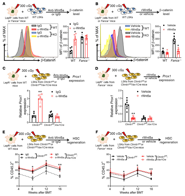
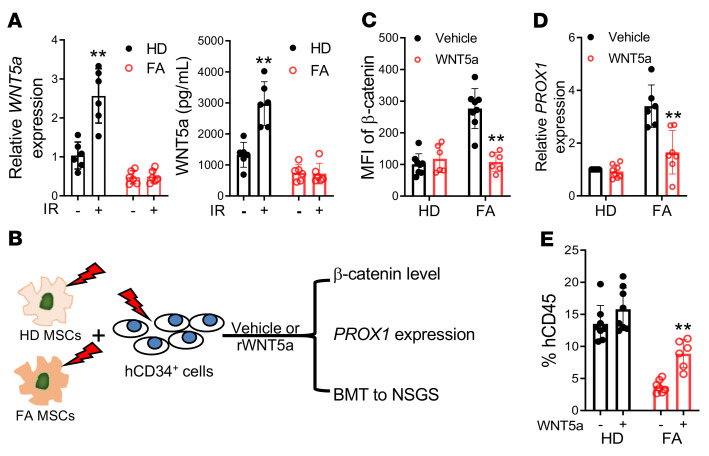
The crosstalk between the BM microenvironment (niche) and hematopoietic stem cells (HSCs) is critical for HSC regeneration. Here, we show that in mice, deletion of the Fanconi anemia (FA) genes Fanca and Fancc dampened HSC regeneration through direct effects on HSCs and indirect effects on BM niche cells. FA HSCs showed persistent upregulation of the Wnt target Prox1 in response to total body irradiation (TBI). Accordingly, lineage-specific deletion of Prox1 improved long-term repopulation of the irradiated FA HSCs. Forced expression of Prox1 in WT HSCs mimicked the defective repopulation phenotype of FA HSCs. WT mice but not FA mice showed significant induction by TBI of BM stromal Wnt5a protein. Mechanistically, FA proteins regulated stromal Wnt5a expression, possibly through modulating the Wnt5a transcription activator Pax2. Wnt5a treatment of irradiated FA mice enhanced HSC regeneration. Conversely, Wnt5a neutralization inhibited HSC regeneration after TBI. Wnt5a secreted by LepR+CXCL12+ BM stromal cells inhibited β-catenin accumulation, thereby repressing Prox1 transcription in irradiated HSCs. The detrimental effect of deregulated Wnt5a/Prox1 signaling on HSC regeneration was also observed in patients with FA and aged mice. Irradiation induced upregulation of Prox1 in the HSCs of aged mice, and deletion of Prox1 in aged HSCs improved HSC regeneration. Treatment of aged mice with Wnt5a enhanced hematopoietic repopulation. Collectively, these findings identified the paracrine Wnt5a/Prox1 signaling axis as a regulator of HSC regeneration under conditions of injury and aging.

Keywords: Cell Biology; Hematology; Hematopoietic stem cells.


Figures

Figure 1
Figure 1. FA deficiency compromises hematologic recovery after irradiation.
(A) PB WBC, neutrophil (NEU), and lymphocyte (LYMPH) counts in WT, Fanca–/–, or Fancc–/– mice at the indicated time points after 500 cGy TBI (n = 12/group). (B) Left: representative microscopic H&E images (20× original magnification) of BM cellularity in mice described in A on day +15. Right: mean BM cell counts per femur for each group. Results are mean ± SEM of 3 independent experiments (n = 12/group). (C) Left: representative flow cytometry analysis of percentages of BM LSK (LinSca1+ckit+) cells in the mouse groups shown on day +15. Right: mean numbers of BM LSK cells in each group on day +15 (n = 9). (D) Left: representative flow cytometry analysis of percentages of BM SLAM (LSKCD48CD150+) cells in the mouse groups shown on day +15. Right: mean numbers of BM SLAM cells in each group on day +15 (n = 9). (E) Mean numbers of BM colony forming cells (CFCs) in the groups shown on day +15 after 500 cGy TBI (n = 12). (F) Percentage survival of the mouse groups shown through day +30 after 500 cGy TBI (P < 0.01 for WT versus Fanca–/– and Fancc–/– mice; log-rank test for survival analysis; WT: n = 10; Fanca–/– and Fancc–/–: n = 15). Statistics were performed in the indicated groups: 2-tailed, paired t test (parametric). *P < 0.05; **P < 0.01.
Figure 2
Figure 2. FA deficiency dampens HSC regeneration after irradiation.
(A) Donor (CD45.2+) cell engraftment at 16 weeks in recipient CD45.1+ mice that were transplanted with 100 BM SLAM cells from nonirradiated and irradiated WT, Fanca–/–, and Fancc–/– mice, along with 2 × 10⁵ competing CD45.1+ WT BM cells. Representative flow cytometry analysis (left) and quantification (right) are shown (WT TBI–: n = 6; WT TBI+: n = 8; Fanca–/– TBI–: n = 6; Fancc–/– TBI+: n = 7; Fancc–/–: n = 6). (B) Donor myeloid (Mac1/Gr1), B cell (B220), and T cell (CD3ε) engraftment levels at 16 weeks are shown (WT TBI–: n = 6; WT TBI+: n = 8; Fanca–/– TBI–: n = 6; Fancc–/– TBI+: n = 7; Fancc–/–: n = 6). (C) Donor HSC engraftment at 16 weeks. Mean percentages of CD45.2+ SLAM cells are shown for each group (WT TBI–: n = 6; WT TBI+: n = 8; Fanca–/– TBI–: n = 6; Fancc–/– TBI+: n = 7; Fancc–/–: n = 6). (D and E) Mean levels of donor CD45.2+ cell (D) and lineage engraftment (E) in secondary recipient CD45.1+ mice at 16 weeks after competitive transplantation with BM cells from the primary mice in A (WT TBI–: n = 8; WT TBI+: n = 10; Fanca–/–: n = 8; Fancc–/–: n = 8 in D; WT TBI–: n = 8; WT TBI+: n = 10; Fanca–/–: n = 8; Fancc–/–: n = 8 in E). Statistics were performed in the indicated groups: 2-tailed, paired t test (parametric). *P < 0.05; **P < 0.01.
Figure 3
Figure 3. Deletion of Prox1 improves long-term repopulation of irradiated FA HSCs.
(A) FA HSCs show persistent upregulation of Prox1 in response to TBI. RNA extracted from SLAM cells isolated from the indicated mice at different time points after 500 cGy TBI was analyzed with qPCR. Samples were normalized to the level of WT Gapdh mRNA at day 0 (n = 6). (B) Schematic diagram of experimental design. (C) Deletion of Prox1 improves repopulation of irradiated FA HSCs. LSK (LinSca1+c-kit+) cells from the indicated mice were subjected to irradiation and transplanted along with competitor cells into BoyJ recipients. Donor-derived chimera were determined at 16 weeks after BMT. Prox1fl/fl: n = 8; Prox1fl/flVav1-Cre: n = 9; Fanca–/–;Prox1fl/fl: n = 8; Fanca–/–;Prox1fl/flVav1-Cre: n = 8; Fancc–/–;Prox1fl/fl: n = 9; Fanc9–/–;Prox1fl/flVav1-Cre: n = 9. (D) Loss of Prox1 improves long-term repopulation capacity of FA HSCs. WBMCs from recipients described in C were transplanted into BoyJ recipients. Prox1fl/fl: n = 8; Prox1fl/flVav1-Cre: n = 9; Fanca–/–;Prox1fl/fl: n = 8; Fanca–/–;Prox1fl/flVav1-Cre: n = 8; Fancc–/–;Prox1fl/fl: n = 10; Fanc9–/–;Prox1fl/flVav1-Cre: n = 10. (E) Schematic presentation of experimental design. (F) Forced expression of Prox1 mimics FA HSC phenotype in transplanted recipients. LSK cells from WT mice were transduced with lentiviral vector expressing GFP or GFP-Prox1. Sorted GFP+ cells were subjected to 300 cGy irradiation and transplanted into BoyJ recipients. Vector, TBI–: n = 8; vector, TBI+: Prox1 TBI–: n = 7; Prox1 TBI+: n = 6. (G) Ectopic overexpression of Prox1 compromises long-term reconstitution of WT HSCs. WBMCs from the recipients described in F were transplanted into sublethally irradiated BoyJ recipients (n = 9). Statistics were performed in the indicated groups: 2-tailed, paired t test (parametric). *P < 0.05; **P < 0.01; ***P < 0.001.
Figure 4
Figure 4. Wnt5a from LepR+CXCL12+ cells regulates hematopoietic recovery and HSC regeneration after irradiation.
(A) Wnt5a expression in LepR+ cells at 24 hours after 500 cGy TBI (n = 6). (B) Wnt5a protein levels in the BM supernatants of the indicated mice at 24 hours after TBI (n = 6). (C) Mouse PB parameters at day 22 after TBI. Mice were administered 500 cGy TBI and treated i.p. with rWnt5a (50 μg/kg) or vehicle (saline). WT, vehicle: n = 7; WT rWnt5a: n = 9; Fanca–/–, vehicle: n = 6; Fanca–/– rWnt5a: n = 6. (D) Total BM cells (left) and SLAM cells (right) in mice described in C. WT, vehicle: n = 7; WT rWnt5a: n = 9; Fanca–/–, vehicle: n = 6; Fanca–/– rWnt5a: n = 6. (E) First (n = 8) and second (n = 10) BMT with cells from mice described in C (first, 8; second, 10). (F) Effect of anti-Wnt5a neutralization on HSC expansion. WT SLAM cells were subjected to 300 cGy irradiation and cocultured with WT LepR+ cells and anti-Wnt5a (2 μg/mL) or control IgG for 5 days (IgG: n = 9; α-Wnt5a: n = 6). (G) Neutralization of Wnt5a dampens the repopulation capacity of the irradiated WT HSCs. 1000 progeny cells from the cocultures described in F were subjected to serial BMT (n = 9). (H) Deletion of Wnt5a delays PB recovery after irradiation. WT: n = 9; Wnt5a–/–, 0: n = 7; Wnt5a–/–, 5, 10, 15: n = 6. (I) Mean total BM cells (left) and SLAM cells (right) for the mice described in H at day 15 after TBI. WT: n = 9; Wnt5a–/–: n = 6. (J) Deletion of Wnt5a dampens HSC repopulation after irradiation. BM SLAM cells from the mice described in H were subjected to BMT (n = 9). Statistics were performed in the indicated groups: 2-tailed, paired t test (parametric). *P < 0.05; **P < 0.01.
Figure 5
Figure 5. LepR+ niche cell–derived Wnt5a inhibits β-catenin accumulation and represses Prox1 expression in irradiated HSPCs.
(A) Wnt5a neutralization increases β-catenin accumulation in cocultured HSPCs. LSK cells and LepR+ cells were subjected to 300 cGy irradiation and cocultured with anti-Wnt5a (2 μg/mL) or control IgG for 5 days. Levels of β-catenin in suspension cells were determined. IgG: n = 6; α-Wnt5a: n = 8. (B) Wnt5a treatment reduces β-catenin accumulation in cocultured HSPCs. WT LSK cells and LepR+ cells were subjected to 300 cGy irradiation and cocultured with rWnt5a (100 ng/mL) or vehicle (saline) for 5 days. IgG: n = 6; α-Wnt5a: n = 8. (C) Deletion of Ctnnb1 abrogates the effect of Wnt5a neutralization on Prox1 expression in cocultured HSPCs. LSK cells from Ctnnb1fl/fl or Ctnnb1fl/flVav1-Cre mice were cocultured with WT LepR+ cells and anti-Wnt5a (2 μg/mL) or control IgG for 5 days after 300 cGy irradiation. Prox1 expression was determined. Ctnnb1fl/flVav1-Cre, IgG: n = 9; others: n = 6. (D) Deletion of Ctnnb1 abolishes the effect of rWnt5a. LSK cells were cocultured with LepR+ cells in the presence of rWnt5a (100 ng/mL) or vehicle for 5 days after 300 cGy irradiation. Ctnnb1fl/fl, vehicle: n = 8; Ctnnb1fl/fl, rWnt5a: n = 9; Ctnnb1fl/flVav1-Cre, vehicle: n = 6; Ctnnb1fl/flVav1-Cre, rWnt5a: n = 7. (E) Ctnbb1 deletion abrogates the dampening effect of Wnt5a neutralization to 1000 progeny cells from cocultures were transplanted into BoyJ recipients. Ctnnb1fl/flVav1-Cre, α-Wnt5a: n = 12; others, n = 10. (F) Ctnbb1 deletion abolishes the promoting effect of rWnt5a to 1000 progeny cells from cocultures of were transplanted into BoyJ recipients. Ctnnb1fl/fl, vehicle; Ctnnb1fl/flVav1-Cre, rWnt5a: n = 10; Ctnnb1fl/fl, rWnt5a; Ctnnb1fl/flVav1-Cre, vehicle: n = 12. Statistics were performed in the indicated groups: 2-tailed, paired t test (parametric). *P < 0.05; **P < 0.01.
Figure 6
Figure 6. Effect of Wnt5a/Prox1 signaling on HSC regeneration and hematopoietic recovery in aged mice.
(A) Deletion of Prox1 improves CFC recovery in aged mice after irradiation. Mean numbers of BM CFCs in nonirradiated and irradiated (15 days after 500 cGy TBI) young and old Prox1fl/fl or Prox1fl/flVav1-Cre mice (n = 6). (B) Deletion of Prox1 improves BM and HSC recovery in aged mice after irradiation. Mean numbers of total BM cells (left) and BM SLAM cells (right) in nonirradiated and radiated young and old mice (n = 6). (C) Ablation of Prox1 increases survival of irradiated aged mice (young Prox1fl/fl: n = 15; young Prox1fl/flVav1-Cre: n = 15; old Prox1fl/fl: n = 14; old Prox1fl/flVav1-Cre: n = 15). Old Prox1fl/fl versus Prox1fl/flVav1-Cre mice: P = 0.0174. (D) Wnt5a improves regeneration of aged HSCs after irradiation. SLAM cells from young and old mice were irradiated at 300 cGy and cultured in the presence of rWnt5a (100 ng/mL) or vehicle (saline) for 5 days. 500 progeny cells from the cultures were transplanted into BoyJ recipients (n = 12). (E) Mean levels of donor CD45.2+ cell engraftment in secondary recipients at 16 weeks following transplantation with BM cells from the primary mice in D (old vehicle: n = 10; others: n = 12). (F) Systemic administration of rWnt5a improves hematopoietic recovery in aged mice after irradiation. Total BM cells (left) and SLAM cells (right) in young and old mice on day 22 after 500 cGy TBI. Mice were subjected to 500 cGy TBI and treated i.p. with rWnt5a (50 μg/kg) or vehicle (saline) (old vehicle: n = 10; others: n = 12). Statistics were performed in the indicated groups: 2-tailed, paired t test (parametric). *P < 0.05; **P < 0.01.
Figure 7
Figure 7. Dysregulated paracrine WNT5a/PROX1 axis in patients with FA.
(A) MSCs from healthy donors (HDs) but not patients with FA show significant induction of Wnt5a in response to irradiation. MSCs from HDs or patients with FA were cultured in MSC culture medium. The levels of mRNA (left) and protein (right) of WNT5a were measured by qPCR and ELISA, respectively (n = 6). (B) Schematic presentation of experimental design. Healthy BM hCD34+ cells cocultured with irradiated MSCs from HDs or patients with FA followed by β-catenin staining, qPCR analysis, or BMT. (C) Recombinant WNT5a reduces β-catenin accumulation in cocultured human HSPCs. Healthy hCD34+ cells and MSCs from HDs were subjected to 300 cGy irradiation and then cocultured for 5 days in the presence of rWNT5a or vehicle control; β-catenin levels were determined in the suspension cells by flow cytometry analysis. MFI of β-catenin shown (HD vehicle: n = 8; HD WNT5a: n = 6; FA vehicle: n = 8; FA WNT5a: n = 6). (D) rWNT5a represses PROX1 expression in hCD34+ cells cocultured on FA MSCs. Healthy hCD34+ cells and MSCs cells from HDs or patients with FA were subjected to 300 cGy irradiation followed by coculture for 5 days in the presence of recombinant WNT5a or vehicle control. Suspension cells were collected for RNA extract and qPCR analysis for PROX1 expression (HD vehicle: n = 8; HD WNT5a: n = 8; FA vehicle: n = 6; FA WNT5a: n = 7). (E) rWNT5a improves repopulating capacity of the progenies of hCD34+ cells cocultured on FA MSC in NSGS recipients. Ten thousand progeny cells after coculture in the presence of recombinant WNT5a or vehicle control for 5 days were transplanted into sublethally irradiated NSGS mice. Human engraftment at 16 weeks after BMT were determined by flow cytometry. Statistics were performed in the indicated groups: 2-tailed, paired t test (parametric). *P < 0.05; **P < 0.01.
Figure 8
Figure 8. FA deficiency reduces stromal Wnt5a via downregulation of Wnt5a transcription activators.
(A) Downregulation of Wnt5a transcription activators c-Myb and Pax2 in FA MSCs. Western blotting of known Wnt5a transcription activators and repressors in whole cell lysates (WCLs) extracted from irradiated or control WT, Fanca–/–, and Fancc–/– MSCs using the indicated antibodies. (B) Genetic correction of FA deficiency restores both steady-state and irradiation-responsive levels of c-Myb and Pax2. MSCs from WT or Fanca–/– mice were transduced with retroviral vectors expressing eGFP-FANCA or eGFP alone, and the sorted GFP+ cells were subjected to irradiation (300 cGy). WCL from irradiated or control MSCs were subjected to immunoblotting using antibodies against c-Myb, Pax2, or β-actin. (C) Genetic correction of FA deficiency rescues both steady-state and irradiation-induced Wnt5a expression in FA MSCs. The MSCs described in B were subjected to qPCR analysis for Wnt5a expression. Statistics were performed in the indicated groups: 2-tailed, paired t test (parametric). **P < 0.01. WT + eGFP IR (–) versus WT + eGFP IR (+): P = 0.0035; WT + FANCA IR (–) versus WT + FANCA IR (+): P = 0.0030; Fanca–/– + FANCA IR (–) versus Fanca–/– + FANCA IR (+): P = 0.0010. (D) Forced expression of Pax2 restores Wnt5a expression in Fanca–/– MSCs. MSCs from WT and Fanca–/– mice were transduced with retroviral vectors expressing eGFP, c-Myb, or Pax2. Sorted GFP+ cells were subjected to qPCR analysis for Wnt5a expression. Statistics were performed in the indicated groups: 2-tailed, paired t test (parametric). WT + eGFP IR (–) versus WT + eGFP IR (+): P = 0.0057; WT + c-MyB IR (–) versus WT + c-MyB IR (+): P = 0.0020; WT + Pax2 IR (–) versus WT + Pax2 IR (+): P = 0.0016; Fanca–/– + Pax2 IR (–) versus Fanca–/– + Pax2 IR (+): P = 0.0063.

Similar articles

Cited by

References

    1. Himburg HA, et al. Dickkopf-1 promotes hematopoietic regeneration via direct and niche-mediated mechanisms. Nat Med. 2017;23(1):91–99. doi: 10.1038/nm.4251. - DOI - PMC - PubMed
    1. Himburg HA, et al. Distinct bone marrow sources of pleiotrophin control hematopoietic stem cell maintenance and regeneration. Cell Stem Cell. 2018;23(3):370–381. doi: 10.1016/j.stem.2018.07.003. - DOI - PMC - PubMed
    1. Joseph C, et al. Deciphering hematopoietic stem cells in their niches: a critical appraisal of genetic models, lineage tracing, and imaging strategies. Cell Stem Cell. 2013;13(5):520–533. doi: 10.1016/j.stem.2013.10.010. - DOI - PubMed
    1. Zhou BO, et al. Hematopoietic stem and progenitor cells regulate the regeneration of their niche by secreting Angiopoietin-1. Elife. 2015;4:e05521. doi: 10.7554/eLife.05521. - DOI - PMC - PubMed
    1. Zhou BO, et al. Bone marrow adipocytes promote the regeneration of stem cells and haematopoiesis by secreting SCF. Nat Cell Biol. 2017;19(8):891–903. doi: 10.1038/ncb3570. - DOI - PMC - PubMed

Publication types