. 2022 Jul;118(1-2):77-91.
doi: 10.1111/mmi.14953. Epub 2022 Jul 1.

The C-terminal head domain of Burkholderia pseudomallei BpaC has a striking hydrophilic core with an extensive solvent network

Affiliations

The C-terminal head domain of Burkholderia pseudomallei BpaC has a striking hydrophilic core with an extensive solvent network

Andreas R Kiessling et al. Mol Microbiol. 2022 Jul.

Abstract

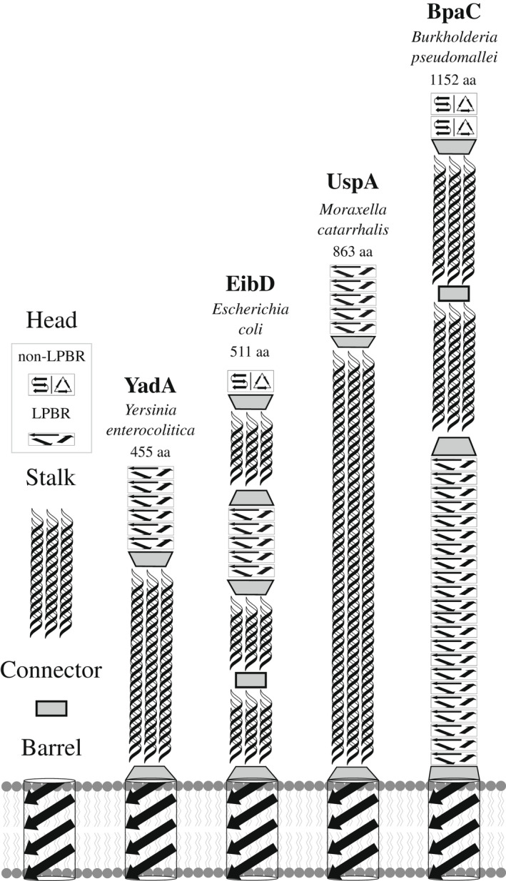
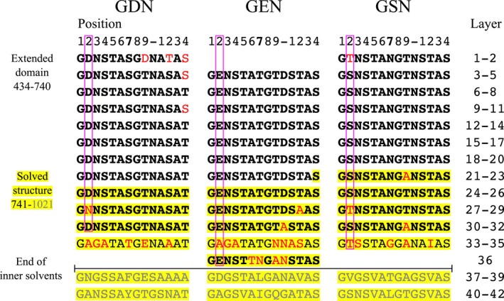
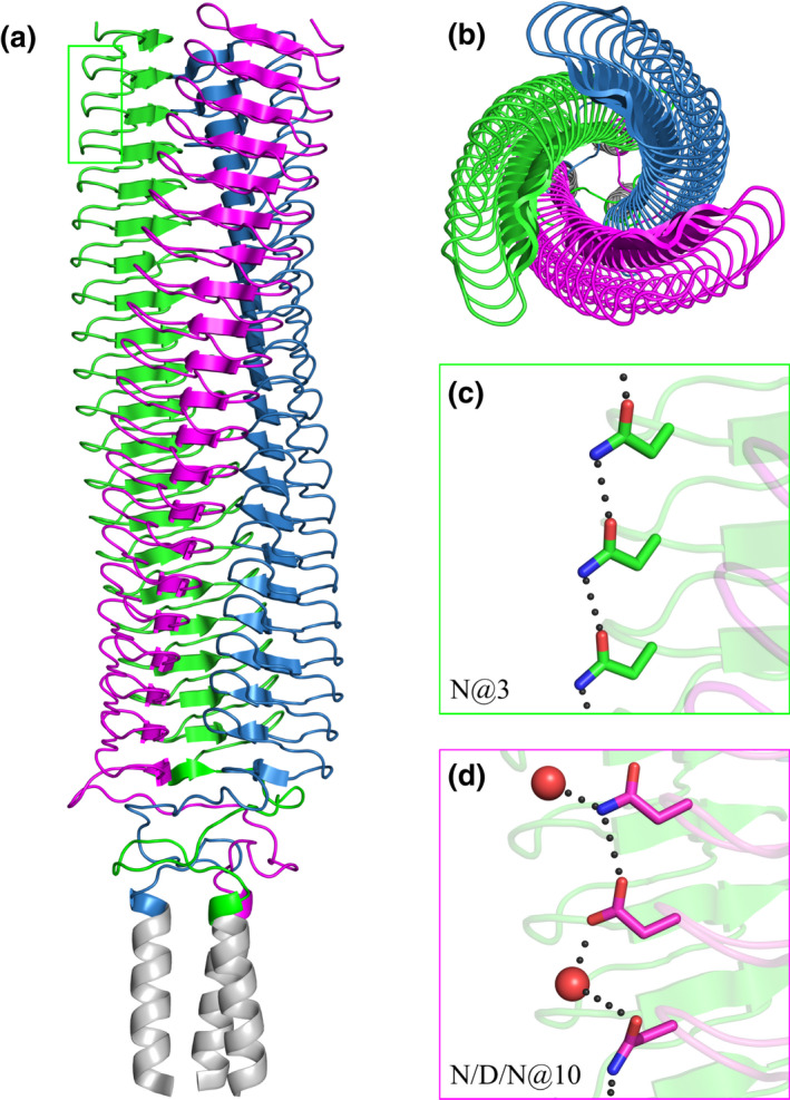
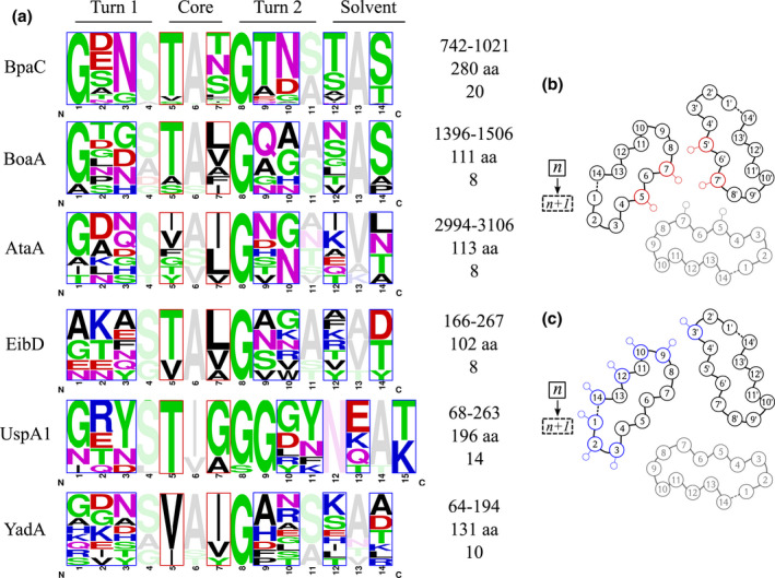
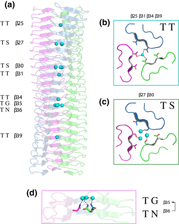
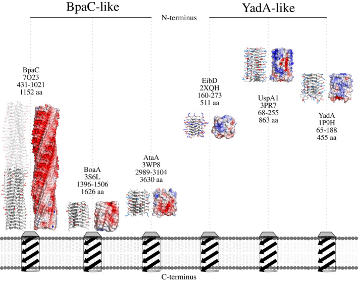
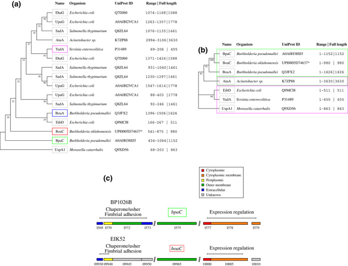
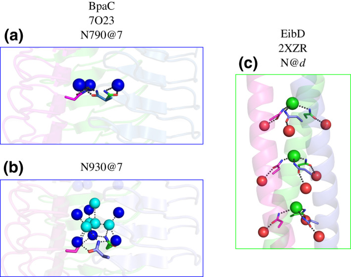
Gram-negative pathogens like Burkholderia pseudomallei use trimeric autotransporter adhesins such as BpaC as key molecules in their pathogenicity. Our 1.4 Å crystal structure of the membrane-proximal part of the BpaC head domain shows that the domain is exclusively made of left-handed parallel β-roll repeats. This, the largest such structure solved, has two unique features. First, the core, rather than being composed of the canonical hydrophobic Ile and Val, is made up primarily of the hydrophilic Thr and Asn, with two different solvent channels. Second, comparing BpaC to all other left-handed parallel β-roll structures showed that the position of the head domain in the protein correlates with the number and type of charged residues. In BpaC, only negatively charged residues face the solvent-in stark contrast to the primarily positive surface charge of the left-handed parallel β-roll "type" protein, YadA. We propose extending the definitions of these head domains to include the BpaC-like head domain as a separate subtype, based on its unusual sequence, position, and charge. We speculate that the function of left-handed parallel β-roll structures may differ depending on their position in the structure.

Keywords: Burkholderia pseudomallei; Type V secretion systems; bacterial adhesin; bacterial outer membrane proteins; melioidosis; protein conformation; β-sheet.


Conflict of interest statement

The authors declare that they have no conflicts of interest.

Figures

FIGURE 1
FIGURE 1
Schematic diagram of solvent‐accessible passenger domains of four different TAAs. Left: Structural modules in TAAs consist of β‐sheet rich head domains, coiled‐coil stalk domains, connector neck motifs, and the membrane‐anchoring β‐barrel domain. Head domain folds can be identified as either left‐handed parallel β‐rolls (LPBR) or non‐LPBR; with the latter predicted as either β‐meander or β‐prism motif (indicated by a split in the illustration). The different locations of the head domains are shown for the passenger domains of Yersinia enterocolitica YadA, Escherichia coli EibD, Moraxella catarrhalis UspA1, and B. pseudomallei BpaC. Connector domains show transition of coiled‐coil stalk domains to head domains or from the β‐barrel to the passenger domain. The length of each TAA is shown (aa).
FIGURE 2
FIGURE 2
Alignment of the BpaC434–1021 region. 14‐residue long LPBR layer repeats are aligned to emphasize similarities between GDN/GEN/GSN families. Bold: identical copies of the consensus sequence in each family. Residues included in the solved structure have a yellow background and D/E/S@2 highlighted by magenta boxes. Residues that do not match the consensus sequence are colored in red. Layers 37–42 deviate too much from the GDN/GEN/GSN family assignment and are excluded (grey). The end of the inner solvent channel is indicated, as this correlates with the end of the GDN/GEN/GSN family assignments.
FIGURE 3
FIGURE 3
Overall view of the C‐terminal head domain of BpaC741–1054. The structure covers 20 of the predicted 42 LPBR head repeats, each layer consisting of 14 amino acids. The three monomers are colored magenta, blue, and green, apart from the added GCN4 leucine zipper (grey). (a) Side view. (b) Top view from the N‐terminus. (c) N@3 in the first 10 repeats form a continuous network of stabilizing hydrogen bonds (only three shown). (d) N/D/N@10 also forms a network of stabilizing hydrogen bonds with interspersed solvent molecules (red spheres).
FIGURE 4
FIGURE 4
Solvent channels and interactions in BpaC741–1021. Water molecules are color‐coded: outer (red), inner (blue), and central (cyan). The three monomers are shown in magenta and blue, with hydrogen bonds in black dots. (a) Side view of the different solvent channels of BpaC741–1021 from N‐ to C‐terminus. (b) Top view of BpaC741–1021 showing the different solvent channels. (c) Schematic of a 14‐residue layer of BpaC741–1021 indicating the location of the various solvent channels alongside the Cβ atoms of residues 5 and 7 to help distinguish the different channels. (d) Outer solvent channel: stabilizing chiefly by interactions between backbone atoms in layer “n“, and the γOH of T@5 in layer “n+1” between two monomers. (e) Inner solvent channel: stabilized by interactions of hydrophilic sidechains S/T/N@7, and supported by backbone carbonyls. The γOH of S/T@7 connects to the inner solvent channel from monomer “A” (cyan spheres) while the interactions of N@7 contribute from the adjacent monomer “B” (green). (f) The central solvent molecule, with the only interaction from an adjacent T972@7 γOH. (g) The central solvent channel stabilized by a S846@7 γOH and further connections to the inner solvent channel.
FIGURE 5
FIGURE 5
Residue frequencies for TAA left‐handed parallel β‐roll repeats, in which the height represents the frequency of residues in each layer. The G@8 is completely conserved. Each layer consists of 14 residues except for the mostly 15‐residue repeats in UspA1, and rare loop insertions or deletions that were excluded prior to logo creation (see Figure S4). These occur between Pos14/14 of layer “n” and Pos1/14 of layer “n + 1”. (a) Sequence logos of residue frequencies. Amino acids are colored by side‐chain properties. Residues that face into the chain interior are shown transparently for better visibility. (b) Cβ positions of amino acids contributing to core interactions are boxed in both frequency plot and sketch (red). (c) Solvent‐facing Cβ positions are highlighted in both plot and sketch (blue). There is one exception to the completely conserved G@8: UspA1 S105 (PDB: 3PR7; Agnew et al., 2011). We ascribe this to an error in the sequence or the structure of the protein, as the ϕ and φ angles are disallowed for serine and there is no sidechain density even at Cβ in the 2F o ‐F c map (data not shown).
FIGURE 6
FIGURE 6
Varying arrangement of central solvent molecules in BpaC741–1021. (a) Side view of BpaC741–1021 showing LPBR layers containing solvent molecules (cyan spheres) within the trimer core. Layers with shown motifs are numbered; motifs without solvent molecules are not included in this Figure. (b) Top view of a T@7 layer. Hydrogen bonds (black) are indicated between three γOH sharing a single solvent molecule. (c) Top view of hydrogen bonds is shown between three S@7 γOH and three solvent molecules. (d) Side view of hydrogen bonds displayed between three N@7 δNH and four solvent molecules arranged in a tetragonal arrangement in the layer above. The missing sidechain of the G@7 creates a large cavity for these solvent molecules.
FIGURE 7
FIGURE 7
Surface charge distribution of selected LPBR head structures. The structures were trimmed to contain just the LPBR layers to show the distribution of solvent‐accessible side chain charges (APBS plugin, PyMOL). Structures are sorted by surface charge (negative to positive, red to blue). Structures are labeled with name, PDB ID, residues used for APBS map, and overall size of the whole TAA. The positions of the head domains relative to the membrane and TAA termini are indicated. Models not to scale. BpaC is divided into a known structure (741–1021, grey) and model inferred by the identity of repeats (431–740, transparent). LPBR structures are divided into BpaC‐like and YadA‐like depending on their relative position and their surface charge profile.
FIGURE 8
FIGURE 8
Evolutionary relationship of TAA LPBRs and genetic environment of bpaC and boaC. (a) Alignments of LPBRs with the BpaC (green) are produced based on the conserved G@8. A phylogenetic tree is shown with TAA names, organism name, UniProt ID, the range of residues included in the final alignment, and the total length of the TAA. “Adhesin YadA‐like” or BoaC (red) from B. oklahomensis is given with UniParc ID (*). The BoaA head domain which was used for molecular replacement for the structure in this publication is highlighted (blue). UspA1 is a separate branch, probably due to the unusual 15‐residue repeats. YadA as the canonical LPBR is highlighted to show the distance to BpaC (magenta). (b) Alignment of full‐length sequences focussing on TAAs that include the LPBR domains analyzed in the previous structural comparison. Clade assignment was split into BpaC‐like (green box with solid line) and YadA‐like (magenta box with solid line) supporting our previous subclassification evolving out of the structural comparison of LPBRs. AtaA is assigned to BpaC‐like and YadA‐like (dashed lines), as the full sequence contains both an N‐terminal and a C‐terminal LPBR head domain. (c) Genome island surrounding bpaC (green) and boaC (blue). Locus tag of genes adjacent to bpaC of B. pseudomallei 1026b and boaC of B. oklahomensis LMG 23618 is displayed in shortened form (BP1026B_X and EIK52_X). Localization of gene product is shown.
FIGURE 9
FIGURE 9
Comparison of N@d motif in EibD with BpaC N790 and N930 interactions—(a) Solvent interactions of the inner channel (blue spheres) are displayed with N790@7 as the example for other layers like this in BpaC. (b) A complex solvent network involving central solvent molecules (cyan spheres) and the inner channel (blue spheres) are being supported by N930@7 pointing toward the layer above which has G@7 that creates a cavity for these solvent molecules to fill in. (c) The N@d motif in the coiled‐coil of EibD (2XZR; Leo et al., 2011) sequesters chloride ions (green spheres) and interacts with adjacent solvent molecules (red spheres).

Similar articles

Cited by

References

    1. Adams, P.D. , Afonine, P.V. , Bunkóczi, G. , Chen, V.B. , Echols, N. , Headd, J.J. et al. (2011) The Phenix software for automated determination of macromolecular structures. Methods, 55(1), 94–106. 10.1016/j.ymeth.2011.07.005 - DOI - PMC - PubMed
    1. Agnew, C. , Borodina, E. , Zaccai, N.R. , Conners, R. , Burton, N.M. , Vicary, J.A. et al. (2011) Correlation of in situ mechanosensitive responses of the Moraxella catarrhalis adhesin UspA1 with fibronectin and receptor CEACAM1 binding. Proceedings of the National Academy of Sciences of the United States of America, 108(37), 15174–15178. 10.1073/pnas.1106341108 - DOI - PMC - PubMed
    1. Altschul, S.F. , Gish, W. , Miller, W. , Myers, E.W. & Lipman, D.J. (1990) Basic local alignment search tool. Journal of Molecular Biology, 215(3), 403–410. 10.1016/S0022-2836(05)80360-2 - DOI - PubMed
    1. Altschul, S.F. , Madden, T.L. , Schäffer, A.A. , Zhang, J. , Zhang, Z. , Miller, W. et al. (1997) Gapped BLAST and PSI‐BLAST: a new generation of protein database search programs. Nucleic Acids Research, 25(17), 3389–3402. 10.1093/nar/25.17.3389 - DOI - PMC - PubMed
    1. Balder, R. , Lipski, S. , Lazarus, J.J. , Grose, W. , Wooten, R.M. , Hogan, R.J. et al. (2010) Identification of Burkholderia mallei and Burkholderia pseudomallei adhesins for human respiratory epithelial cells. BMC Microbiology, 10(1), 250. 10.1186/1471-2180-10-250 - DOI - PMC - PubMed

Publication types

MeSH terms