. 2023 Mar 31;119(2):571-586.
doi: 10.1093/cvr/cvac096.

Myocardial brain-derived neurotrophic factor regulates cardiac bioenergetics through the transcription factor Yin Yang 1

Affiliations

Myocardial brain-derived neurotrophic factor regulates cardiac bioenergetics through the transcription factor Yin Yang 1

Xue Yang et al. Cardiovasc Res. .

Abstract

Aims: Brain-derived neurotrophic factor (BDNF) is markedly decreased in heart failure patients. Both BDNF and its receptor, tropomyosin-related kinase receptor (TrkB), are expressed in cardiomyocytes; however, the role of myocardial BDNF signalling in cardiac pathophysiology is poorly understood. Here, we investigated the role of BDNF/TrkB signalling in cardiac stress response to exercise and pathological stress.

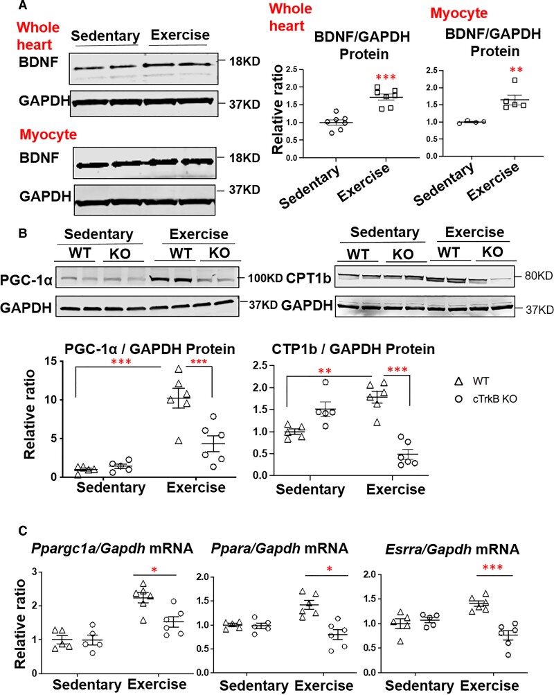
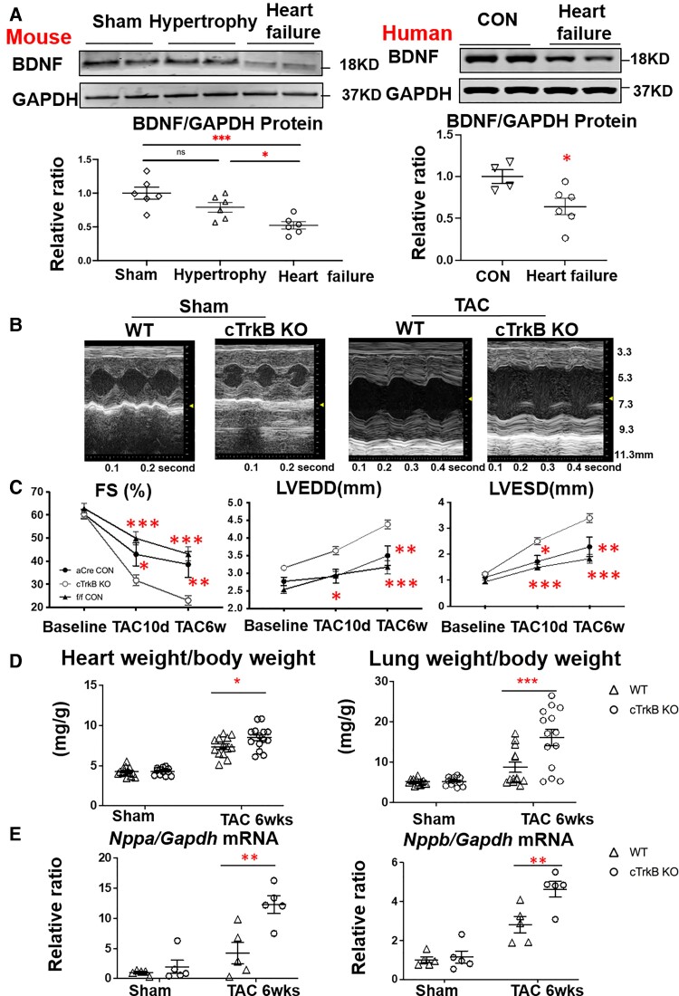
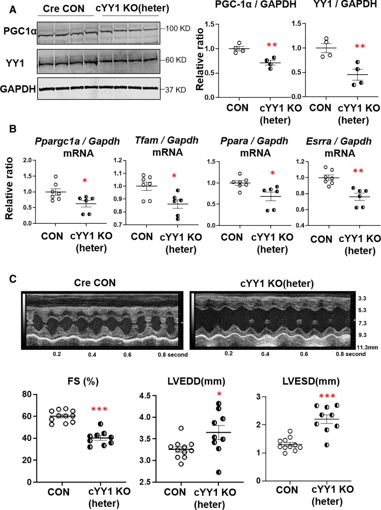
Methods and results: We found that myocardial BDNF expression was increased in mice with swimming exercise but decreased in a mouse heart failure model and human failing hearts. Cardiac-specific TrkB knockout (cTrkB KO) mice displayed a blunted adaptive cardiac response to exercise, with attenuated upregulation of transcription factor networks controlling mitochondrial biogenesis/metabolism, including peroxisome proliferator-activated receptor gamma coactivator 1 alpha (PGC-1α). In response to pathological stress (transaortic constriction, TAC), cTrkB KO mice showed an exacerbated heart failure progression. The downregulation of PGC-1α in cTrkB KO mice exposed to exercise or TAC resulted in decreased cardiac energetics. We further unravelled that BDNF induces PGC-1α upregulation and bioenergetics through a novel signalling pathway, the pleiotropic transcription factor Yin Yang 1.

Conclusion: Taken together, our findings suggest that myocardial BDNF plays a critical role in regulating cellular energetics in the cardiac stress response.

Keywords: BDNF; PGC-1α; YY1; cardiac energetics; heart failure.


Conflict of interest statement

Conflict of interest: None declared.

Figures

Graphical abstract
Graphical abstract
Figure 1
Figure 1
The cTrkB KO mice display blunted adaptive response to exercise. (A) BDNF expression (n = 7) was increased in the mouse hearts with swimming exercise for one week (upper section). BDNF expression (n = 4–5) was upregulated in isolated cardiomyocytes from hearts with swimming exercise (bottom section). (B) The PGC-1α and its downstream target CPT1b protein level were increased with swimming exercise, but the upregulation was blunted in cTrkB KO mice (n = 6). (C) Similarly, the upregulation of PGC-1α mRNA (Ppargc1a), PPARα (Ppara) mRNA, and ERRα (Esrra) mRNA were attenuated in cTrkB KO mice (n = 6) subjected to swimming exercise. Data are shown as means ± SEM. Student’s t-test was used to assess means between two groups in (A). Two-way ANOVA with Tukey’s test was used to assess means between multiple groups in panel (B) and (C). *P < 0.05, **P < 0.01, ***P < 0.001.
Figure 2
Figure 2
The fatty acid oxidation rate and cardiac function are impaired in cTrkB KO mice with swimming exercise. (A) Compared to WT controls, the fatty acid oxidation rate, measured in isolated working heart with H3-labelled oleic acid, was decreased in cTrkB KO mice with 2 weeks of swimming exercise, whereas there was no significant change in glucose oxidation (n = 6–7). (B) Compared to WT controls, the contractility of hearts from cTrkB KO mice with exercise, which was measured by dP/dT in the isolated working heart system, was significantly reduced (n = 6–7). (C) Representative echocardiography and data summary of WT and cTrkB KO mice subjected to swimming exercise. Compared to WT mice, cTrkB KO mice showed decreased fractional shortening, increased LV end systolic or diastolic dimension (LVEDD or LVESD) (n = 9). Data are shown as means ± SEM. Student’s t-test was used to assess means between two groups. *P < 0.05, *P < 0.01, ***P < 0.001.
Figure 3
Figure 3
cTrkB KO mice display exacerbated heart failure progression post TAC. (A) In contrast with exercise, there was no significant change in myocardial BDNF level in concentric cardiac hypertrophy (2 weeks after TAC), but the BDNF was significantly decreased in failing hearts (6 weeks after TAC) (n = 6). The level of myocardial BDNF was also significantly reduced in patients with non-ischemic cardiomyopathy (n = 4–6). (B) Representative echocardiography of WT and cTrkB KO mice subjected to sham or TAC surgery. (C) Compared to WT mice and αMHC-Cre controls, cTrkB KO mice showed decreased fractional shortening (WT 43.1 ± 2.8% or αMHC-Cre 38.7 ± 5.2% vs cTrkB KO 23.1 ± 2.1%), increased LV end diastolic dimension (LVEDD), and LV end systolic dimension (LVESD). (WT n = 12, αMHC-Cre n = 9, cTrkB KO n = 15). (D) TAC cTrkB KO mice heart weight and lung weight were increased relative to WT controls (WT n = 12, cTrkB KO n = 9). (E) ANP (Nppa) and BNP (Nppb) were increased in cTrkB KO mice subjected to TAC (n = 6). Data are shown as means ± SEM. One-way ANOVA test was used to assess means between multiple groups and Student’s t-test was used to assess means between two groups in (A). Two-way ANOVA with Tukey’s test was used to assess means in (C), (D), and (E).*P < 0.05, **P < 0.01, ***P < 0.001.
Figure 4
Figure 4
PGC-1α expression, mitochondrial proteins, mitoDNA copy numbers, and mitochondrial OCR are decreased in cTrkB KO mice subjected to TAC. (A and B) The PGC-1α mRNA (Ppargc1a) and protein level were significantly lower in cTrkB KO mice subjected to TAC compared to WT controls (n = 5–6). (C) OXPHOS cocktail western blot showed that mitochondrial proteins including NDUFB8 (complex I), SDHB (complex II), UQCRC2 (complex III), ATP5A (complex IV), and COXII (complex V) were decreased in TAC cTrkB KO mice, compared to WT controls (n = 6). (D) Mitochondrial DNA copy number, assessed by MT-ND2/18 s RNA gene and MT-ATP6/18 s RNA gene, was reduced in cTrkB KO mice subjected to TAC (WT n = 5, cTrkB n = 5). (E) OCR of complex II and complex IV was impaired in TAC cTrkB KO hearts relative to WT controls (n = 6). Data are shown as means ± SEM. Two-way ANOVA with Tukey’s test was used to assess means in (A), (B), and (D). Student’s t-test was used to assess means between two groups in (C) and (E). *P < 0.05, **P < 0.01, ***P < 0.001.
Figure 5
Figure 5
Myocardial BDNF regulates PGC-1α expression through AKT-mTOR-YY1 pathway. (A) Isoproterenol (10μM) induced BDNF expression in NRCMs (n = 8). (B) PGC-1α and transcription factor Yin-Yang 1 (YY1) expression, and the level of phosphorylated AKT were increased with isoproterenol stimulation, and the upregulation was attenuated in TrkB siRNA transfected NRCMs (n = 5–6). (C) Overexpression of BDNF increased PGC-1α and YY1 expression, and the BDNF induced upregulation of PGC-1α and YY1 was attenuated by AKT inhibitor LY294002 or mTOR inhibitor rapamycin (n = 4). (D and E) The level of phosphorylated AKT and YY1 expression were elevated in WT mice responding to swimming exercise or TAC (n = 5), but the elevation was blunted in cTrkB KO mice (n = 6–8). (F) Chromatin immunoprecipitation assay (ChIP) demonstrated the binding of YY1 to PGC-1α promoter was significantly decreased in NRCMs with TrkB knockdown with the incubation of isoproterenol (n = 4, left section). ChIP assay showed the binding of YY1 to PGC-1α promoter was reduced in TAC cTrkB KO mice (n = 5, right section). (G and H) YY1 knockdown decreased PGC-1α expression in NRCMs with or without isoproterenol (n = 4–6), and the overexpression of YY1 in NRCMs recovered the downregulation of PGC-1α expression in NRCMs with TrkB knockdown (n = 7). Data are shown as means ± SEM. Student’s t-test was used to assess means between two groups in (A). One-way ANOVA test was used to assess means between multiple groups in (C), (F), and (H). Two-way ANOVA with Tukey’s test was used to assess means in (B), (D), (E), and (G). P < 0.05, **P < 0.01, ***P < 0.001.
Figure 6
Figure 6
TrkB or YY1 knockdown in NRCMs impairs mitochondrial function. (A) While there is no significant difference in oxidative respiratory capacity without isoproterenol stimulation between control and NRCMs with TrkB knockdown, the basal respiratory function was significantly decreased with TrkB knockdown in the presence of isoproterenol (n = 8). (B) NRCMs transfected with YY1 siRNA displayed both impaired basal and maximum respiration, at both baseline and with isoproterenol incubation (n = 8). Data are shown as means ± SEM. Two-way ANOVA with Tukey’s test was used to assess means. *P < 0.05, **P < 0.01, ***P < 0.001.
Figure 7
Figure 7
The expression of PGC-1α and cardiac function are decreased in heterozygous cYY1 KO mice. (A) The PGC-1α protein level (n = 4), (B) as well as the mRNA expression of PGC-1α (Ppargc1a), PPARα (ppara), ERRα (esrra), and TFAM were decreased in heterozygous cYY1 KO mice (n = 6–7). (C) Compared to age matched αMHC-Cre controls, heterozygous cYY1 KO mice exhibited decreased fractional shortening and increased left ventricular end systolic and diastolic dimension (LVESD and LVEDD, n = 9). Student’s t-test was used to assess means between two groups in (A), (B) and (C). *P < 0.05, **P < 0.01, ***P < 0.001.

References

    1. Lear SA, Hu W, Rangarajan S, Gasevic D, Leong D, Iqbal R, Casanova A, Swaminathan S, Anjana RM, Kumar R, Rosengren A, Wei L, Yang W, Chuangshi W, Huaxing L, Nair S, Diaz R, Swidon H, Gupta R, Mohammadifard N, Lopez-Jaramillo P, Oguz A, Zatonska K, Seron P, Avezum A, Poirier P, Teo K, Yusuf S. The effect of physical activity on mortality and cardiovascular disease in 130 000 people from 17 high-income, middle-income, and low-income countries: the PURE study. Lancet 2017;390:2643–2654. - PubMed
    1. Mandsager K, Harb S, Cremer P, Phelan D, Nissen SE, Jaber W. Association of cardiorespiratory fitness with long-term mortality among adults undergoing exercise treadmill testing. JAMA Netw Open 2018;1:e183605. - PMC - PubMed
    1. Stamatakis E, Gale J, Bauman A, Ekelund U, Hamer M, Ding D. Sitting time, physical activity, and risk of mortality in adults. J Am Coll Cardiol 2019;73:2062–2072. - PubMed
    1. O’Connor CM, Whellan DJ, Lee KL, Keteyian SJ, Cooper LS, Ellis SJ, Leifer ES, Kraus WE, Kitzman DW, Blumenthal JA, Rendall DS, Miller NH, Fleg JL, Schulman KA, McKelvie RS, Zannad F, Pina IL, Investigators H-A. Efficacy and safety of exercise training in patients with chronic heart failure: HF-ACTION randomized controlled trial. JAMA 2009;301:1439–1450. - PMC - PubMed
    1. Piepoli MF, Davos C, Francis DP, Coats AJ, ExTra MC. Exercise training meta-analysis of trials in patients with chronic heart failure (ExTraMATCH). BMJ 2004;328(189). - PMC - PubMed

Publication types

MeSH terms

Substances