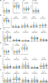
Peripheral blood immune cell profiling of acute corneal transplant rejection
- PMID: 35704290
- PMCID: PMC9796948
- DOI: 10.1111/ajt.17119
Peripheral blood immune cell profiling of acute corneal transplant rejection
Abstract
Acute rejection (AR) of corneal transplants (CT) has a profound effect on subsequent graft survival but detailed immunological studies in human CT recipients are lacking. In this multi-site, cross-sectional study, clinical details and blood samples were collected from adults with clinically diagnosed AR of full-thickness (FT)-CT (n = 35) and posterior lamellar (PL)-CT (n = 21) along with Stable CT recipients (n = 177) and adults with non-transplanted corneal disease (n = 40). For those with AR, additional samples were collected 3 months later. Immune cell analysis was performed by whole-genome microarrays (whole blood) and high-dimensional multi-color flow cytometry (peripheral blood mononuclear cells). For both, no activation signature was identified within the B cell and T cell repertoire at the time of AR diagnosis. Nonetheless, in FT- but not PL-CT recipients, AR was associated with differences in B cell maturity and regulatory CD4+ T cell frequency compared to stable allografts. These data suggest that circulating B cell and T cell subpopulations may provide insights into the regulation of anti-donor immune response in human CT recipients with differing AR risk. Our results suggest that, in contrast to solid organ transplants, genetic or cellular assays of peripheral blood are unlikely to be clinically exploitable for prediction or diagnosis of AR.
Keywords: biomarker; corneal transplantation/ophthalmology; flow cytometry; rejection: acute; translational research/science.
© 2022 The Authors. American Journal of Transplantation published by Wiley Periodicals LLC on behalf of The American Society of Transplantation and the American Society of Transplant Surgeons.
Figures


References
-
- Gupta N, Vashist P, Tandon R, Gupta SK, Dwivedi S, Mani K. Prevalence of corneal diseases in the rural Indian population: the corneal opacity rural epidemiological (CORE) study. Br J Ophthalmol. 2015;99(2):147‐152. - PubMed
-
- Pascolini D, Mariotti SP. Global estimates of visual impairment: 2010. Br J Ophthalmol. 2012;96(5):614‐618. - PubMed
-
- Spinozzi D, Miron A, Bruinsma M, et al. New developments in corneal endothelial cell replacement. Acta Ophthalmol. 2020;99:712‐729. - PubMed
Publication types
MeSH terms
LinkOut - more resources
Full Text Sources
Molecular Biology Databases
Research Materials

