. 2022 Jun 15;14(649):eaba4380.
doi: 10.1126/scitranslmed.aba4380. Epub 2022 Jun 15.

Calreticulin mutant myeloproliferative neoplasms induce MHC-I skewing, which can be overcome by an optimized peptide cancer vaccine

Affiliations

Calreticulin mutant myeloproliferative neoplasms induce MHC-I skewing, which can be overcome by an optimized peptide cancer vaccine

Mathieu Gigoux et al. Sci Transl Med. .

Abstract

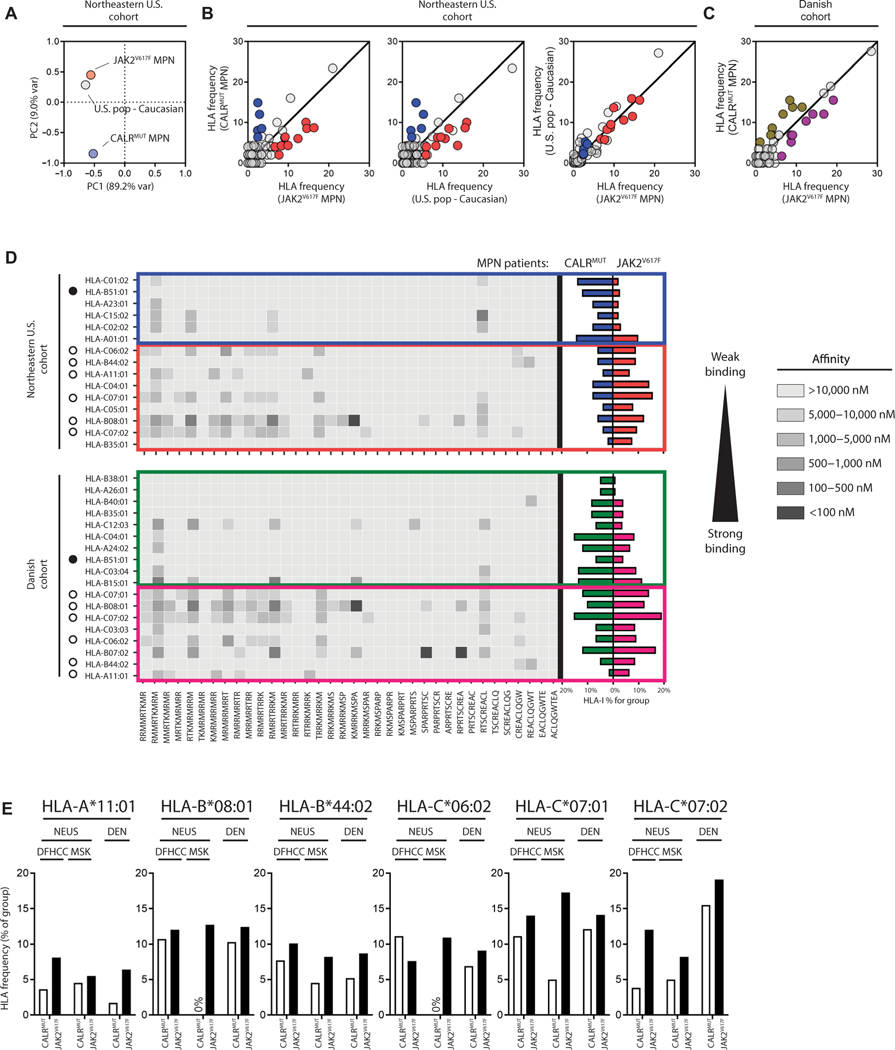
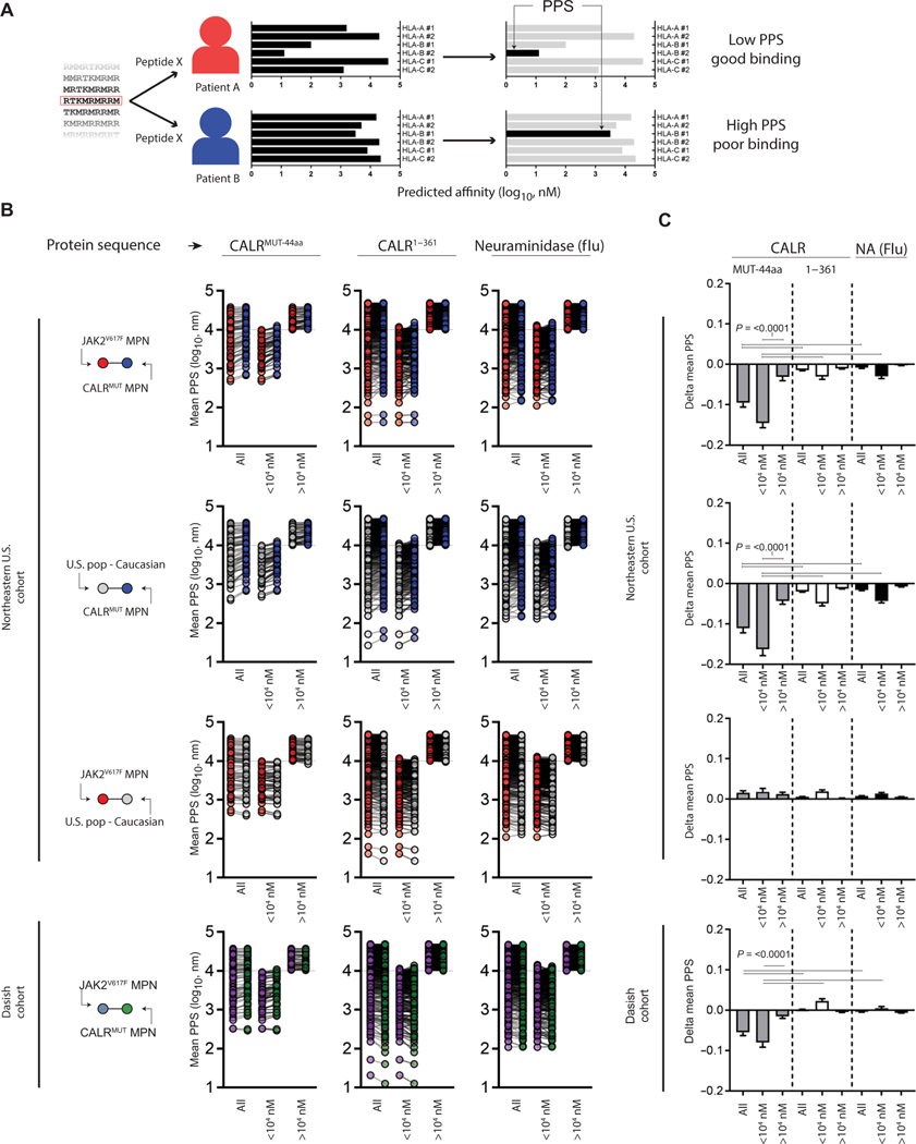
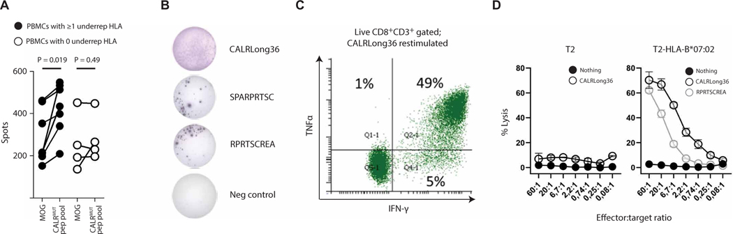
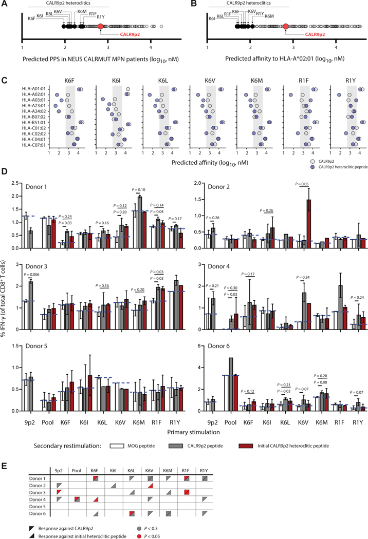
The majority of JAK2V617F-negative myeloproliferative neoplasms (MPNs) have disease-initiating frameshift mutations in calreticulin (CALR), resulting in a common carboxyl-terminal mutant fragment (CALRMUT), representing an attractive source of neoantigens for cancer vaccines. However, studies have shown that CALRMUT-specific T cells are rare in patients with CALRMUT MPN for unknown reasons. We examined class I major histocompatibility complex (MHC-I) allele frequencies in patients with CALRMUT MPN from two independent cohorts. We observed that MHC-I alleles that present CALRMUT neoepitopes with high affinity are underrepresented in patients with CALRMUT MPN. We speculated that this was due to an increased chance of immune-mediated tumor rejection by individuals expressing one of these MHC-I alleles such that the disease never clinically manifested. As a consequence of this MHC-I allele restriction, we reasoned that patients with CALRMUT MPN would not efficiently respond to a CALRMUT fragment cancer vaccine but would when immunized with a modified CALRMUT heteroclitic peptide vaccine approach. We found that heteroclitic CALRMUT peptides specifically designed for the MHC-I alleles of patients with CALRMUT MPN efficiently elicited a CALRMUT cross-reactive CD8+ T cell response in human peripheral blood samples but not to the matched weakly immunogenic CALRMUT native peptides. We corroborated this effect in vivo in mice and observed that C57BL/6J mice can mount a CD8+ T cell response to the CALRMUT fragment upon immunization with a CALRMUT heteroclitic, but not native, peptide. Together, our data emphasize the therapeutic potential of heteroclitic peptide-based cancer vaccines in patients with CALRMUT MPN.


Figures

Fig. 1.
Fig. 1.. MHC-I alleles with predicted binding to CALRMUT-derived peptides are less frequent in CALRMUT MPNs.
(A) Principal components analysis of MHC-I allele frequencies from patients with CALRMUT MPN, patients with JAK2V617F MPN, and U.S. Caucasian population. (B) Comparison of MHC-I allele frequencies from patients with CALRMUT MPN, patients with JAK2V617F MPN, and U.S. Caucasian population compared to each other in the NEUS cohort and (C) the Danish cohort. Frequencies are expressed as percentages. MHC-I alleles that are overrepresented in patients with CALRMUT are in blue (NEUS cohort) and green (Danish cohort), whereas MHC-I alleles that are underrepresented in patients with CALRMUT are in red (NEUS cohort) and pink (Danish cohort). For the NEUS cohort, only MHC-I alleles differentially expressed between patients with CALRMUT MPN compared to both patients with JAK2V617F MPN and U.S. Caucasian population were considered for further analysis. (D) Heatmap of predicted binding of each CALRMUT-derived peptide to each MHC-I allele from (B) (left). MHC-I alleles that are underrepresented or overrepresented in patients with CALRMUT MPN in both cohorts are noted with white or black circles, respectively. Actual MHC-I allele frequencies in CALRMUT and JAK2V617F MPN patient populations are also noted (right). (E) Cohort breakdown of CALRMUT MPN MHC-I allele frequencies of individual institution consisting of the NEUS cohort for the six less frequent MHC-I alleles.
Fig. 2.
Fig. 2.. MHC-I bias is selective against the CALRMUT fragment.
(A) Schema depicting calculation of patient:peptide score (PPS). Briefly, single peptides derived from longer sequences were applied to NetMHCpan v3 to predict class-I HLA binding against any given patient’s six possible class-I HLAs. In each patient-peptide combination, the strongest affinity was registered as the PPS. (B) Mean PPS of individual peptides derived from indicated protein sequence (9- to 10-mers) from patients with CALRMUT and JAK2V617F MPN and pseudopopulation created from expected frequencies of U.S. Caucasian population (NEUS only). Protein sequences are the 44–amino acid CALRMUT sequence (CALRMUT−44aa), the wild-type CALR sequence upstream of CALRMUT−44aa (CALR1–361), and the irrelevant foreign antigen neuraminidase (NA) from influenza. Also shown are peptides broadly subdivided from predicted possible binding (<104 nM) and predicted nonbinding (>104 nM) peptides. (C) Difference in mean PPS of each group from (B). The Student’s t test was performed to calculate significance. The horizontal bars represent the combination where the statistical test resulted in the noted P value.
Fig. 3.
Fig. 3.. Underrepresented MHC-I alleles potentiate response to CALRMUT peptides.
(A) IFN-γ ELISpot of PBMCs from healthy donors that had either at least one (black circles) or zero (white circles) underrepresented MHC-I allele expanded with a peptide pool (15-mers) derived from the entire CALRMUT fragment and restimulated after 10 days with either the irrelevant peptide (MOG) or the same CALRMUT fragment peptide pool. (B) Reactivity of CALRLong36-specific CD8+ T cells derived from an HLA-B*07:02+ patient with post–essential thrombocythemia myelofibrosis. Cells restimulated with different CALRMUT peptides in an IFN-γ ELISpot, (C) intracellular IFN-γ and TNFα cytokine staining on the CALRLong36-specific CD8+ T cells stimulated with CALRLong36 (D) target cell lysis of wild-type or HLA-B*07:02–transduced T2 cells pulsed with different CALRMUT peptides.
Fig. 4.
Fig. 4.. Human CD8+ T cells activated with heteroclitic CALR9p2 peptides cross-react with CALR9p2 peptide.
(A) Predicted mean PPS in patients with CALRMUT MPN of all single amino acid substitution of CALR9p2. (B) Predicted affinity of all single amino acid substitution of CALR9p2 to HLA-A*02:01. Seven heteroclitic peptides were chosen for further testing and are identified here only by their amino acid substitution in the CALR9p2 peptide. (C) Binding affinity of CALR9p2 and indicated heteroclitic peptide to the most common MHC-I in the CALRMUT MPN patient cohorts. Shadowed area indicates predicted binding affinity range of 5000 to 500 nM. (D) Percent IFN-γ+ CD8+ T cells after primary in vitro stimulation of PBMCs with CALR9p2 or CALR9p2 heteroclitic peptides (noted on x axis) followed by secondary restimulation with either control (MOG, median represented by dotted blue line) peptide, CALR9p2, or initial CALR9p2 heteroclitic peptide (noted by color of bar). (E) Summary of responding donor PBMCs to CALR9p2 or CALR9p2 heteroclitic peptides.
Fig. 5.
Fig. 5.. CALRMUT sequence is not immunogenic in C57BL/6 J mice.
(A) The predicted binding affinity of CALRMUT-derived peptides (8- to 10-mers shown) against all available murine MHC-I alleles. The strongest binding peptide CALR9p2 is identified in red. (B) H-2Kb stabilization assay using TAP-deficient RMA/S cells was performed for CALR9p2 in the presence and absence of serum. The chicken ovalbumin (OVA)–derived peptide SIINFEKL was used as a positive control. (C) Timeline of DNA immunization schedule and CD8+ T cell collection for the experiment in (D). (D) IFN-γ ELISpot depicting secondary reactivity of CD8+ T cells isolated from draining lymph nodes of mice DNA immunized with full-length CALRWT, CALRMUT, and OVA. (E) Timeline of peptide immunization and CD8+ T cell collection for the experiment in (F). (F) IFN-γ ELISpot depicting secondary reactivity of CD8+ T cells isolated from draining lymph nodes of mice peptide immunized with adjuvant and DMSO, CALR9p2, or SIINFEKL. Data shown represent results from one repeat of experiments performed at least three times.
Fig. 6.
Fig. 6.. Heteroclitic CALR9p2 peptide vaccine elicits cross-reactive CD8+ T cell response against CALR9p2 and controls tumor growth in mice.
(A) Predicted binding affinity to H-2Kb of all single amino acid substitution variants of CALR9p2. Top predicted peptide CALR9p2(T5F) is shown. (B) Cartoon of the expected effect of T5F substitution in CALR9p2 peptide conformational binding into H-2Kb. Known dominant anchor sites and minor anchor sites are depicted in red and blue, respectively. (C) MHC-I stabilization assay using TAP-deficient RMA/S cells was performed for CALR9p2 and CALR9p2(T5F) in the absence of serum. SIINFEKL was used as a positive control. Representative results from one repeat of an experiment performed at least three times. (D) IFN-γ ELISpot depicting secondary reactivity of CD8+ T cells isolated from draining lymph nodes of mice peptide immunized with adjuvant and DMSO, CALR9p2, or CALR9p2(T5F). Representative results from one repeat of an experiment performed at least three times. The Student’s t test was performed to calculate significance. (E) Killing assay of peptide-pulsed B16 cells by CD8+ T cells isolated from peptide-immunized mice. Representative results from one repeat of an experiment performed at least three times. The Student’s t test was performed to calculate statistical significance. (F) Timeline of peptide immunization prior to tumor implantation for prophylactic vaccine. (G) RMA/SpER-CALR9p2 tumor growth over time after prophylactic peptide immunization for individual tumors or (H) average growth until the second mouse reached the end point of a tumor size of 400 mm2. (I) Survival of mice after the prophylactic vaccine. (J) Timeline of peptide immunization and tumor implantation for therapeutic vaccine and in combination with anti–PD-1 therapy. ip, intraperitoneally. (K) Tumor growth over time after therapeutic vaccine for individual tumors or (L) averaged tumor growth until the second mouse reached the end point. (M) Survival of mice after the therapeutic vaccine, with or without anti–PD-1 therapy. Data from tumor growth experiments represent results from one repeat of experiments performed twice. Significance for tumor growth experiments was calculated by performing a Student’s t test on the area under the curve of each tumor. Significance for survival was calculated by performing a log-rank test.

References

    1. Levine RL, Gilliland DG, Myeloproliferative disorders. Blood 112, 2190–2198 (2008). - PMC - PubMed
    1. Campbell PJ, Green AR, The myeloproliferative disorders. N. Engl. J. Med. 355, 2452–2466 (2006). - PubMed
    1. Rampal R, Al-Shahrour F, Abdel-Wahab O, Patel JP, Brunel JP, Mermel CH, Bass AJ, Pretz J, Ahn J, Hricik T, Kilpivaara O, Wadleigh M, Busque L, Gilliland DG, Golub TR, Ebert BL, Levine RL, Integrated genomic analysis illustrates the central role of JAK-STAT pathway activation in myeloproliferative neoplasm pathogenesis. Blood 123, e123–e133 (2014). - PMC - PubMed
    1. Nangalia J, Massie CE, Baxter EJ, Nice FL, Gundem G, Wedge DC, Avezov E, Li J, Kollmann K, Kent DG, Aziz A, Godfrey AL, Hinton J, Martincorena I, Van Loo P, Jones AV, Guglielmelli P, Tarpey P, Harding HP, Fitzpatrick JD, Goudie CT, Ortmann CA, Loughran SJ, Raine K, Jones DR, Butler AP, Teague JW, O’Meara S, McLaren S, Bianchi M, Silber Y, Dimitropoulou D, Bloxham D, Mudie L, Maddison M, Robinson B, Keohane C, Maclean C, Hill K, Orchard K, Tauro S, Du MQ, Greaves M, Bowen BJP Huntly CN Harrison NC Cross P, Ron D, Vannucchi AM, Papaemmanuil, Campbell PJ, Green AR, Somatic CALR mutations in myeloproliferative neoplasms with nonmutated JAK2. N. Engl. J. Med. 369, 2391–2405 (2013). - PMC - PubMed
    1. Klampfl T, Gisslinger H, Harutyunyan AS, Nivarthi H, Rumi E, Milosevic JD, Them NC, Berg T, Gisslinger B, Pietra D, Chen D, Vladimer GI, Bagienski K, Milanesi C, Casetti IC, Sant’Antonio E, Ferretti V, Elena C, Schischlik F, Cleary C, Six M, Schalling M, Schonegger A, Bock C, Malcovati L, Pascutto C, Superti-Furga G, Cazzola M, Kralovics R, Somatic mutations of calreticulin in myeloproliferative neoplasms. N. Engl. J. Med. 369, 2379–2390 (2013). - PubMed

Publication types