. 2022 Jun 14;39(11):110942.
doi: 10.1016/j.celrep.2022.110942.

Inflammatory adipose activates a nutritional immunity pathway leading to retinal dysfunction

Affiliations

Inflammatory adipose activates a nutritional immunity pathway leading to retinal dysfunction

Jacob K Sterling et al. Cell Rep. .

Abstract

Age-related macular degeneration (AMD), the leading cause of irreversible blindness among Americans over 50, is characterized by dysfunction and death of retinal pigment epithelial (RPE) cells. The RPE accumulates iron in AMD, and iron overload triggers RPE cell death in vitro and in vivo. However, the mechanism of RPE iron accumulation in AMD is unknown. We show that high-fat-diet-induced obesity, a risk factor for AMD, drives systemic and local inflammatory circuits upregulating interleukin-1β (IL-1β). IL-1β upregulates RPE iron importers and downregulates iron exporters, causing iron accumulation, oxidative stress, and dysfunction. We term this maladaptive, chronic activation of a nutritional immunity pathway the cellular iron sequestration response (CISR). RNA sequencing (RNA-seq) analysis of choroid and retina from human donors revealed that hallmarks of this pathway are present in AMD microglia and macrophages. Together, these data suggest that inflamed adipose tissue, through the CISR, can lead to RPE pathology in AMD.

Keywords: CP: Immunology; IL-1β; age-related macular degeneration; high fat diet; iron; macrophage; microglia; neuroinflammation; nutritional immunity; visceral adipose.


Conflict of interest statement

Declaration of interests The authors declare no competing interests.

Figures

Figure 1.
Figure 1.. High-fat diet induces iron-dependent RPE dysfunction
(A) Mass in grams of mice fed a low-fat diet (LFD) or high-fat diet (HFD) alone or in combination with iron chelator deferiprone (DFP) (n = 20/group). (B) Visceral adipose tissue (VAT) and subcutaneous adipose tissue (SAT) hypertrophy were dependent on diet but not DFP treatment (n = 10/group). (C) Diet and DFP treatment had no effect on glycemic control measured with intraperitoneal glucose tolerance test (n = 10/group). (D) HFD mice accumulate RPE iron, measured using inductively coupled plasma mass spectrometry, which can be prevented with DFP treatment (n = 10/group). (E) HFD mice accumulate lipid oxidation product MDA in RPE, which is rescued by treatment with DFP (n = 10/group). (F) HFD mice RPE upregulate Hmox1, measured by qPCR, in an iron-dependent manner (n = 5/group). (G) HFD mice exhibit c-wave amplitude deficits, measured by electroretinography, which can be partially rescued by iron chelation with DFP (n = 10/group). Data indicate mean and, where error bars are shown, ± SD. *p < 0.05, **p < 0.01, ***p < 0.001, ****p < 0.0001, by 2-way repeated-measures ANOVA (A and C) or 2-way ANOVA (B and D–G) with Tukey’s honestly significant difference (HSD) post hoc test.
Figure 2.
Figure 2.. IL-1β is necessary for HFD-induced RPE iron accumulation and dysfunction
(A) Human fetal RPE (fRPE) cultures were treated with either transferrin-bound ferric iron (Tf·55Fe3+) or ferrous iron ascorbate (55Fe2+) alone or in the presence of recombinant IL-1β (rIL-1β). Intracellular iron accumulation in fRPE lysates was measured during 8 h of treatment using a scintillation counter to quantify counts per minute (CPM) (n = 3/group). (B) fRPE cultures were treated with either media (vehicle) or recombinant IL-1β (rIL-1β) for 8 h before cells were harvested for qPCR analysis of iron transport/ regulatory genes (n = 6/group). (C) Mass in grams of WT and Il1b−/− (KO) mice fed an LFD or an HFD (n = 20/group). (D) VAT and SAT hypertrophy were dependent on diet but not genotype (n = 10/group). (E) Diet and genotype had no effect on glycemic control measured with intraperitoneal glucose tolerance test (n = 10/group). (F) RPE gene expression measured by qPCR following 12 weeks of either LFD or HFD (n = 5/group). (G) Retinal CD11b+ cell gene expression following 12 weeks of either LFD or HFD (n = 5/group). (H) HFD WT mice accumulate RPE iron, measured using inductively coupled plasma mass spectrometry, which is rescued in the HFD Il1b−/− mice (n = 10/group). (I) HFD WT mice accumulate lipid oxidation product MDA in RPE, which is rescued in HFD Il1b−/− mice (n = 10/group). (J) HFD WT mice RPE upregulate Hmox1 in an Il1b-dependent manner (n = 5/group). (K) HFD WT mice exhibit c-wave amplitude deficits, measured by electroretinography, which is rescued in HFD Il1b−/− mice (n = 10/group). Data indicate mean and, where error bars are shown, ± SD. *p < 0.05, **p < 0.01, ***p < 0.001, ****p < 0.0001, by 1-way ANOVA with multiple comparisons (A), unpaired Student’s t test (B), or 2-way repeated-measures ANOVA (C and E) or 2-way ANOVA (D and F–K) with Tukey’s HSD post hoc test.
Figure 3.
Figure 3.. Activation of systemic and local inflammatory circuits contributes to IL-1β production
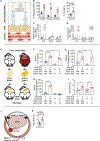
(A) Schematic of RPE exposed apically to the neurosensory retina behind the blood-retina barrier, and basolaterally to the fenestrated choriocapillaris. (B) IL-1β ELISA performed on epididymal white adipose tissue (eWAT), retina, choroid, and serum harvested from WT mice fed an LFD or an HFD diet for 12 weeks (n = 10/group). (C and D) qPCR analysis of Il1b, Nlrp3, and Casp1 gene expression in retinal (C) and choroidal (D) CD11b+ cells (n = 5/group). (E) Schematic of eWAT transplant. (F–H) IL-1β ELISA performed on serum (F), choroid (G), or retina (H) from WT mice fed an LFD or an HFD that underwent a sham transplant surgery (indicated by NA for transplant Il1b genotype and diet ) or Il1b−/− mice fed an HFD that received eWAT donor tissue from HFD-fed WT or Il1b−/− mice (n = 10/group). (I) Inductively coupled plasma mass spectrometry (iCP-MS) performed on RPE isolated from WT mice fed an LFD or an HFD that underwent a sham transplant surgery (indicated by NA for transplant Il1b genotype and diet) or Il1b−/− mice fed an HFD that received eWAT donor tissue from HFD-fed WT or Il1b−/− mice (n = 10/ group). (J) Schematic of intravitreal injection. (K) WT mice received an intravitreal injection of PBS or recombinant IL-1β (rIL-1β). Eight hours after injection, RPE was harvested, and intracellular iron was quantified using iCP-MS (n = 10/group). Data indicate mean. *p < 0.05, **p < 0.01, and ****p < 0.0001 by unpaired student’s t test (B–D and K) or 2-way ANOVA (F–I) with Tukey’s HSD post hoc test.
Figure 4.
Figure 4.. Hallmarks of IL-1β-dependent CISR are observed in AMD patients
(A) Human choroidal single-cell RNA sequencing data. Cells expressing at least one read of IL1B, NLRP3, and CASP1 (i.e., triple-positive cells) are colored in red, while the remaining cells are colored in gray. A total of 58.6% of macrophages expressed IL1B, NLRP3, and CASP1. (B) Violin plots of IL1B, NLRP3, and CASP1 expression by cell type indicate macrophage-specific expression of these transcripts. (C) Using the same single-cell RNA sequencing data, differential expression analysis was performed between macrophages originating from AMD donors(n = 2) versus control donors (n = 4). Differential expression results are displayed in a volcano plot, where positive log fold changes indicate enrichment in AMD samples. Results for IL1B, CASP1, and NLRP3 are highlighted. (D) Visualization of healthy human neurosensory retina single-cell clusters using t-distributed stochastic neighbor embedding (t-SNE) sequencing data from normal retinas. Cells expressing at least one read of IL1B, NLRP3, and CASP1 (i.e., triple-positive cells) are colored in red, while the remaining cells are colored in gray. Enrichment in these three transcripts is specific to microglia. (E) Visualization of healthy human neurosensory retina single-cell clusters using t-SNE sequencing data from normal retinas. Cells expressing at least one read of HAMP (protein name hepcidin) are colored in red, while the remaining cells are colored in gray. Enrichment in HAMP is specific to microglia. (F) Bulk neurosensory retina RNA sequencing on normal (NL), early AMD, and late AMD macular retina or peripheral retina showing expression of IL1B, NLRP3, and CASP1 across disease progression in a region-specific manner. Color scale corresponds to log2 of expression measured in fragments per kilobase of transcript per million mapped reads (FPKM). (G) IL-1β ELISA performed on neurosensory retina from eyes without AMD (MGS1, n = 6) and with early (MGS2, n = 6) or intermediate (MGS3, n = 6) AMD. See Table S1 for donor information. **p < 0.01 by unpaired Student’s t test (J).

References

    1. Adams MKM, Simpson JA, Aung KZ, Makeyeva GA, Giles GG, English DR, Hopper J, Guymer RH, Baird PN, and Robman LD (2011). Abdominal obesity and age-related macular degeneration. Am. J. Epidemiol 173, 1246–1255. 10.1093/aje/kwr005. - DOI - PubMed
    1. Age-Related Eye Disease Study 2 Research Group (2013). Lutein + zeaxanthin and omega-3 fatty acids for age-related macular degeneration: the Age-Related Eye Disease Study 2 (AREDS2) randomized clinical trial. JAMA 309, 2005–2015. 10.1001/jama.2013.4997. - DOI - PubMed
    1. Ambati J, Atkinson JP, and Gelfand BD (2013). Immunology of age-related macular degeneration. Nat. Rev. Immunol 13, 438–451. 10.1038/nri3459. - DOI - PMC - PubMed
    1. Andriessen EM, Wilson AM, Mawambo G, Dejda A, Miloudi K, Sennlaub F, and Sapieha P (2016). Gut microbiota influences pathological angiogenesis in obesity-driven choroidal neovascularization. EMBO Mol. Med 8, 1366–1379. 10.15252/emmm.201606531. - DOI - PMC - PubMed
    1. Beatty S, Koh H, Phil M, Henson D, and Boulton M (2000). The role of oxidative stress in the pathogenesis of age-related macular degeneration. Surv. Ophthalmol 45, 115–134. 10.1016/s0039-6257(00)00140-5. - DOI - PubMed

Publication types