. 2022 Jun 14;39(11):110961.
doi: 10.1016/j.celrep.2022.110961.

Absence of microglia promotes diverse pathologies and early lethality in Alzheimer's disease mice

Affiliations

Absence of microglia promotes diverse pathologies and early lethality in Alzheimer's disease mice

Sepideh Kiani Shabestari et al. Cell Rep. .

Abstract

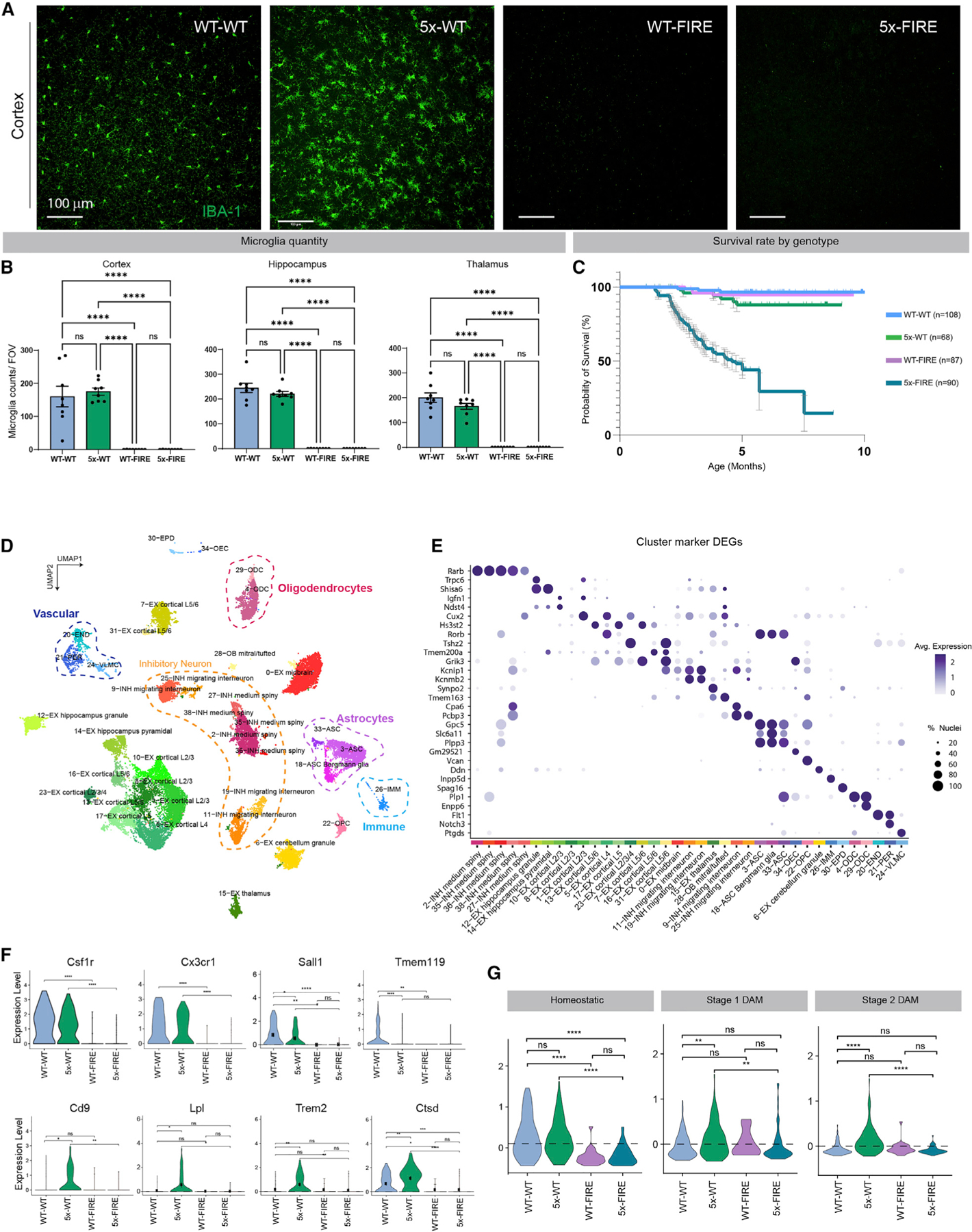
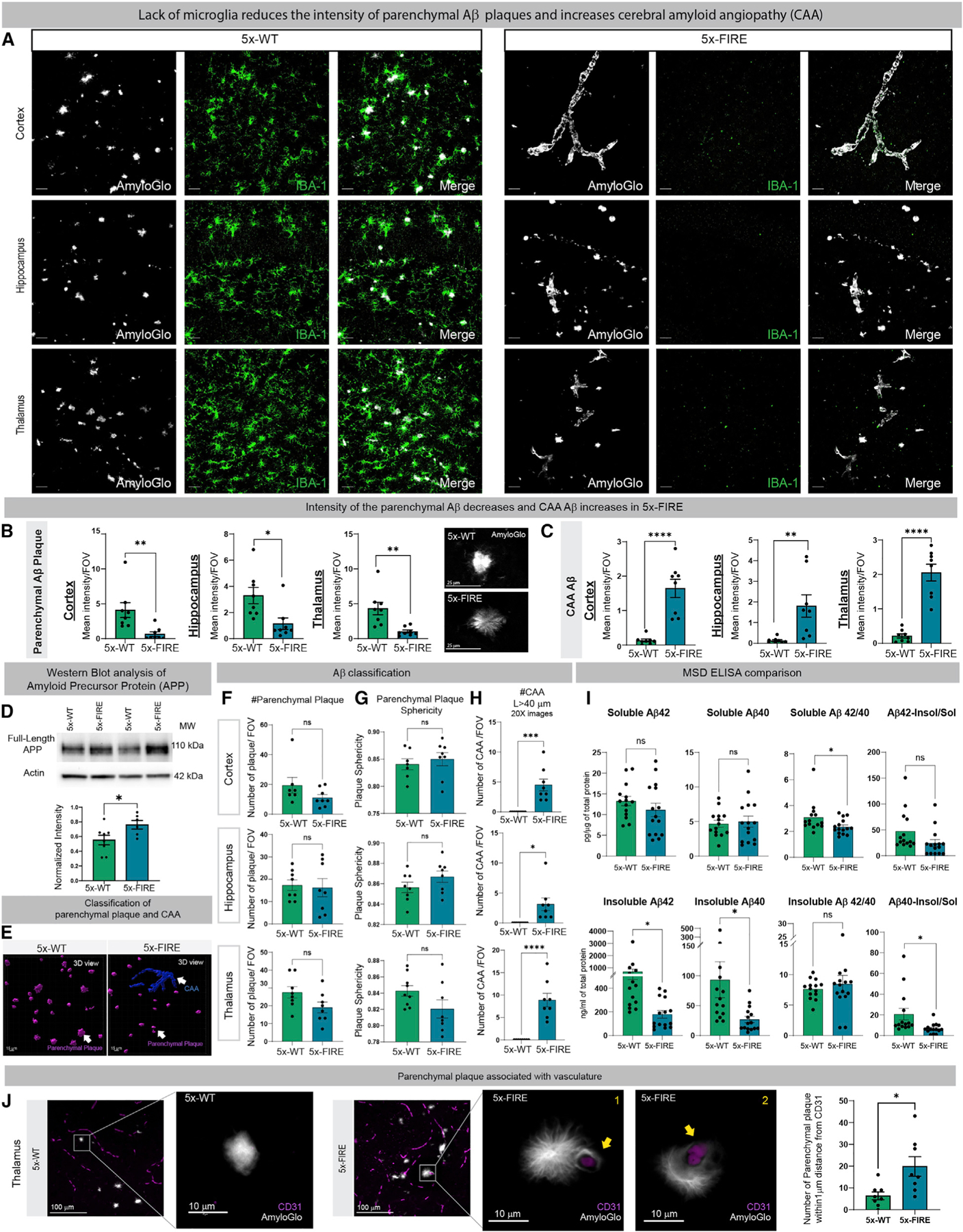
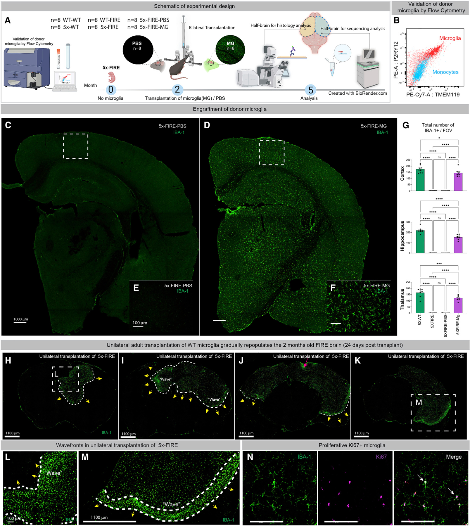
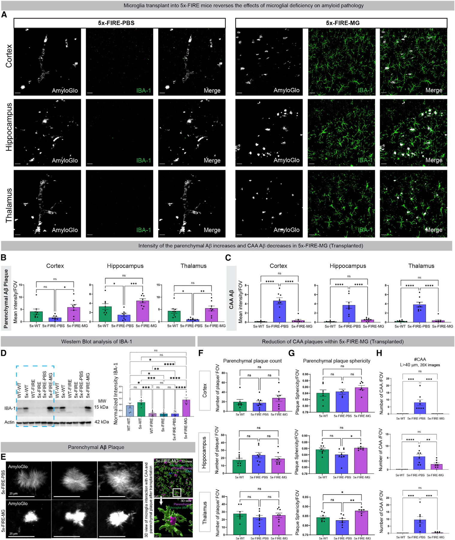
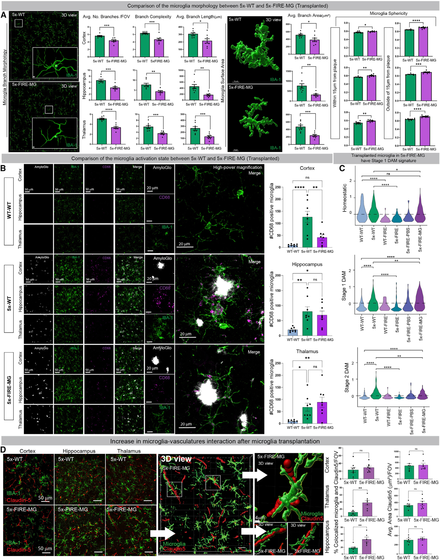
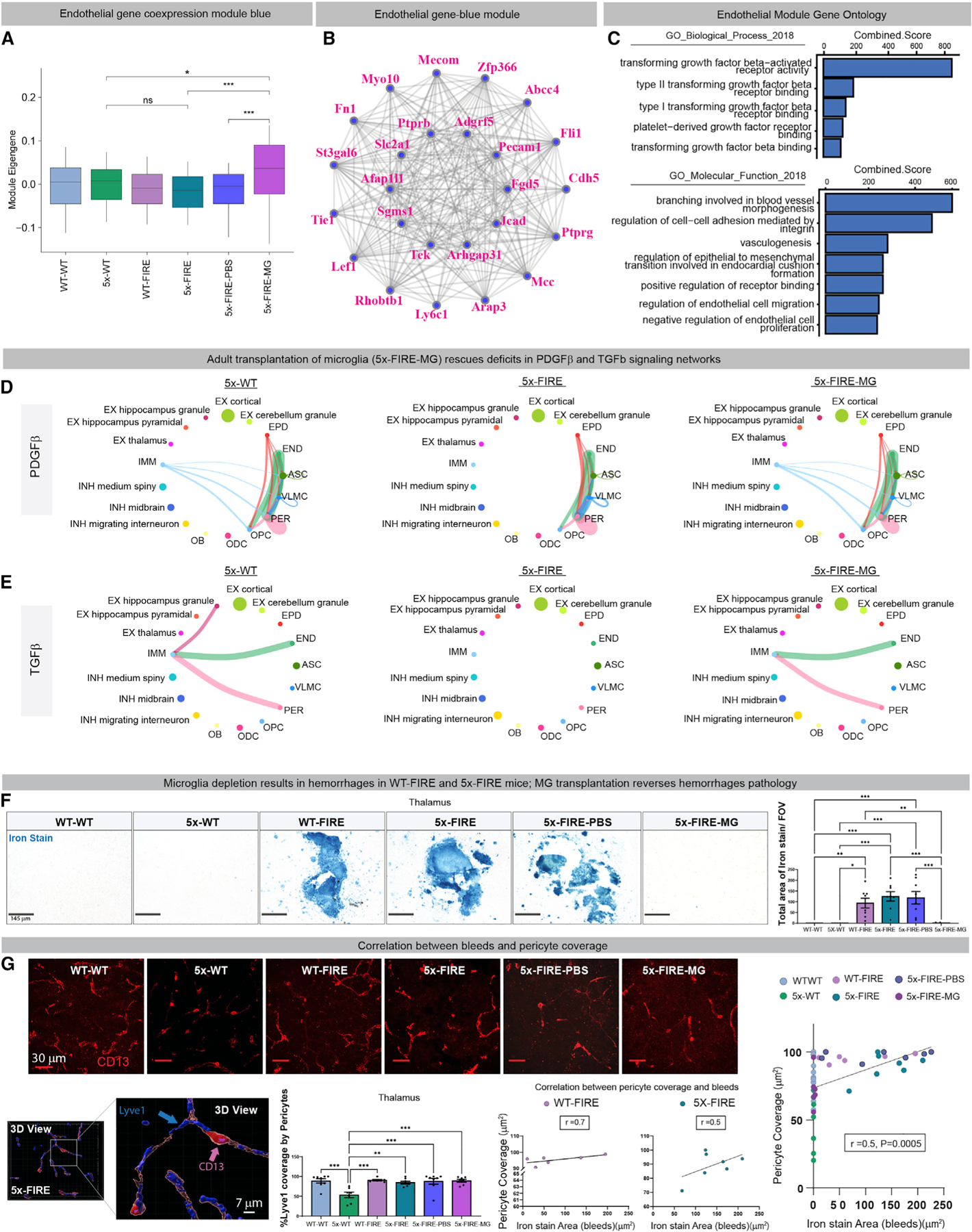
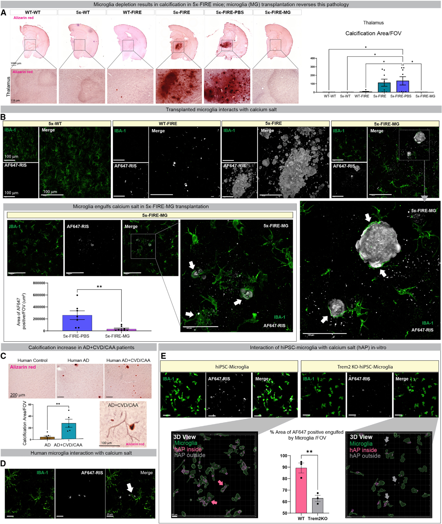
Microglia are strongly implicated in the development and progression of Alzheimer's disease (AD), yet their impact on pathology and lifespan remains unclear. Here we utilize a CSF1R hypomorphic mouse to generate a model of AD that genetically lacks microglia. The resulting microglial-deficient mice exhibit a profound shift from parenchymal amyloid plaques to cerebral amyloid angiopathy (CAA), which is accompanied by numerous transcriptional changes, greatly increased brain calcification and hemorrhages, and premature lethality. Remarkably, a single injection of wild-type microglia into adult mice repopulates the microglial niche and prevents each of these pathological changes. Taken together, these results indicate the protective functions of microglia in reducing CAA, blood-brain barrier dysfunction, and brain calcification. To further understand the clinical implications of these findings, human AD tissue and iPSC-microglia were examined, providing evidence that microglia phagocytose calcium crystals, and this process is impaired by loss of the AD risk gene, TREM2.

Keywords: Alzheimer’s disease; Alzheimer’s disease co-pathologies; CP: Neuroscience; TREM2; brain calcification; cerebral amyloid angiopathy; hemorrhage; iPSC-microglia; microglia; mortality; neurovascular.


Conflict of interest statement

Declaration of interests M.B.-J. is a co-inventor of patent application WO/2018/160496, related to the differentiation of pluripotent stem cells into microglia, and co-founder of NovoGlia. All other authors declare no competing interests.

Figures

Figure 1.
Figure 1.. Genetic absence of microglia in AD mice induces premature lethality and alters cell-specific transcriptional states
(A) Brains form 5–6-month-old mice (n = 8/group) were stained with IBA-1 (green). Representative confocal images from the cortex demonstrate a homeostatic distribution of IBA-1 immunoreactive microglia in WT-WT mice, a more activated clustering of microglia in 5x-WT mice, and absence of microglia in WT-FIRE and 5x-FIRE mice. (B) FIRE mice lack microglia throughout the brain as quantified within the cortex, hippocampus, and thalamus. (C) Kaplan-Meier survival analysis reveals early lethality in 5x-FIRE mice, with fewer than 29% of mice remaining alive at 6 months of age and only 15% remaining alive at 7.5 months. In contrast, WT-WT, 5x-WT, and WT-FIRE mice exhibit minimal lethality. (D) Uniform manifold approximation and projection (UMAP) of snRNA-seq analysis of 5–6-month-old mice (n = 8/group) provides transcriptomic evidence of 37 distinct clusters, including multiple neuronal subtypes, several astrocyte and oligodendrocyte subtypes, and endothelial and immune cell clusters. (E) A dot plot of the highest expressed gene for each cluster; size of dots indicates percent of cells expressing that gene; color indicates relative expression levels. (F) The absence of microglia in WT-FIRE and 5x-FIRE mice is further confirmed by lack of CSF1R, CX3CR1, SALL1, and TMEM119 gene expression, among others. In addition, increased expression of several disease-associated microglial (DAM) transcripts, including CD9, LPL, TREM2, and CTSD, is observed within the 5x-WT group but not within 5x-FIRE mice. (G) Whereas 5x-WT mice exhibit induction of both stage 1 and 2 DAM module genes, 5x-FIRE show no such induction. Scale bars, 100 µm in (A). All data presented as mean ± SEM. *p ≤ 0.05, **p ≤ 0.01, ***p ≤ 0.001, ****p ≤ 0.0001.
Figure 2.
Figure 2.. Microglial-deficient AD mice exhibit reduced intensity of parenchymal plaques and diminished insoluble Aβ but a robust induction of cerebral amyloid angiopathy (CAA)
(A) The 5–6-month-old 5x-WT mice exhibit parenchymal plaque deposition (Amylo-Glo, white) and clustering of IBA-1 microglia (green). (B and C) In contrast, 5x-FIRE mice exhibit diminished plaque intensity and more diffuse morphology (B) and a robust induction of CAA (C) within all three brain regions examined. (D) Western blots reveal a small but significant increase in human APP protein expression in 5x-FIRE mice. (E–G) (E) Imaris image analysis was used to further classify parenchymal versus vascular amyloid pathology, revealing no significant differences in plaque number (F) or sphericity (G). (H) However, the number of CAA deposits (H) was significantly increased in 5x-FIRE mice. (I) ELISA analysis further reveals significantly reduced levels of insoluble Aβ40 and Aβ42. (J) Whereas 5x-WT plaques are only occasionally observed adjacent to CD31+ blood vessels, 5x-FIRE plaques are more frequently associated with blood vessels. Scale bars, 25 µm in (A) and (B); 15 µm in (E); and 100 µm and 10 um in (J). All data presented as mean ± SEM. *p ≤ 0.05, **p ≤ 0.01, ***p ≤ 0.001, ****p ≤ 0.0001.
Figure 3.
Figure 3.. Microglial transplantation leads to brain-wide repopulation of the microglial niche
(A) A schematic illustration of the design of adult microglial transplantation studies. (B) Donor wild-type haplotype-matched microglia (red) were examined by flow cytometry in comparison with murine blood-derived monocytes (blue) and found to express high levels of the homeostatic markers P2RY12 and TMEM119. (C–G) (C and E) IBA-1 labeling of 5x-FIRE-PBS mice show very little immunoreactivity. In contrast, 3 months post bilateral transplantation of 160,000 total wild-type microglia, the brains of 5x-FIRE-MG mice are almost fully repopulated, with IBA-1+ donor microglia (D and F), quantified in (G). (H–N) Pilot studies were performed to examine the repopulation kinetics 24 days after unilateral transplantation of 80,000 microglia. The red arrowhead in (J) marks the injection site from which microglia migrate and expand into the unoccupied niche. (L and M) Higher-power views of the boxed regions in H and M show a dense wavefront of microglia. (N) Immunolabeling with the mitotic marker Ki67 shows increased levels of proliferation within these wavefront microglia. Scale bars, 1000 µm in (C) and (D), 100 µm in (E) and (F), 1100 µm in (H–K) and (M), and 100 µm in (L) and (N). All data presented as mean ± SEM. *p ≤ 0.05, **p ≤ 0.01, ***p ≤ 0.001, ****p ≤ 0.0001.
Figure 4.
Figure 4.. Adult transplantation of wild-type donor microglia prevents the effects of microglial deficiency on amyloid pathology
(A–C) (A) Microglial repopulation leads to a reversal of the previously observed changes in amyloid distribution, increasing parenchymal Aβ intensity back to 5X-WT levels (B), while concurrently decreasing CAA (C) in comparison with PBS-injected control 5x-FIRE mice. (D) Western blot analysis of IBA-1 further demonstrates the loss of microglia in FIRE mice and the return of IBA-1 signal following microglial transplantation. (E) Representative high-power images of parenchymal plaques further demonstrated a shift in morphology from more diffuse filamentous plaques in 5x-FIRE-PBS mice toward more compact, intense morphology in 5x-FIRE-MG mice. (F) Transplantation of microglia has no effect on total plaque numbers. (G and H) In contrast, plaque sphericity within both the hippocampus and thalamus was enhanced by microglial transplantation (G) and the number of CAA deposits in all three brain regions was significantly reduced (H). Scale bars, 25 µm in (C); 25 µm, 20 µm, and 4 µm in (G). All data presented as mean ± SEM. *p ≤ 0.05, **p ≤ 0.01, ***p ≤ 0.001, ****p ≤ 0.0001.
Figure 5.
Figure 5.. Transplanted microglia exhibit differential morphology, altered CD68 expression, and increased association with blood vessels
(A) Imaris analysis of microglial branch number, complexity, length, and area reveals reductions in each of these measures in transplanted microglia versus endogenous 5x-WT microglia. Transplanted microglia also exhibit reduced branch complexity and increase sphericity, both adjacent to (≤15 µm) and more distant from (>15 µm) Aβ plaques. (B) Immunolabeling for CD68 revealed a significant reduction in transplanted microglia within the cortex, but no changes within the hippocampus or thalamus. (C) snRNA-seq demonstrates that transplanted microglia adopt both a DAM1 and DAM2 transcriptomic response in 5x-FIRE-MG mice. (D) Examination of Claudin-5 and IBA-1 immunoreactivity reveal an increased association between transplanted microglia and blood vessel endothelial cells within the thalamus and hippocampus (thalamus shown in Imaris 3D view) but no change in Claudin-5 immunoreactive blood vessel area. Scale bars, 1 µm and 10 µm in (A), 50 µm and 20 µm in (B), and 50 µm and 4 µm in (D). Graphical data presented as mean ± SEM.*p ≤ 0.05, **p ≤ 0.01, ***p ≤ 0.001.
Figure 6.
Figure 6.. Microglial deficiency induces changes in endothelial gene expression and promotes brain hemorrhages, which can be prevented by adult microglial transplantation
(A and B) Co-expression network analysis of snRNA-seq data from 5–6-month-old mice (n = 8/group) reveals significant changes in endothelial cell gene expression between 5x-FIRE mice transplanted with microglia versus PBS-injected 5x-FIRE controls, untranslated 5x-FIRE mice, and 5x-WT mice. (C) Gene ontology analysis further highlights several important pathways that are altered in endothelial cells, including PDGF-β and TGF-β receptor signaling, blood vessel branching, and regulation of endothelial cell proliferation and migration. (D) CellChat analysis was utilized to identify signaling networks between differing cell types, revealing PDGFβ-related connectivity between the IMM microglia and pericyte clusters that was abolished in 5x-FIRE mice but fully restored with microglial transplantation. (E) Analysis of TGF-β signaling likewise exhibited a strong connectivity with both pericytes and endothelial cells that was lost in 5x-FIRE mice but restored by transplantation. (F) Prussian blue iron staining revealed clear evidence of hemorrhages in WT-FIRE, 5x-FIRE, and 5x-FIRE-PBS mice that was completely prevented by adult microglial transplantation. (G) Given our CellChat identification of pericytes as microglial signaling recipients, pericyte coverage of endothelial cells was examined via CD13 labeling (red) of pericytes adjacent to Lyve1+ blood vessels (blue). This analysis confirmed the previously reported reduction in AD transgenic mice pericytes but revealed no differences between WT-WT and FIRE groups. However, regression analysis comparing pericyte coverage to Prussian blue+ hemorrhages revealed a positive correlation between these measures. Scale bars, 145 µm in F, 30 µm in (G), and 7 µm in the 3D view in (G). Data in (A), (F), and (G) presented as mean ± SEM.*p ≤ 0.05, **p ≤ 0.01, ***p ≤ 0.001.
Figure 7.
Figure 7.. Brain calcification occurs in 5x-FIRE mice and a subset of human AD subjects, and loss of TREM2 impairs microglial phagocytosis of calcium crystals
(A) To determine whether other brain vascular pathologies might impact cerebral hemorrhages, we next examined calcium accumulation with alizarin red, revealing a striking increase in calcification within the thalamus of 5x-FIRE mice and 5x-FIRE-PBS mice that was fully resolved in microglial-transplanted 5x-FIRE mice. (B) Excessive calcium accumulates in the form of hydroxyapatite calcium phosphate crystals, which can be detected with the fluorescent indicator AF657-VIS. Confocal imaging of AF657-VIS (white) revealed a complete lack of calcium accumulation in 5x-WT mice, a small increase in hydroxyapatite in WT-FIRE mice, and a substantial increase in 5x-FIRE mice. In contrast, microglial transplanted FIRE-5x mice exhibited greatly decreased hydroxyapatite labeling with remaining calcium crystals (white) often surrounded by IBA-1+ microglia (green, arrows) and evidence of microglial phagocytic engulfment of hydroxyapatite observed. (C) To determine the potential implications of this finding for human AD, we examined the relationship between brain calcium accumulation and Aβ plaque pathology in AD cases. (D) This analysis revealed a significant increase in brain calcification within AD patients that exhibit vascular pathologies, with alizarin red calcification often observed adjacent to blood vessels, and microglia (D, IBA-1, green) observed adjacent to AF657-VIS+ calcification (white). (E) Lastly, to determine whether microglial expression of TREM2 might influence calcification, WT and TREM2 knockout iPSC-microglia (GFP, green) were exposed to hydroxyapatite calcium phosphate crystals (HAp, AF647-RIS, gray) and percent internalization determined, revealing a failure of Trem2 KO microglia to efficiently phagocytose calcium crystals. Scale bars, 1000 µm and 118 µm in (A), 100 µm in (B), 200 µm and 100 µm in (C), 25 µm in (D), and 100 µm and 20 µm in (E), as labeled within the images. All graphs presented as mean ± SEM. *p ≤ 0.05, **p ≤ 0.01

References

    1. Arnold TD, Niaudet C, Pang M-F, Siegenthaler J, Gaengel K, Jung B, Ferrero GM, Mukouyama Y.s., Fuxe J, Akhurst R, et al. (2014). Excessive vascular sprouting underlies cerebral hemorrhage in mice lacking αVβ8-TGFβ signaling in the brain. Dev. Camb. Engl 141, 4489–4499. 10.1242/dev.107193. - DOI - PMC - PubMed
    1. Arvanitakis Z, Leurgans SE, Wang Z, Wilson RS, Bennett DA, and Schneider JA (2011). Cerebral amyloid angiopathy pathology and cognitive domains in older persons. Ann. Neurol 69, 320–327. 10.1002/ana.22112. - DOI - PMC - PubMed
    1. Ayrignac X, Nicolas G, Carra-Dallière C, Hannequin D, and Labauge P (2017). Brain calcifications in adult-onset genetic leukoencephalopathies: a review. JAMA Neurol 74, 1000. 10.1001/jamaneurol.2017.1062. - DOI - PubMed
    1. Bell RD, and Zlokovic BV (2009). Neurovascular mechanisms and blood-brain barrier disorder in Alzheimer’s disease. Acta Neuropathol. 118, 103–113. 10.1007/s00401-009-0522-3. - DOI - PMC - PubMed
    1. Bennett RE, Bryant A, Hu M, Robbins AB, Hopp SC, and Hyman BT (2018). Partial reduction of microglia does not affect tau pathology in aged mice. J. Neuroinflammation 15, 311. 10.1186/s12974-018-1348-5. - DOI - PMC - PubMed

Publication types