. 2023 Apr;72(4):671-685.
doi: 10.1136/gutjnl-2021-326500. Epub 2022 Jun 15.

Protective and aggressive bacterial subsets and metabolites modify hepatobiliary inflammation and fibrosis in a murine model of PSC

Affiliations

Protective and aggressive bacterial subsets and metabolites modify hepatobiliary inflammation and fibrosis in a murine model of PSC

Muyiwa Awoniyi et al. Gut. 2023 Apr.

Abstract

Objective: Conflicting microbiota data exist for primary sclerosing cholangitis (PSC) and experimental models.

Goal: define the function of complex resident microbes and their association relevant to PSC patients by studying germ-free (GF) and antibiotic-treated specific pathogen-free (SPF) multidrug-resistant 2 deficient (mdr2-/- ) mice and microbial profiles in PSC patient cohorts.

Design: We measured weights, liver enzymes, RNA expression, histological, immunohistochemical and fibrotic biochemical parameters, faecal 16S rRNA gene profiling and metabolomic endpoints in gnotobiotic and antibiotic-treated SPF mdr2-/- mice and targeted metagenomic analysis in PSC patients.

Results: GF mdr2-/- mice had 100% mortality by 8 weeks with increasing hepatic bile acid (BA) accumulation and cholestasis. Early SPF autologous stool transplantation rescued liver-related mortality. Inhibition of ileal BA transport attenuated antibiotic-accelerated liver disease and decreased total serum and hepatic BAs. Depletion of vancomycin-sensitive microbiota exaggerated hepatobiliary disease. Vancomycin selectively decreased Lachnospiraceae and short-chain fatty acids (SCFAs) but expanded Enterococcus and Enterobacteriaceae. Antibiotics increased Enterococcus faecalis and Escherichia coli liver translocation. Colonisation of GF mdr2-/- mice with translocated E. faecalis and E. coli strains accelerated hepatobiliary inflammation and mortality. Lachnospiraceae colonisation of antibiotic pretreated mdr2-/- mice reduced liver fibrosis, inflammation and translocation of pathobionts, and SCFA-producing Lachnospiraceae and purified SCFA decreased fibrosis. Faecal Lachnospiraceae negatively associated, and E. faecalis/ Enterobacteriaceae positively associated, with PSC patients' clinical severity by Mayo risk scores.

Conclusions: We identified novel functionally protective and detrimental resident bacterial species in mdr2-/- mice and PSC patients with associated clinical risk score. These insights may guide personalised targeted therapeutic interventions in PSC patients.

Keywords: antibiotics; cholestatic liver diseases; intestinal microbiology; primary sclerosing cholangitis; short chain fatty acids.


Conflict of interest statement

Competing interests: RBS has consulted for and received grant support from Takeda, Janssen, Second Genome, Vedanta, BiomX, Biomica, SERES and Artizan; JRH has served on advisory boards and/or given lectures for Orkla Health, Novartis, Amgen and Roche, and received research support from Biogen, all unrelated to the present study.

Figures
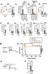
Figure 1
Figure 1
Clinical, biochemical and liver histological indicators of germ-free (GF) mdr2−/− mice with or without faecal microbiotal transplant compared with control mice. Mouse body weight (A) and serum alkaline phosphatase (AP), (B) of 6–8 week old GF (n=6–10) and SPF (n=20) mdr2−/− mice. Longitudinal total bilirubin (TB) (C) or total serum bile acids (TBA) (D) assessment in GF mdr2−/− mice from 4–5 week old (n=7) to 6–8 week old (n=8), compared 6–8 week old SPF mdr2−/− and C57/BL6 WT GF mice and older (26–70 week old) mdr2+/− ±. Total liver bile acids (Bas) in mdr2−/− or WT mice in GF or SPF environments (E, n=5–9/mice per group). Liver cyp7a1 expression (F) and liver FGF-15 expression (G). Relative expression (fold change) of liver cyp7a1, terminal Ileal FGF-15, Rorγt and tnf-α (F–I) of 6–8 week old GF (n=5–7) and SPF (n=5–7) mdr2−/− mice. Hepatic collagen deposition expressed as total hepatic hydroxyproline (HYP) expressed in ug HYP/whole liver, calculated by multiplying individual liver weight with relative HYP content (J). Relative expression (fold change) of collagen I alpha-1 (col1α1) and tissue inhibitor of metalloproteinases (TIMP) 1 (K–L) in livers of 6–8 week old GF (n=5–12) and SPF (n=5–8) mdr2−/− mice. Experimental design of autologous faecal microbiota transplant (FMT) study (M) Kaplan-Meyer survival curves of pooled SP mdr2−/ donor stool orally gavaged into GF mdr2−/− C57Bl/6 mice twice in the first 2 days at age 3–4 weeks (n=8) or 5.5–7 weeks (n=6) compared with untreated GF (n=23) or SPF (n=14) mdr2−/− mice monitored for survival for 42 weeks (N). Experimental design of GF mdr2−/− given autologous GF (n=7) or SPF donor (n=6) stool for 14 days (O) monitoring serum TB (P). Results are expressed as means±SEM. Survival data are analysed by log-rank (Mantel-Cox) test, group or pairwise comparisons performed by analysis of variance or Student’s t-test, respectively. Welch correction was applied to histological scoring analysis. *P<0.05, **p<0.01, ***p<0.001, ****p<0.0001. SPF, specific pathogen free.
Figure 2
Figure 2
Histological impact of absent and reconstitution of resident microbes in GF mdr2−/− mice. Representative photomicrographs of 6–8 week old GF mdr2−/− mice±faecal microbiota transplant at 3–4 week old compared with age-matched SPF mdr2−/− mice with blinded scoring for liver inflammation (A and B), macrophages (4-4/80 immunohistochemical staining) (C and D), ductular reaction (CK-19 immunohistochemical staining) (E and F) and fibrosis (G and H) stained by H&E (200×), antibodies to F4/80 (200×) and CK-19 (200×), and Sirius Red (100×), respectively. Arrows indicate foci of hepatocyte degeneration and necrosis bordered by inflammation (bile infarct). CK-19 staining in panels D and F count positive cells in five high powered fields/ slide. Results are expressed as means±SEM. Group or pairwise comparisons were performed by analysis of variance or Student’s t-test, respectively. Welch correction was applied to histological scoring analysis. *P<0.05, **p<0.01, ***p<0.001, ****p<0.0001. CV, central vein; PV, portal vein.
Figure 3
Figure 3
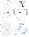
Faecal microbiome and metabolite profiles of preantibiotic/postantibiotic treated mdr2−/− and wild-type (WT) mice on C57BL/6 background. Pooled baseline untreated SPF mdr2−/− and WT C57BL/6 16S sequencing faecal beta diversity (Permanova test) (A) and linear discriminant analysis plot showing differential enrichment of taxa (B). Experimental design of broad-spectrum antibiotic (Abx) study, 3–5 week old SPF mdr2−/− or WT mice exposed ad libitum to broad-spectrum antibiotics (metronidazole: 30 mg/mL; vancomycin: 0.5 mg/mL; and neomycin: 1 mg/mL) in drinking H2O or H2O alone (n=10 mice/group) for 14 days (pooled from three experiments) (C). Effect of 14 days of Abx assessing pooled baseline alpha diversity (Faith’s PD) and faecal universal 16S qCR (expressed in ΔΔCT differences) in mdr2−/− (D and E) and WT BL/6 (F and G) mice, respectively. Principal coordinates analysis (PcoA) plots assessing beta diversity (Permanova test) antibiotic exposure between genotypes (WT and mdr2−/− mice) (H) and antibiotic exposure in mdr2−/− mice (I). 3D-PCA plot shows the separation of cecal (J) and serum (K) metabolites from mdr2−/− with and without antibiotics. Mummichog pathway cloud plot of potential pathway differences with exposure of broad antibiotics in SPF mdr2−/− mice (L). The radius of each circle represents the number of metabolites relative to the number of metabolites represented by other circles. Darker circles mean more pathways are represented (mdr2−/− no Abx: n=13, mdr2−/− with Abx: n=25). Group or pairwise comparisons performed by analysis of variance or Student’s t-test, respectively. *P<0.05, **p<0.01, ***p<0.001, ****p<0.0001. SPF, specific pathogen free.
Figure 4
Figure 4
Clinical, biochemical and liver histological indicators of broad-spectrum antibiotic treated mdr2−/− mice. Experimental design of broad-spectrum antibiotic (Abx) study, 3–5 week old SPF mdr2−/− mice exposed ad libitum to broad-spectrum antibiotics in drinking H2O (n=15) or H2O alone (n=13) for 14 days (pooled from three experiments) (A). Effect of Abx exposure on various outcomes including relative abundance of selected bacterial phyla expressed as unadjusted raw average operational taxonomic unit (OTU) relative abundance (B), colonic/TI permeability (WT & mdr2−/− mice) (C), 14 days body weight change (D), serum ALP, TB (E and F), RNA expression of liver tnf-α (G), col1α1 (I) and timp-1 (J) and hepatic collagen deposition expressed as ug HYP/whole liver (H). Representative photomicrographs and blinded composite histological scoring murine liver stained with H&E (K–L), CK-19 (M–N) and Sirius Red (O–P) of untreated (n=15) versus Abx-treated SPF mdr2−/− mice (n=15). Results are expressed as means±SEM. Pairwise comparisons were performed by Student’s t-test for biochemical and molecular studies; Welch correction was applied to histological scoring analysis. Unadjusted raw average OTU relative abundance and SEs of bacterial groups against the variables detected with significant effects by analysis of composition of microbes). *P<0.05, **p<0.01, ***p<0.001. ALP, alkaline phosphatase; SPF, specific pathogen free; TB, total bilirubin.
Figure 5
Figure 5
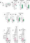
Bile acid homeostasis and hepatobiliary injury assessed in Abx treated mdr2−/− mice following ASBT blockage. Experimental design of ad libitum exposure in drinking H2O of ASBT inhibitor (GSK23306, 10 mg/kg) for 14-day concomitant with 7-day broad antibiotic pretreatment in 4–5 week old SPF mdr2−/− mice versus no ASBTi (water only) group (n=5–7 mice/group) (A). Effect of ASBT inhibition on: weight change (B), TB (C), serum and liver total BA (D–E), liver C4 (F, metabolite of Cyp7a1 activation), histological liver inflammation and fibrosis (G and H). Assessment of longitudinal faecal bile acid homeostasis at times 0, 7 and 14 days following initiation of ASBT inhibition evaluating TBA, cholic acid (CA) and α+β MCA (I–K). Group or pairwise comparisons performed by analysis of variance or Student’s t-test, respectively. *P<0.05, **p<0.01, ***p<0.001, ****p<0.0001. ASBTi, apical sodium-dependent bile acid inhibitor; SPF, specific pathogen free; TB, total bilirubin.
Figure 6
Figure 6
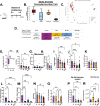
Differential effects of selective antibiotics on mdr2−/− microbial, clinical, biochemical and histological outcomes. Experimental design of selective antibiotic treatment (A): SPF mdr2−/− mice were given individual antibiotics (vancomycin (V; 0.5 mg/mL, n=16), neomycin (N; 1 mg/mL, n=12) or metronidazole (M; 30 mg/mL, n=16) in autoclaved drinking water ad libitum and control mdr2−/− mice received autoclaved water alone (n=16) for 14 days. Alpha diversity, beta diversity (Permanova test) and differential abundance (centred log-ratio (CLR) transformation from CoDA methods) of Clostridiaceae and Lachnospiraceae from 14d faecal samples (B–D). Measured parameters include: 14-day weight change (E), serum ALP (F) and serumtotal bile acids (TBA) (G), and liver RNA expression of tnf-α (H); along with blinded composite histologic scoring murine liver stained with H&E (I) and CK-19 (J). Fibrosis readouts include hepatic collagen deposition (K), RNA expression of col1α1 (L) and timp-1 (M) along with Sirius Red composite staining (N) cyp7a1 liver expression (O) as well as pooled cecal content bile acids (BAs) differences in surrogate bile salt hydrolase activity indicator based on ratios of total unconjugated/conjugated BA (P) and total BA (R) of selective antibiotic treated mdr2−/− compared with H2O control. (V=9, N=8, M=8, H2O=8). CK-19+ represented of 5HPF/liver/mouse. Group or pairwise comparisons performed by analysis of variance or Student’s t-test, respectively. Welch correction was applied to histological scoring analysis. Unadjusted raw average OTU relative abundance and SEs of bacterial groups against the variables detected with significant effects by analysis of composition of microbes. *P<0.05, **p<0.01, ***p<0.001, ****p<0.0001. ALP, alkaline phosphatase; SPF, specific pathogen free.
Figure 7
Figure 7
Lachnospiraceae phenotypic and metabolic effects on dysbiosis in mdr2−/− mice. Using the 7-day broad-spectrum antibiotic pretreatment model, followed by a 1-day washout, 21 Lachnospiraceae strains (Lachno) versus H2O were administered to SPF mdr2−/− mice on days 8, 10 and 12 (A). Changes following Lachno treatment included Δweight over 7 days (B), serum ALP (C), liver RNA col1α1 expression (D), blinded histological inflammation, ductal reaction and hepatic fibrosis scoring (E–G). Differential abundance expression of 16S rRNA analysis (H) (N, mdr2−/− untreated=24, Abx=36, H2O=11, Lachnospiraceae=13, pooled from two experiments. Presence of universal 16S, Enterococcus and Klebsiella pneumoniae (K. pneum) faecal DNA by qPCR (I–K) (one of two representative experiment, n=6 mice/group). Hepatic bacterial translocation (L), % of mice with Enterococcus faecalis liver translocation (M). Experimental design of treatment of 3–4 week old SPF mdr2−/− mice treated with broad-spectrum antibiotic cocktail (vancomycin, neomycin and metronidazole) for 7 days followed by a 1-day washout, then inoculated with mdr2 /− resident Ec/Ef isolates, versus Ec/Ef+23-Lachno combination or water only controls (N) assessing translocated bacteria cultured from homogenised liver (O) (n=4 mice/group). Experiment design (P) and Kaplan-Meyer survival curves (Q) of orally inoculated 108 pooled Ec/Ef hepatic isolates±Lachnospiraceae (Lachno) or H2O controls in GF mdr2−/− mice (H2O: n=12; Ec/Ef: n=7; Lachno: n=6). GF mdr2−/− mice were administered 108 pooled Ec/Ef hepatic isolates and euthanized at 5 weeks in order to measure translocated bacteria to liver, spleen or blood (S–U). Longitudinal measurements of serum TBA and bilirubin conducted in GF mdr2−/− ±Ec/Ef hepatic isolates (V and W). Group or pairwise comparisons performed by analysis of variance or Student’s t-test, respectively. *P<0.05, **p<0.01, ***p<0.001, ****p<0.0001. GF, germ free; SPF, specific pathogen free.
Figure 8
Figure 8
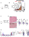
Effect of short-chain fatty acids (SCFAs) on mdr2−/− antibiotic model. Experimental design of 14-day selective antibiotic treatment (A) with measurement of cecal content SCFA in SPF mdr2−/− mice (B). Design of SPF mdr2−/− mice treated with vancomycin (0.5 mg/mL in drinking water, n=10) with or and without SCFA (67.5 mM acetate, 25.9 mM propionate, 40 mM butyrate and 3% sucrose, n=13) ad libitum (C), pooled from two experiments with representative Sirius Red photomicrographs of vancomycin versus vancomycin+SCFA SPF mdr2−/− mice, arrows highlight bridging fibrosis (D), blinded histologic fibrosis scoring (E) along with liver RNA col1α1 and timp1 expression (F–G). Three relatively low and high SCFA-producing (Lo strain #s: 8, 9 and 21; Hi #s: 52, 60 and 70) Lachnospiraceae strains selected from 21 consortium strains were identified by mass spectrometry (H). Experimental design of accelerated antibiotic pretreatment of SPF mdr2−/− mice for 7 days and following a 1 day washout, administering Hi or Lo SCFA-producing Lachnospiraceae strains by gavage on days 8, 9 and 10, then followed for 6 days (I) with measurement of histologic hepatic fibrosis (J) and inflammation (K) in Hi compared with Lo SCFA-producing strains. Group or pairwise comparisons performed by analysis of variance or Student’s t-test, respectively. *P<0.05, **p<0.01, ***p<0.001, ****p<0.0001.
Figure 9
Figure 9
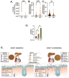
Selective faecal bacterial profile in PSC patients and effect of antibiotic exposure. Relative abundance of faecal Enterococcus faecalis (E. faecalis) (A), Enterobacteriaceae (B) and Lachnospiraceae (C) in heathy controls (HCs; n=158) versus PSC patients (n=136). (D) Prevalence of faecal E. faecalis in HC (irrespective of antibiotics use) versus PSC patients exposed to antibiotics (n=24) and no antibiotics exposure (n=112) the last 6 months. Data in figure parts A–C compared with Mann–Whitney U test, (D) with Fisher’s exact test. Graphical summary depicting the effect of antibiotic-induced dysbiosis of SPF mdr2−/− mice to potentiate hepatobiliary disease with hepatic translocation of E. faecalis and Escherichia coli, increased bile salt hydrolase activity (conjugated/unconjugated BA), hepatic bile acid pool size, and decreased SCFA production (E). *P<0.05, **p<0.01, ***p<0.001, ****p<0.0001. BA, bile acid; PSC, primary sclerosing cholangitis; SCFA, short-chain fatty acid.

Comment in

References

    1. Weismüller TJ, Trivedi PJ, Bergquist A, et al. . Patient age, sex, and inflammatory bowel disease phenotype associate with course of primary sclerosing cholangitis. Gastroenterology 2017;152:e8:1975–84. 10.1053/j.gastro.2017.02.038 - DOI - PMC - PubMed
    1. Nakamoto N, Sasaki N, Aoki R, et al. . Gut pathobionts underlie intestinal barrier dysfunction and liver T helper 17 cell immune response in primary sclerosing cholangitis. Nat Microbiol 2019;4:492–503. 10.1038/s41564-018-0333-1 - DOI - PubMed
    1. Liwinski T, Zenouzi R, John C, et al. . Alterations of the bile microbiome in primary sclerosing cholangitis. Gut 2020;69:665–72. 10.1136/gutjnl-2019-318416 - DOI - PubMed
    1. Kummen M, Thingholm LB, Rühlemann MC, et al. . Altered gut microbial metabolism of essential nutrients in primary sclerosing cholangitis. Gastroenterology 2021;160:1784–98. 10.1053/j.gastro.2020.12.058 - DOI - PMC - PubMed
    1. Kunzmann LK, Schoknecht T, Poch T, et al. . Monocytes as potential mediators of pathogen-induced T-helper 17 differentiation in patients with primary sclerosing cholangitis (PSC). Hepatology 2020;72:1310–26. 10.1002/hep.31140 - DOI - PubMed

Publication types