

Abnormal sensory perception masks behavioral performance of Grin1 knockdown mice
- PMID: 35705513
- PMCID: PMC9744498
- DOI: 10.1111/gbb.12825
Abnormal sensory perception masks behavioral performance of Grin1 knockdown mice
Abstract
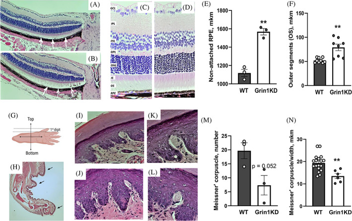
The development and function of sensory systems require intact glutamatergic neurotransmission. Changes in touch sensation and vision are common symptoms in autism spectrum disorders, where altered glutamatergic neurotransmission is strongly implicated. Further, cortical visual impairment is a frequent symptom of GRIN disorder, a rare genetic neurodevelopmental disorder caused by pathogenic variants of GRIN genes that encode NMDA receptors. We asked if Grin1 knockdown mice (Grin1KD), as a model of GRIN disorder, had visual impairments resulting from NMDA receptor deficiency. We discovered that Grin1KD mice had deficient visual depth perception in the visual cliff test. Since Grin1KD mice are known to display robust changes in measures of learning, memory, and emotionality, we asked whether deficits in these higher-level processes could be partly explained by their visual impairment. By changing the experimental conditions to improve visual signals, we observed significant improvements in the performance of Grin1KD mice in tests that measure spatial memory, executive function, and anxiety. We went further and found destabilization of the outer segment of retina together with the deficient number and size of Meissner corpuscles (mechanical sensor) in the hind paw of Grin1KD mice. Overall, our findings suggest that abnormal sensory perception can mask the expression of emotional, motivational and cognitive behavior of Grin1KD mice. This study demonstrates new methods to adapt routine behavioral paradigms to reveal the contribution of vision and other sensory modalities in cognitive performance.
Keywords: GRIN1; Morris water maze; anxiety; autism; elevated plus maze; learning & memory; mice; puzzle box; schizophrenia; tactile function; vision.
© 2022 The Authors. Genes, Brain and Behavior published by International Behavioural and Neural Genetics Society and John Wiley & Sons Ltd.
Conflict of interest statement
A.J.R. is a paid consultant to CureGRIN Research Foundation. The authors declare no potential conflict of interest.
Figures


Similar articles
-
ENU-mutagenesis mice with a non-synonymous mutation in Grin1 exhibit abnormal anxiety-like behaviors, impaired fear memory, and decreased acoustic startle response.BMC Res Notes. 2013 May 21;6:203. doi: 10.1186/1756-0500-6-203. BMC Res Notes. 2013. PMID: 23688147 Free PMC article.
-
Preweaning sensorimotor deficits and adolescent hypersociability in Grin1 knockdown mice.Dev Neurosci. 2012;34(2-3):159-73. doi: 10.1159/000337984. Epub 2012 May 8. Dev Neurosci. 2012. PMID: 22571986 Free PMC article.
-
NMDA receptor-deficient mice display sexual dimorphism in the onset and severity of behavioural abnormalities.Genes Brain Behav. 2014 Nov;13(8):850-62. doi: 10.1111/gbb.12183. Epub 2014 Nov 12. Genes Brain Behav. 2014. PMID: 25327402
-
5-HT1B receptor knock-out mice exhibit increased exploratory activity and enhanced spatial memory performance in the Morris water maze.J Neurosci. 1999 Jul 15;19(14):6157-68. doi: 10.1523/JNEUROSCI.19-14-06157.1999. J Neurosci. 1999. PMID: 10407051 Free PMC article.
-
Impact of postnatal blockade of N-methyl-D-aspartate receptors on rat behavior: a search for a new developmental model of schizophrenia.Neuroscience. 2008 Jun 2;153(4):1370-9. doi: 10.1016/j.neuroscience.2008.03.016. Epub 2008 Mar 20. Neuroscience. 2008. PMID: 18434025
Cited by
-
Discovering functional interactions among schizophrenia-risk genes by combining behavioral genetics with cell biology.Neurosci Biobehav Rev. 2024 Dec;167:105897. doi: 10.1016/j.neubiorev.2024.105897. Epub 2024 Sep 14. Neurosci Biobehav Rev. 2024. PMID: 39278606 Free PMC article. Review.
-
PDE4B Missense Variant Increases Susceptibility to Post-traumatic Stress Disorder-Relevant Phenotypes in Mice.J Neurosci. 2024 Oct 23;44(43):e0137242024. doi: 10.1523/JNEUROSCI.0137-24.2024. J Neurosci. 2024. PMID: 39256048 Free PMC article.
-
Valence-Driven Cognitive Flexibility: Neurochemical and Circuit-Level Insights from Animal Models and Their Relevance to Schizophrenia.Biomolecules. 2025 Aug 11;15(8):1154. doi: 10.3390/biom15081154. Biomolecules. 2025. PMID: 40867599 Free PMC article. Review.
-
Identification and validation of integrated stress-response-related genes as biomarkers for age-related macular degeneration.Front Mol Biosci. 2025 Jul 16;12:1583237. doi: 10.3389/fmolb.2025.1583237. eCollection 2025. Front Mol Biosci. 2025. PMID: 40740189 Free PMC article.
-
Modulation of the thermosensory system by oxytocin.Front Mol Neurosci. 2023 Jan 9;15:1075305. doi: 10.3389/fnmol.2022.1075305. eCollection 2022. Front Mol Neurosci. 2023. PMID: 36698777 Free PMC article. Review.
References
-
- Chisholm K, Lin A, Abu‐Akel A, Wood SJ. The association between autism and schizophrenia spectrum disorders: a review of eight alternative models of co‐occurrence. Neurosci Biobehav Rev. 2015;55:173‐183. - PubMed
-
- Zhou H‐Y, Cai X‐L, Weigl M, Bang P, Cheung EFC, Chan RCK. Multisensory temporal binding window in autism spectrum disorders and schizophrenia spectrum disorders: a systematic review and meta‐analysis. Neurosci Biobehav Rev. 2018;86:66‐76. - PubMed
-
- Tomchek SD, Dunn W. Sensory processing in children with and without autism: a comparable study using the short sensory profile. Am J Occup Ther. 2007;61:190‐200. - PubMed
Publication types
MeSH terms
Substances
LinkOut - more resources
Full Text Sources
