. 2022 Jun 15;12(1):10008.
doi: 10.1038/s41598-022-13584-5.

TSP, a virulent Podovirus, can control the growth of Staphylococcus aureus for 12 h

Affiliations

TSP, a virulent Podovirus, can control the growth of Staphylococcus aureus for 12 h

Rabia Tabassum et al. Sci Rep. .

Abstract

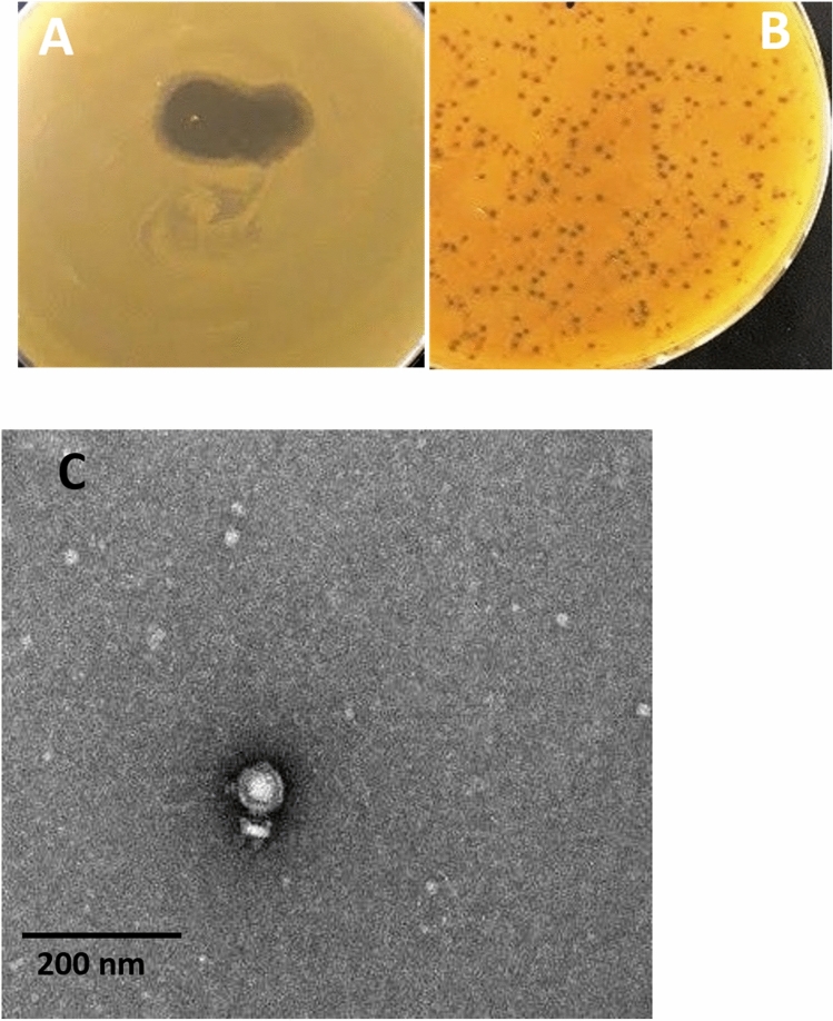
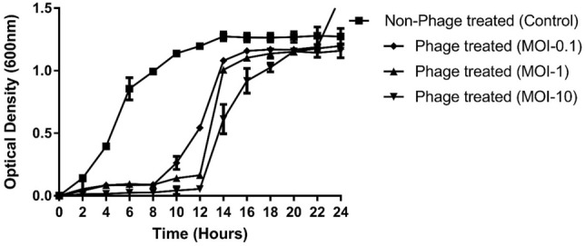
Methicillin-resistant Staphylococcus aureus (MRSA) is a prevailing nosocomial pathogen that is increasingly isolated in community settings. It shows resistance against all beta-lactam drugs and has acquired mechanisms to resist other groups of antibiotics. To tackle this emerging issue of MRSA, there is an urgent need for antibiotic alternatives, and utilizing lytic bacteriophages is one of the most promising therapeutic approaches. In the present study, a lytic bacteriophage TSP was isolated from hospital wastewater against MRSA. The phage efficiently inhibited bacterial growth for up to 12 h at MOI of 1 and 10. TSP phage showed activity against various isolates of MRSA and MSSA, isolated from different clinical samples, with variable antibiotic susceptibility patterns. The bacteriophage TSP showed stability at varying temperatures (25 °C, 37 °C) and pH values (5-9), while its maximum storage stability was observed at 4 °C. It had a short latent period (20 min) and burst size of 103 ± 5pfu/infected cells. TSP genome sequence and restriction analysis revealed that its genome has a linear confirmation and length of 17,987 bp with an average GC content of 29.7%. According to comparative genomic analysis and phylogenetic tree,TSP phage can be considered a member of genus "P68viruses". The strong lytic activity and short latent period in addition to its lytic nature makes it a good candidate for phage therapy against MRSA infections, if it proves to be effective in in-vivo studies.


Conflict of interest statement

The authors declare no competing interests.

Figures

Figure 1
Figure 1
(A) The bacteriophage TSP spot on the lawn of MR10. (B) Small, circular and lytic plaques of TSP phage obtained by plaque assay (C). Transmission electron microscopy of the TSP phage showed its resemblance to the Podoviridae family. The scale bar represents 200 nm and image was taken at 25,000X magnification.The uncroped Fig. (A) and (B) are shown in Fig. S3 in supplementary data.
Figure 2
Figure 2
Determination of the invitro bacteriolytic activity of TSP bacteriophage. Phage-treated group: MRSA strain cocultured in logarithmic phase with TSP phage at an MOI of 0.1, MOI-1 and MOI-10. Control group: MRSA culture without phage TSP. The OD of the control and phage-treated groups was measured at 600 nm after an interval of 2 h for up to 24 h. The results are shown as the mean values with standard deviation.
Figure 3
Figure 3
TSP bacteriophage stability assay. (A) Effect of different temperatures (25, 37, 45, 50 and 60 °C) on the stability of the TSP phage. (B) TSP phage was treated over a wide pH range (4, 5, 6, 7, 8, 9 and 10) for 1 h. (C) Storage stability of TSP phage at different temperatures (4, 25,  − 20 and -80 °C) showed maximum survival ability at 4 °C. The experiment was performed three times, and phage titers are expressed as the mean ± standard deviation.
Figure 4
Figure 4
(A) TSP phage adsorption kinetics. (B) One-step growth curve analysis of bacteriophage TSP infecting MR10 at 37 °C. The results were obtained from three independent experiments.
Figure 5
Figure 5
Agarose gel analysis of TSP phage DNA double digested with NcoI and EcoRI. Lane 1: Restricted TSP phage DNA, Lane 2: Unrestricted TSP phage DNA and Lane 3: Lambda phage DNA HindIII digested marker (Cat#302,005 Bioron). An uncroped agrose gel of the above figure isshown in Fig. S4.
Figure 6
Figure 6
Linear genome map of TSP phage. The direction of ORFs is depicted via arrows. Four functional groups are present in the TSP phage genome, and genes in each functional group are represented by different colors: structural genes (yellow), regulatorygenes (blue), host lysis genes (red), and DNApackaginggenes (green). Hypothetical ORFs are indicated in purple.
Figure 7
Figure 7
(A) Comparative genomic analysis of S. aureus phages. Whole genome sequences were used to construct a phylogenetic tree in Victor, an online tool. The red tilted square box highlights the phage TSP. Phylogenetic analysis of TSP based on the amino acid sequences of major capsid (B) and DNA polymerase (C). Phylogenetic trees for phage proteins were constructed with the alignment tool UPGMA with a bootstrap value of 2000. Pseudomonas phage ZC08 and Streptococcus phage C1 act as an outgroup. The scale bar represents 0.5 and 0.1 fixed mutations per amino acid position.The dark dot highlights newly isolated phage TSP.
Figure 8
Figure 8
Computational analysis of endolysin structural features from TSP, SLPW and vB_SauP-436A phages. (A) Surface model of peptidoglycan ligand (cyan) docked with TSP endolysin (orange), (B) SLPW phage endolysin (blue) and (C) vB_SauP-436A phage endolysin (green). The surface model of SLPW and vB_SauP-436A phage endolysin showing different conformations, with variation in the position of the docked ligand (pink). (D) Most of the residues (orange) involved in binding with ligand (cyan) are located at the C-terminus of TSP endolysin, while (E), the SLPW and vB_SauP-436A phage endolysin showed that the N-terminal residues are involved in binding with peptidoglycan ligand (pink) (G) The superimposed model of TSP, SLPW and vB_SauP-436A phage endolysin showing different conformations with an RMSD 11–13.5A°.

References

    1. Shukla I, Tiwari R, Agrawal M. Prevalence of extended spectrum-lactamase producing Klebsiella pneumoniae in a tertiary care hospital. Indian J. Med. Microbiol. 2004;22(2):87. doi: 10.1016/S0255-0857(21)02886-3. - DOI - PubMed
    1. Kovalskaya NY, et al. Antimicrobial activity of bacteriophage derived triple fusion protein against Staphylococcus aureus. AIMS Microbiol. 2019;5(2):158. doi: 10.3934/microbiol.2019.2.158. - DOI - PMC - PubMed
    1. Ali T, et al. Mutation-based antibiotic resistance mechanism in methicillin-resistant staphylococcus aureus clinical isolates. Pharmaceuticals. 2021;14(5):420. doi: 10.3390/ph14050420. - DOI - PMC - PubMed
    1. Foster TJ. Antibiotic resistance in Staphylococcus aureus. Current status and future prospects. FEMS Microbiol. Rev. 2017;41(3):430–449. doi: 10.1093/femsre/fux007. - DOI - PubMed
    1. Shoaib NF, et al. Effect of ablution on Methicillin-resistant Staphylococcus aureus (MRSA) nasal colonisation in healthcare workers. JPMA J. Pak. Med. Assoc. 2021;71(5):1472–1475. - PubMed

Publication types

Substances