. 2022 Jun 15;12(1):9919.
doi: 10.1038/s41598-022-12980-1.

Prospective intraoperative and histologic evaluation of cavernous sinus medial wall invasion by pituitary adenomas and its implications for acromegaly remission outcomes

Affiliations

Prospective intraoperative and histologic evaluation of cavernous sinus medial wall invasion by pituitary adenomas and its implications for acromegaly remission outcomes

Ahmed Mohyeldin et al. Sci Rep. .

Abstract

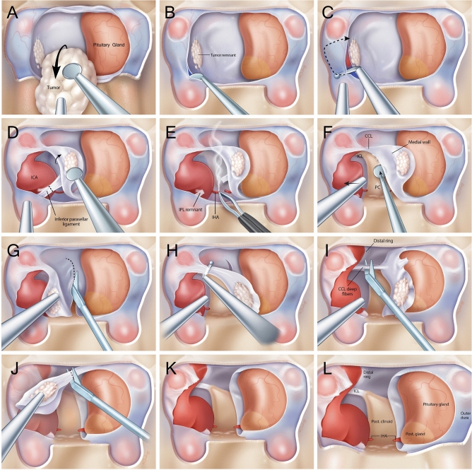
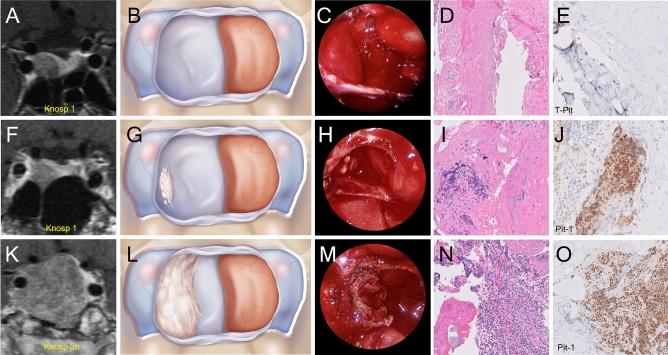
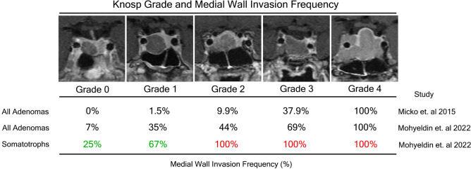
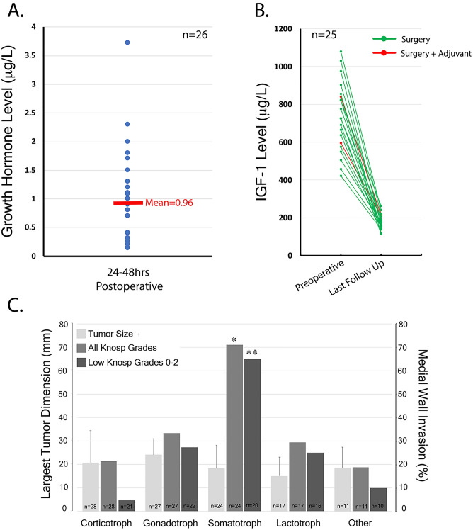
Recurrence and biochemical remission rates vary widely among different histological subtypes of pituitary adenoma. In this prospective study, we evaluated 107 consecutive primary pituitary adenomas operated on by a single neurosurgeon including 28 corticotroph, 27 gonadotroph, 24 somatotroph, 17 lactotroph, 5 null-cell and 6 plurihormonal. In each case, we performed direct endoscopic intraoperative inspection of the medial wall of the cavernous sinus, which was surgically removed when invasion was visualized. This was performed irrespective of tumor functional status. Medial wall resection was performed in 47% of pituitary adenomas, and 39/50 walls confirmed pathologic evidence of invasion, rendering a positive predictive value of intraoperative evaluation of medial wall invasion of 78%. We show for the first-time dramatic disparities in the frequency of medial wall invasion among pathological subtypes. Somatotroph tumors invaded the medial wall much more often than other adenoma subtypes, 81% intraoperatively and 69% histologically, followed by plurihormonal tumors (40%) and gonadotroph cell tumors (33%), both with intraoperative positive predictive value of 100%. The least likely to invade were corticotroph adenomas, at a rate of 32% intraoperatively and 21% histologically, and null-cell adenomas at 0%. Removal of the cavernous sinus medial wall was not associated with permanent cranial nerve morbidity nor carotid artery injury, although 4 patients (all Knosp 3-4) experienced transient diplopia. Medial wall resection in acromegaly resulted in the highest potential for biochemical remission ever reported, with an average postoperative day 1 GH levels of 0.96 ug/L and surgical remission rates of 92% based on normalization of IGF-1 levels after surgery (mean = 15.56 months; range 3-30 months). Our findings suggest that tumor invasion of the medial wall of the cavernous sinus may explain the relatively low biochemical remission rates currently seen for acromegaly and illustrate the relevance of advanced intradural surgical approaches for successful and durable outcomes in endonasal pituitary surgery for functional adenomas.


Conflict of interest statement

The authors declare no competing interests.

Figures

Figure 1
Figure 1
Medical illustrations demonstrating the sequential steps of resecting the medial wall of the cavernous sinus. Wide exposures of the anterior cavernous sinus and paraclinoid carotid artery allow for adequate visualization and safe removal of the medial wall. (A) Pituitary adenoma is resected under high resolution endoscopy with bimanual technique preserving the normal pituitary gland (B) The medial wall is adequately visualized with high resolution angled endoscopes once the bulk of the adenoma has been removed, tumor remnants that are intraoperatively viewed to adhere strongly to the medial wall suggests invasion necessitating resection. The anterior cavernous sinus is entered by developing a plane between the 2 layers of dura of the intercavernous sinus (C) Using a right angled blunt tipped feather blade, the anterior cavernous sinus wall is cut and hemostasis is achieved (D) The inferior parasellar ligament (IPL) is one of the first ligaments encountered and is cut to allow mobilization of the medial wall away from the carotid artery (E) The inferior hypophyseal artery is often encountered next and is coagulated and cut to avoid any evulsions off the carotid artery (F) the dura overlying the base of the posterior clinoid forms the posterior wall of the cavernous sinus and is dissected off the clinoid till the surgeon encounters the horizontal fibers of the carticoclinoidal ligament (CCL) (G) Microscissors are used to cut the dura covering the dorsum sella to deatch the meial wall from its sellar attachments (H) Using the right angled feather blade, the CCL fibers are cut to begin detaching the medial wall from the carotid artery (I) There are often deep fibers of the CCL that require further transection to completely untether the medial wall from the carotid artery (J) Once all the fibers of the CCL are cut, the medial wall is now completely free from any attachments to the carotid artery and the only remaining cut are dural attachments that make up the proximal dural ring (K) The medial wall is now completely free from attachments and often sent en bloc if it is not severely distorted by tumor invasion leaving behind and open cavernous sinus with an exposed carotid artery. (L) The remaining view should show the medial surface of the carotid artery in the cavernous sinus with visualization of the superior compartment of the cavernous sinus and the interclinoidal ligament (ICL).
Figure 2
Figure 2
We hypothesized from our intraoperative experience with pituitary tumors that some pituitary adenoma subtypes have a predilection for invading the medial wall of the cavernous sinus more so than others. Case examples of various pituitary adenomas with varying Knosp grade and invasion into the medial wall of the cavernous as viewed on preoperative MRI (T1 with gadolinium), through medical illustrations, from an endoscopic intraoperative view, and microscopically with histological H&E staining and immunocytochemistry. (A) Preoperative coronal MRI demonstrating a Knosp grade 1 corticotroph adenoma (B) A medical illustration of the intraoperative view from an endoscopic transsphenoidal approach after the adenoma has been removed and the pituitary gland remains displaced the left side of the field exposing an intact medial of the cavernous sinus with no evidence of invasion (C) An intraoperative view from an endoscopic transsphenoidal approach after the adenoma has been removed and the pituitary gland remains displaced the left side of the field exposing an intact right medial wall of the cavernous sinus with no evidence of invasion (D) Histological images of H&E slides of a resected medial wall with no evidence of invading pituitary adenoma cells (E) Histological images of immunohistochemistry slides of a resected medial wall with no evidence of invading pituitary adenoma cells (staining for T-pit transcription factor) (F) Preoperative coronal MRI demonstrating a Knosp grade 1 somatotroph adenoma (G) A medical illustration of the intraoperative view from an endoscopic transsphenoidal approach after the adenoma has been removed and the pituitary gland remains displaced to the left side of the field exposing the right medial wall of the cavernous sinus with subtle evidence of tumor invasion (H) An intraoperative view from an endoscopic transsphenoidal approach after the adenoma has been removed and the pituitary gland remains displaced the left side of the field exposing the right medial wall of the cavernous sinus with subtle evidence of tumor invasion (I) Histological images of H&E slides of the resected medial wall with evidence of invading pituitary adenoma cells (J) Histological images of immunohistochemistry slides of the resected medial wall in the case with evidence of invading pituitary adenoma cells (staining for Pit-1 transcription factor) (K) Preoperative coronal MRI demonstrating a Knosp grade 3 somatotroph adenoma (L) A medical illustration of the intraoperative view from an endoscopic transsphenoidal approach after the adenoma has been removed and the pituitary gland remains displaced to the left side of the field exposing the right medial wall of the cavernous sinus with frank evidence of tumor invasion (M) An intraoperative view from an endoscopic transsphenoidal approach after the adenoma has been removed and the pituitary gland remains displaced the left side of the field exposing the right medial wall of the cavernous sinus with frank evidence of tumor invasion (N) Histological images of H&E slides of the resected medial wall in this case with evidence of invading pituitary adenoma cells O. Histological images of immunohistochemistry slides of the resected medial wall in this case with evidence of invading pituitary adenoma cells (staining for Pit-1 transcription factor).
Figure 3
Figure 3
Knosp grade and medial wall invasion frequency as reported by Knosp and colleagues compared to invasion frequencies exhibited in the current study among all adenomas and then among somatotroph adenomas. Representative MRI images from case examples of somatotrophs in our current series stratified by Knosp grade. Knosp criteria were applied based on carotid tangents and each grade is represented by a coronal T1 MRI scan with gadolinium capturing the extent of cavernous sinus invasion.
Figure 4
Figure 4
(A) Mean postoperative GH nadir 24-48hrs after surgical resection in acromegaly cohort (n=26) (B) Preoperative and postoperative (3-12 months) IGF-1 levels in acromegaly cohort (n=25), two-tailed unpaired t-test demonstrates statistically significance between preoperative and postoperative IGF-1 levels (p<0.0001) (C) A graphical visual summary of the important findings from Table 1, 2 and 3 and demonstrates that somatotroph adenomas have a predilection to invade the cavernous sinus irrespective of tumor size. Using a Chi square test, we demonstrate that the effect that somatotrophs have on predicting cavernous sinus invasion was present across all Knosp grades 0-4 (*p<0.02) and even present across low Knosp grades 0-2 (**p<.05).

References

    1. Lopes MBS. The 2017 world health organization classification of tumors of the pituitary gland: a summary. Acta. Neuropathol. 2017;134:521–35. doi: 10.1007/s00401-017-1769-8. - DOI - PubMed
    1. Melmed S. Pituitary-tumor endocrinopathies. N. Engl. J. Med. 2020;382:937–50. doi: 10.1056/NEJMra1810772. - DOI - PubMed
    1. WHO classification of tumours of endocrine organs, 4th edn. Lyon, France: IARC Press 2017.
    1. Lonser RR, Ksendzovsky A, Wind JJ, Vortmeyer AO, Oldfield EH. Prospective evaluation of the characteristics and incidence of adenoma-associated dural invasion in Cushing disease. J. Neurosurg. 2012;116:272–9. doi: 10.3171/2011.8.JNS11456. - DOI - PMC - PubMed
    1. Micko AS, Wohrer A, Wolfsberger S, Knosp E. Invasion of the cavernous sinus space in pituitary adenomas: endoscopic verification and its correlation with an MRI-based classification. J. Neurosurg. 2015;122:803–11. doi: 10.3171/2014.12.JNS141083. - DOI - PubMed