. 2022 Jun 15;12(1):9940.
doi: 10.1038/s41598-022-14064-6.

Decoronation-induced infected alveolar socket defect rat model for ridge preservation

Affiliations

Decoronation-induced infected alveolar socket defect rat model for ridge preservation

Chih-Hsiang Fang et al. Sci Rep. .

Abstract

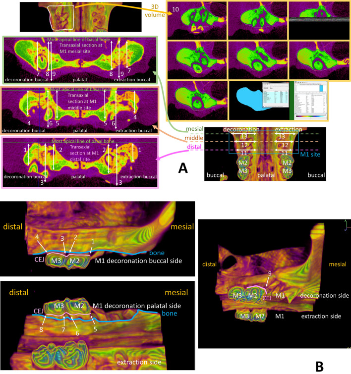
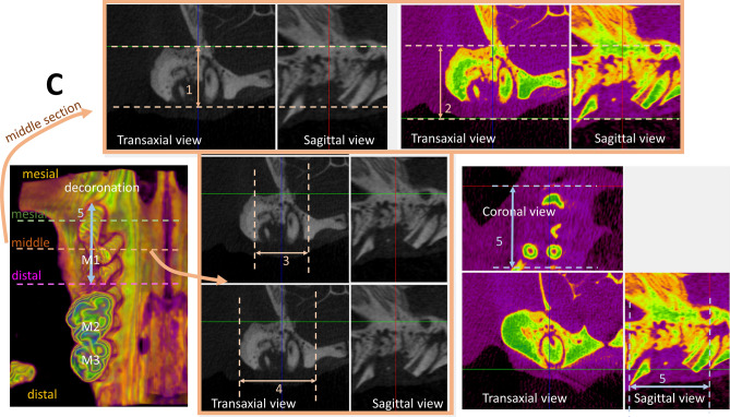
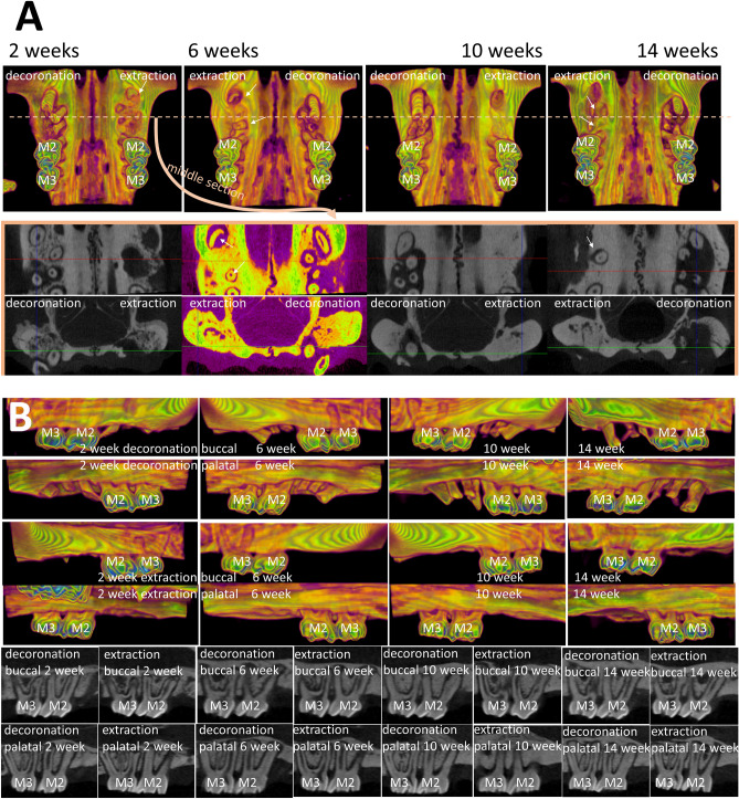
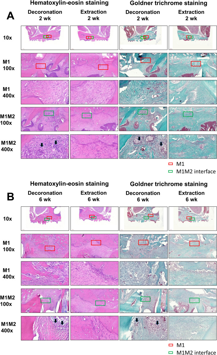
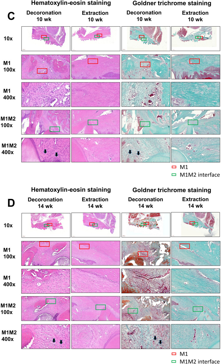
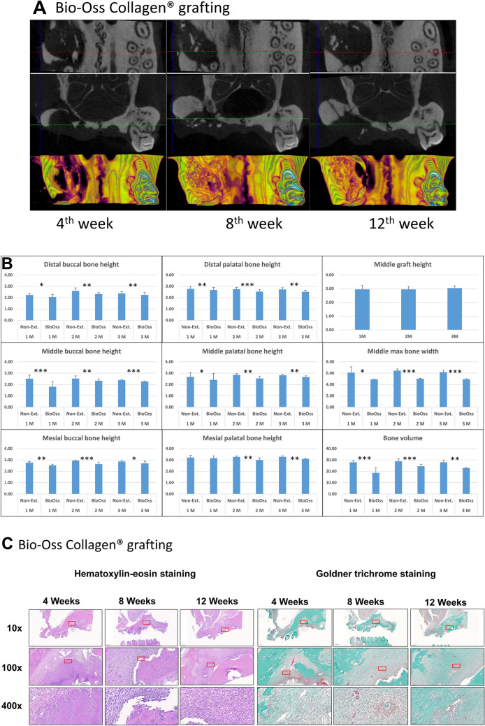
Current rat alveolar ridge preservation models have not been well standardized. In this study, we proposed decoronation-induced infected alveolar socket model of rat. The bilateral maxillary first molars (M1) of twenty-four rats were decoronized or extracted. After 2, 6, 10, and 14 weeks, bone and soft tissue changes at M1 and periodontal conditions of maxillary second (M2) and third molars (M3) were evaluated by micro-computed tomography and histological analysis. Additional eighteen rats with standardized size defects were grafted with Bio-Oss Collagen to compare with unmanipulated contralateral side. Decoronation preserved greater bone and soft tissue dimensions at M1, provided larger three-dimensional (3D) bone contour volume, but also promoted periodontal breakdown of M2 Histological results showed intense inflammatory cell infiltrations and severe bone resorption within M1 socket and at mesial aspect of M2. The critical dimensions to accommodate largest standardized defect at M1 were 2.2-2.3 mm at vertical bone height and 2.8-3.2 mm at alveolar crestal width. Bio-Oss Collagen could not fully preserve buccal or palatal bone height but could be beneficial in preserving ridge width in large alveolar defects. Collectively, if periodontally-involved alveolar bone defect is preferred, we suggest extracting M1 roots 6 weeks after decoronation to allow periodontitis to occur at M2. If standardized critical dimension defect is preferred, we suggest extracting M1 roots 2 weeks after decoronation, and creating defect in the middle of M1 site with size no larger than 2.7 mm diameter to its full depth.


Conflict of interest statement

The authors declare no competing interests.

Figures

Figure 1
Figure 1
Micro-computed tomography assessments of alveolar bone and soft tissue dimension. (A) To evaluate the effectiveness of hard and soft tissue preservation of alveolar ridge dimension associated with the decoronation procedures, thirteen parameters were measured at maxillary first molar (M1) sites including (1) distal buccal bone height, (2) distal palatal bone height, (3) distal maximum soft tissue height, (4) middle buccal bone height, (5) middle palatal bone height, (6) middle maximum soft tissue height, (7) mesial buccal bone height, (8) mesial palatal bone height, (9) mesial maximum soft tissue height, (10) 3D bone contour volume*, (11) distal maximum alveolar ridge width, (12) middle maximum alveolar ridge width, and (13) mesial maximum alveolar ridge width. (B) To evaluate the influence of decoronation on the periodontal conditions of maxillary second molar (M2) and third molar (M3), the distances from marginal bone to cemento-enamel junction (CEJ) including (1) bM2mCEJ: M2 mesiobuccal CEJ to bone; (2) bM2dCEJ: M2 distobuccal CEJ to bone; (3) bM3mCEJ: M3 mesiobuccal CEJ to bone; (4) bM3dCEJ: M3 distobuccal CEJ to bone; (5) pM2mCEJ: M2 mesiopalatal CEJ to bone; (6) pM2dCEJ: M2 distopalatal CEJ to bone; (7) pM3mCEJ: M3 mesiopalatal CEJ to bone; (8) pM3dCEJ: M3 distopalatal CEJ to bone; and (9) the distance from CEJ to mesial furcation of M2 (M2mf) were measured. (C) To provide reference for alveolar critical size defect dimensions in a standardized and discriminative manner, five parameters were measured at the decoronated M1 site, including (1) middle bone height, (2) middle tissue height, (3) middle alveolar crestal width, (4) middle maximum bone width, and (5) mesio-distal socket length. *Three-dimensional (3D) bone contour volume was calculated on either side by adding up consecutive surfaces of interest on the transaxial section mesio-distally throughout the M1 site.
Figure 1
Figure 1
Micro-computed tomography assessments of alveolar bone and soft tissue dimension. (A) To evaluate the effectiveness of hard and soft tissue preservation of alveolar ridge dimension associated with the decoronation procedures, thirteen parameters were measured at maxillary first molar (M1) sites including (1) distal buccal bone height, (2) distal palatal bone height, (3) distal maximum soft tissue height, (4) middle buccal bone height, (5) middle palatal bone height, (6) middle maximum soft tissue height, (7) mesial buccal bone height, (8) mesial palatal bone height, (9) mesial maximum soft tissue height, (10) 3D bone contour volume*, (11) distal maximum alveolar ridge width, (12) middle maximum alveolar ridge width, and (13) mesial maximum alveolar ridge width. (B) To evaluate the influence of decoronation on the periodontal conditions of maxillary second molar (M2) and third molar (M3), the distances from marginal bone to cemento-enamel junction (CEJ) including (1) bM2mCEJ: M2 mesiobuccal CEJ to bone; (2) bM2dCEJ: M2 distobuccal CEJ to bone; (3) bM3mCEJ: M3 mesiobuccal CEJ to bone; (4) bM3dCEJ: M3 distobuccal CEJ to bone; (5) pM2mCEJ: M2 mesiopalatal CEJ to bone; (6) pM2dCEJ: M2 distopalatal CEJ to bone; (7) pM3mCEJ: M3 mesiopalatal CEJ to bone; (8) pM3dCEJ: M3 distopalatal CEJ to bone; and (9) the distance from CEJ to mesial furcation of M2 (M2mf) were measured. (C) To provide reference for alveolar critical size defect dimensions in a standardized and discriminative manner, five parameters were measured at the decoronated M1 site, including (1) middle bone height, (2) middle tissue height, (3) middle alveolar crestal width, (4) middle maximum bone width, and (5) mesio-distal socket length. *Three-dimensional (3D) bone contour volume was calculated on either side by adding up consecutive surfaces of interest on the transaxial section mesio-distally throughout the M1 site.
Figure 2
Figure 2
Comparison of the effectiveness of alveolar ridge preservation at maxillary first molar (M1) and the periodontal condition at maxillary second molar (M2) and third molar (M3) after decoronation or extraction of M1. (A) Comparison of the effectiveness of alveolar ridge preservation with decoronation technique at maxillary first molar (M1) examined by micro-CT. Representative images show the 3D occlusal view (top), the coronal view, and the transaxial section in the middle of M1 site (bottom) at 2, 6, 10, 14 weeks. Fuller bone contour and better bone height were obvious on the decoronation side compared to the contralateral extraction side. Please note that some root fragments remained in the extraction side (white arrow). (B) Comparison of the periodontal condition at maxillary second molar (M2) and third molar (M3) after decoronation or extraction of M1. Representative images show the 3D buccal and palatal views on the decoronation and extraction sides (color images) and 2D sections cut along a profile line through the buccal aspect or palatal aspect of M2 and M3 (black/white images). On the decoronation side, significant more bone resorption may be seen at both buccal and palatal aspects of the M2 teeth, especially at mesial side.
Figure 3
Figure 3
Representative histological sections of the maxillary first molar (M1) to third molar (M3) region of rats after after M1 decoronation or extraction; hematoxylin and eosin (H&E) or Goldner's Trichrome staining. Sections were identified and photographed under 10x, 100x, and 400 × magnifications. Six weeks after M1 extraction, there were still some inflammatory cell infiltrations in the samples; however, these infiltrates decreased a lot at 10th and 14th weeks after extraction. The decoronation samples were dominated by inflammatory granulation tissues, blood vessels with different calibers, and inflammatory cell infiltrations at 6–14 weeks; moreover, intensive alveolar bone resorption activity was characterized by the presence of a border of osteoclasts (arrow) at the M1-M2 junction (green box). With Goldner's Trichrome staining, the collagenous fiber, fibrin and erythrocytes were selectively visualized. The fibrous connective tissue with the presence of granulation tissue with excessive inflammatory cell infiltrations were clearly demonstrated in the decoronation samples.
Figure 3
Figure 3
Representative histological sections of the maxillary first molar (M1) to third molar (M3) region of rats after after M1 decoronation or extraction; hematoxylin and eosin (H&E) or Goldner's Trichrome staining. Sections were identified and photographed under 10x, 100x, and 400 × magnifications. Six weeks after M1 extraction, there were still some inflammatory cell infiltrations in the samples; however, these infiltrates decreased a lot at 10th and 14th weeks after extraction. The decoronation samples were dominated by inflammatory granulation tissues, blood vessels with different calibers, and inflammatory cell infiltrations at 6–14 weeks; moreover, intensive alveolar bone resorption activity was characterized by the presence of a border of osteoclasts (arrow) at the M1-M2 junction (green box). With Goldner's Trichrome staining, the collagenous fiber, fibrin and erythrocytes were selectively visualized. The fibrous connective tissue with the presence of granulation tissue with excessive inflammatory cell infiltrations were clearly demonstrated in the decoronation samples.
Figure 4
Figure 4
Micro-computed tomography (micro-CT) and histo-morphological evaluation of alveolar ridge bone preservation in standardized critical dimension socket defect model. The effectiveness of alveolar ridge bone preservation at maxillary first molar (M1) examined by micro-computed tomography (micro-CT) after creating large alveolar defect 2 weeks after decoronation and grafted with Bio-Oss Collagen. (A) Representative images show the coronal (top) and transaxial (middle) sections in the middle part of M1 site and the 3D occlusal view (bottom) after Bio-Oss Collagen grafting for 4th, 8th, and 12th weeks. Please note that residual grafting material at the defect site was still evident after 12 weeks. (B) Analyses of bone heights, middle maximum bone width, and bone volume at different time points. Graft heights were measured in the middle section of M1 site on the grafted side only. Bone volume was measured on the grafted side by adding up consecutive bone surfaces excluding the graft material on the transaxial sections mesio-distally throughout the M1 site. (C) Representative histological sections of the maxillary first molar (M1) to third molar (M3) region of rats after M1 decoronation, roots extraction and grafting with Bio-Oss Collagen by hematoxylin & eosin (H&E) and Goldner's Trichrome staining at 4th, 8th, and 12th weeks. At 12th week after grafting, the defects grafted with Bio-Oss Collagen showed some bone regeneration in the defect site; however, the rate of new bone regeneration or mineralization of new bone were still not satisfactory at 12th week. Goldner's trichrome staining shows immature bone formation and soft tissue invasion were observed but no angiogenesis found.
Figure 5
Figure 5
The effectiveness of preservation of alveolar ridge dimension associated with the decoronation procedure. (A) Representative image showing effectiveness of preservation of alveolar ridge dimension associated with the decoronation procedure at 6th week. (B) Superimposition of the mirror image of the decoronation side (red) in (A) to the extraction side. (C) Representative photo showing the maxillary first molar (M1) site just after decoronation. The bone and roots were left exposed for the secondary wound healing of the soft tissue. Yellow circles indicate the separated roots. (D) Representative photo showing 2 weeks after M1 decoronation. The soft tissue has grown well onto the decoronated M1 site. However, some roots have perforated through the soft tissue (black arrows). (E) Representative image of micro-CT section cut through the buccal aspect of M2 and M3 showing possible endodontic involvement (yellow arrow) caused by deep caries (white arrow) on the mesial root of M2. (F) Photo of an extracted M1 showing the slender mid-palatal root and the divergent mesial root. (G) In vitro testing of burr hole defects created on sawbone using diameter 2.7 mm tungsten carbide burr.

References

    1. Kim JJ, et al. Ridge preservation of extraction sockets with chronic pathology using Bio-Oss((R)) Collagen with or without collagen membrane: An experimental study in dogs. Clin. Oral Impl. Res. 2017;28:727–733. doi: 10.1111/clr.12870. - DOI - PubMed
    1. Van der Weijden F, Dell'Acqua F, Slot DE. Alveolar bone dimensional changes of post-extraction sockets in humans: A systematic review. J. Clin. Periodontol. 2009;36:1048–1058. doi: 10.1111/j.1600-051X.2009.01482.x. - DOI - PubMed
    1. Schropp L, Wenzel A, Kostopoulos L, Karring T. Bone healing and soft tissue contour changes following single-tooth extraction: A clinical and radiographic 12-month prospective study. Int. J. Periodontics Restor. Dent. 2003;23:313–323. - PubMed
    1. Stoppenbrink D, et al. Dimensional changes of the alveolar ridge contour of the premolar extraction site in adolescents. J. Orofac. Orthop. 2019;80:205–215. doi: 10.1007/s00056-019-00182-w. - DOI - PubMed
    1. Couso-Queiruga E, Stuhr S, Tattan M, Chambrone L, Avila-Ortiz G. Post-extraction dimensional changes: A systematic review and meta-analysis. J. Clin. Periodontol. 2021;48:126–144. doi: 10.1111/jcpe.13390. - DOI - PubMed

Publication types