. 2023 Feb;45(1):141-158.
doi: 10.1007/s11357-022-00598-0. Epub 2022 Jun 15.

The PICLS high-throughput screening method for agents extending cellular longevity identifies 2,5-anhydro-D-mannitol as novel anti-aging compound

Affiliations

The PICLS high-throughput screening method for agents extending cellular longevity identifies 2,5-anhydro-D-mannitol as novel anti-aging compound

Mohammad Alfatah et al. Geroscience. 2023 Feb.

Abstract

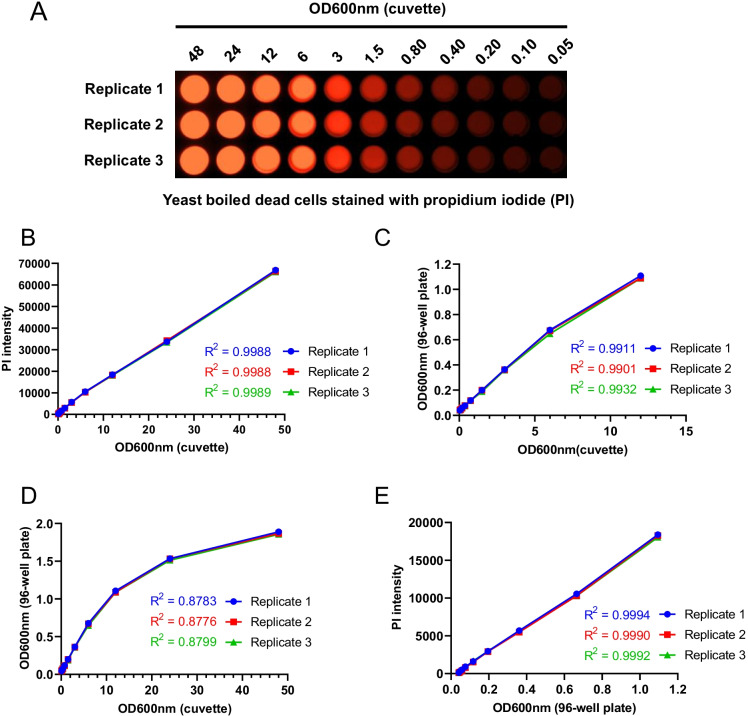
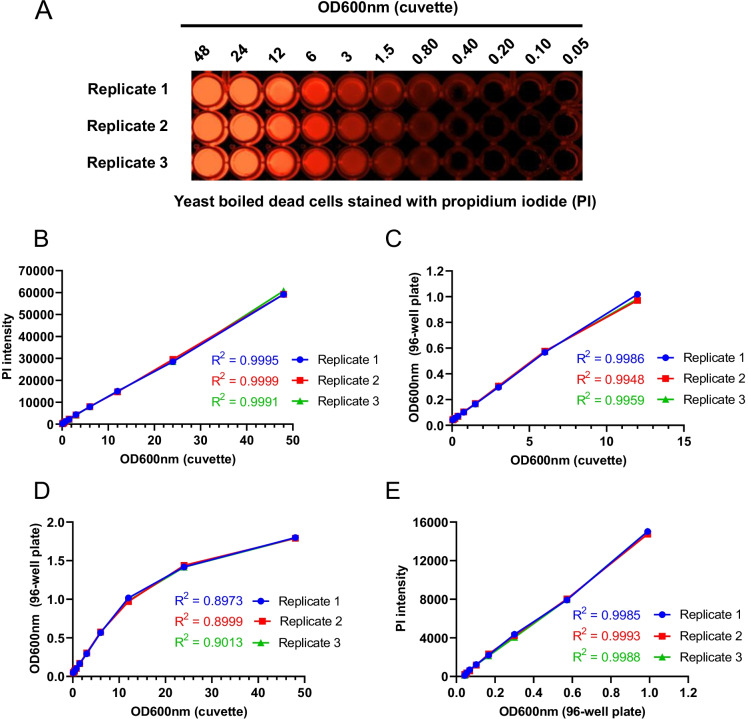
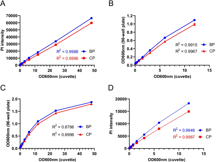
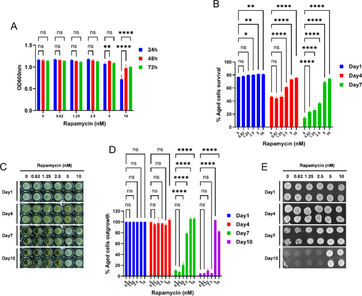
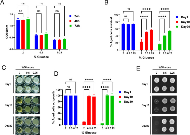
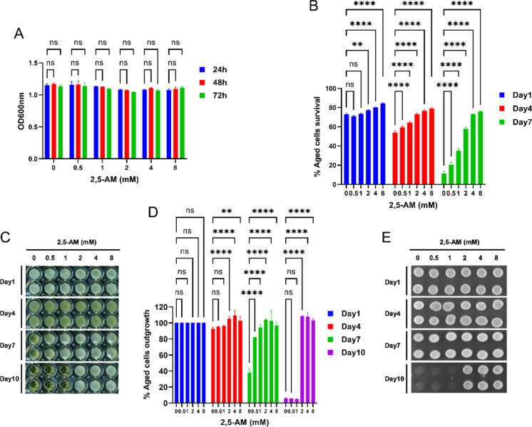
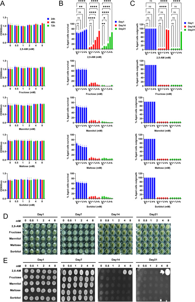
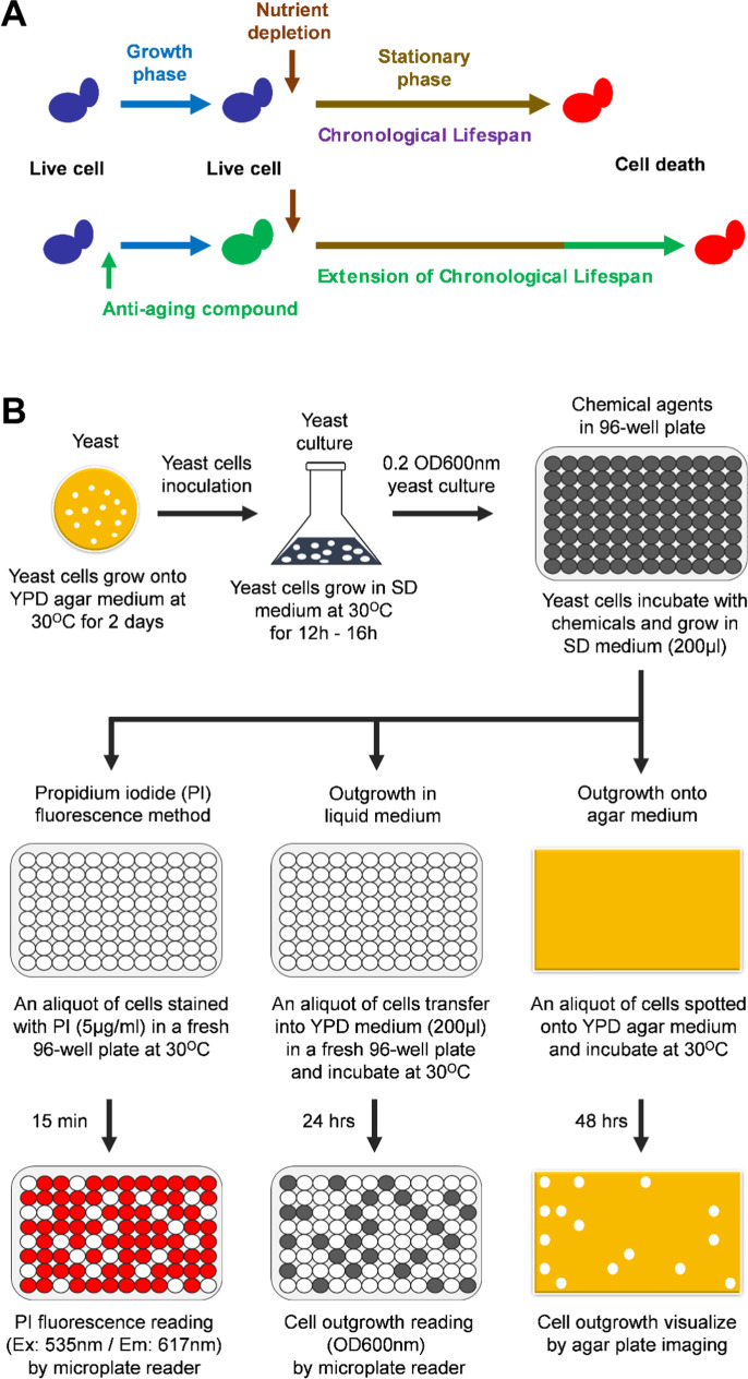
Although aging is the biggest risk factor for human chronic (cancer, diabetic, cardiovascular, and neurodegenerative) diseases, few interventions are known besides caloric restriction and a small number of drugs (with substantial side effects) that directly address aging. Thus, there is an urgent need for new options that can generally delay aging processes and prevent age-related diseases. Cellular aging is at the basis of aging processes. Chronological lifespan (CLS) of yeast Saccharomyces cerevisiae is the well-established model system for investigating the interventions of human post-mitotic cellular aging. CLS is defined as the number of days cells remain viable in a stationary phase. We developed a new, cheap, and fast quantitative method for measuring CLS in cell cultures incubated together with various chemical agents and controls on 96-well plates. Our PICLS protocol with (1) the use of propidium iodide for fluorescent-based cell survival reading in a microplate reader and (2) total cell count measurement via OD600nm absorption from the same plate provides real high-throughput capacity. Depending on logistics, large numbers of plates can be processed in parallel so that the screening of thousands of compounds becomes feasible in a short time. The method was validated by measuring the effect of rapamycin and calorie restriction on yeast CLS. We utilized this approach for chemical agent screening. We discovered the anti-aging/geroprotective potential of 2,5-anhydro-D-mannitol (2,5-AM) and suggest its usage individually or in combination with other anti-aging interventions.

Keywords: 2,5-anhydro-D-mannitol; Anti-aging compound; Chemical screening; Chronological lifespan; Saccharomyces cerevisiae.


Conflict of interest statement

The authors declare no competing interests.

Figures

Fig. 1
Fig. 1
Development of propidium iodide fluorescence–based method for measuring the chronological lifespan. The propidium iodide (PI) fluorescence–based method was developed to measure the CLS of the yeast for screening the chemical agents to identify the anti-aging compounds. A Yeast boiled cells were stained with PI for 15 min at 30 °C. PI-stained cells were diluted (48 to 0.05 OD600nm) in a black 96-well plate and pseudo imaged with a BioRad GelDoc imaging system. Three replicates of each PI-stained sample were imaged and are represented in the figure. B Correlation between PI fluorescence intensity and cell OD600nm of the cuvette. C and D Correlation between cell OD600nm of the 96-well plate and the cuvette. E Correlation between PI fluorescence intensity and cell OD600nm of 96-well plate
Fig. 2
Fig. 2
Determination of the propidium iodide fluorescence–based method for measuring the chronological lifespan in the clear 96-well plate. The propidium iodide (PI) fluorescence–based method for measuring the chronological lifespan (CLS) was determined in the clear 96-well plate for screening the chemical agents to identify the anti-aging compounds. A Yeast-boiled cells were stained with PI for 15 min at 30 °C. PI-stained cells were diluted (48 to 0.05 OD600nm) in a clear 96-well plate and pseudo imaged with a BioRad GelDoc imaging system. Three replicates of each PI-stained sample were imaged and are represented in the figure. B Correlation between PI fluorescence intensity and cell OD600nm of the cuvette. C and D Correlation between cell OD600nm of the clear 96-well plate and the cuvette. E Correlation between PI fluorescence intensity and cell OD600nm of the clear 96-well plate
Fig. 3
Fig. 3
Comparison of the propidium iodide fluorescence–based method for measuring the chronological lifespan in black vs clear 96-well plates. A Comparison of black vs. clear 96-well plates’ correlation between PI fluorescence intensity and cell OD600nm of the cuvette. B and C Comparison of black vs. clear 96-well plates’ correlation between cell OD600nm of the 96-well plate and cuvette. D Comparison of black vs. clear 96-well plates’ correlation between PI fluorescence intensity and cell OD600nm of the 96-well plate
Fig. 4
Fig. 4
Rapamycin drug extends the chronological lifespan of the yeast. The prototrophic yeast strain (CEN.PK113-7D) was grown in the synthetic defined medium with different concentrations of rapamycin in 96-well plates at 30 °C. A Cell growth OD600nm was measured at time points 24 h, 48 h, and 72 h using a microplate reader and graph plotted against different concentrations of rapamycin. B The chronological lifespan (CLS) of different concentrations of rapamycin-incubated cells was determined using the propidium iodide fluorescence–based method. Cell survival at different chronological age points was quantified and the growth time point 72 h was considered as day 1. C The CLS of the aged cells was determined by the outgrowth method in YPD liquid medium. The growth time point 72 h was considered as day 1. At various chronological age points, a 3-μL culture was transferred to a second 96-well plate containing 200μL YPD medium. Outgrowth in 96-well plate was photographed after incubation for 24 h at 30 °C. D Outgrowth OD600nm in YPD liquid medium was measured using a microplate reader. The outgrowth of different chronological age points is plotted relative to day 1. E At various chronological age points, 3-μL cultures were spotted onto the YPD agar plate. Outgrowth was photographed after incubation for 48 h at 30 °C. All data represent as means ± SD.; *P < 0.05, **P < 0.01, and ****P < 0.0001 based on two-way ANOVA followed by Dunnett’s multiple comparisons test (A, B, and D). n.s, non-significant
Fig. 5
Fig. 5
Calorie restriction extends the chronological lifespan of the yeast. The prototrophic yeast strain (CEN.PK113-7D) was grown in the synthetic defined medium containing 2%, 0.5%, and 0.25% glucose in 96-well plates at 30 °C. A Cell growth OD600nm was measured at time points 24 h, 48 h, and 72 h using a microplate reader and graph plotted against different glucose concentrations. B The chronological lifespan (CLS) of the aged cells grown under different glucose concentrations was determined using the propidium iodide fluorescence–based method. Cell survival at different chronological age points was quantified and the growth time point 72 h was considered as day 1. C The CLS of the aged cells grown under different glucose concentrations was determined by the outgrowth method in YPD liquid medium. The growth time point 72 h was considered as day 1. At various chronological age points, a 3-μL culture was transferred to a second 96-well plate containing 200μL YPD medium. Outgrowth in 96-well plate was photographed after incubation for 24 h at 30 °C. D Outgrowth OD600nm in YPD liquid medium was measured using a microplate reader. The graph is plotted relative to day 1. E At various chronological age points, 3-μL cultures were spotted onto the YPD agar plate. Outgrowth was photographed after incubation for 48 h at 30 °C. All data represent as means ± SD.; ****P < 0.0001 based on two-way ANOVA followed by Dunnett’s multiple comparisons test (A, B, and D). n.s, non-significant
Fig. 6
Fig. 6
2,5-Anhydro-D-mannitol is a novel anti-aging compound that extends the chronological lifespan of the yeast. The prototrophic yeast strain (CEN.PK113-7D) was grown in the synthetic defined medium with different concentrations of 2,5-anhydro-D-mannitol (2,5-AM) in 96-well plates at 30 °C. A Cell growth OD600nm was measured at different time points 24 h, 48 h, and 72 h using a microplate reader and graph plotted against different 2,5-AM concentrations. B The chronological lifespan (CLS) of different concentrations of 2,5-AM incubated cells was determined using the propidium iodide fluorescence–based method. Cell survival at different chronological age points was quantified and the growth time point 72 h was considered as day 1. C The CLS of the aged cells was determined by the outgrowth method in YPD liquid medium. The growth time point 72 h was considered as day 1. At various chronological age points, 3-μL cultures were transferred to a second 96-well plate containing 200μL YPD medium. Outgrowth in 96-well plate was photographed after incubation for 24 h at 30 °C. D Outgrowth OD600nm in YPD liquid medium was measured using a microplate reader. The graph is plotted relative to day 1. E At various chronological age points, a 3-μL culture was spotted onto the YPD agar plate. Outgrowth was photographed after incubation for 48 h at 30 °C. All data represent as means ± SD.; **P < 0.01 and ****P < 0.0001 based on two-way ANOVA followed by Dunnett’s multiple comparisons test (A, B, and D). n.s, non-significant
Fig. 7
Fig. 7
Testing the effect of 2,5-anhydro-D-mannitol analogs on chronological lifespan of the yeast. The prototrophic yeast strain (CEN.PK113-7D) was grown in the synthetic defined medium with different concentrations of 2,5-anhydro-D-mannitol (2,5-AM), D-fructose, D-mannitol, D-maltose, and D-sorbitol in 96-well plates at 30 °C. A Cell growth OD600nm was measured at different time points 24 h, 48 h, and 72 h using a microplate reader and graph plotted against different concentrations of 2,5-AM, fructose, mannitol, maltose, and sorbitol. B The chronological lifespan (CLS) of different concentrations of 2,5-AM, fructose, mannitol, maltose, and sorbitol incubated cells was determined using the propidium iodide fluorescence–based method. Cell survival at different chronological age points was quantified and the growth time point 72 h was considered as day 1. C The CLS of the aged cells was determined by the outgrowth method in YPD liquid medium. The growth time point 72 h was considered as day 1. At various chronological age points, a 3-μL culture were transferred to a second 96-well plate containing 200μL YPD medium. Outgrowth OD600nm in YPD liquid medium was measured after incubation for 24 h at 30 °C using a microplate reader. The graph is plotted relative to day 1. D Outgrowth in YPD liquid medium of 96-well plate was photographed after incubation for 24 h at 30 °C. E At various chronological age points, 3-μL cultures were spotted onto the YPD agar plate. Outgrowth was photographed after incubation for 48 h at 30 °C. All data represent as means ± SD.; *P < 0.05, **P < 0.01, and ****P < 0.0001 based on two-way ANOVA followed by Dunnett’s multiple comparisons test (B and C). n.s, non-significant
Fig. 8
Fig. 8
Model for identifying the anti-aging compounds and methods to measuring the chronological lifespan of the yeast. A Schematic representation for determining the anti-aging compounds that extend the chronological lifespan of the yeast. B Flowchart of propidium iodide fluorescence–based PICLS method, traditional outgrowth methods in YPD liquid medium and YPD agar medium (spotting assay) for screening the chemical agents to identify the anti-aging compounds

References

    1. das nações Unidas, O. United Nations, Department of Economic and Social Affairs, Population Division (2019). World Population Prospects, Highlights (ST/ESA/SER.A/423). 2019.
    1. Robine J. Ageing populations: we are living longer lives, but are we healthier? United Nations, Department of Economics and Social Affairs, Population Division, UN DESA/POP/2021/TP/NO. 2. (2021).
    1. Niccoli T, Partridge L. Ageing as a risk factor for disease. Curr Biol. 2012 doi: 10.1016/j.cub.2012.07.024. - DOI - PubMed
    1. Hou Y, et al. Ageing as a risk factor for neurodegenerative disease. Nat Rev Neurol. 2019 doi: 10.1038/s41582-019-0244-7. - DOI - PubMed
    1. Harman D. The aging process: Major risk factor for disease and death. Proc Natl Acad Sci U S A. 1991 doi: 10.1073/pnas.88.12.5360. - DOI - PMC - PubMed

Publication types

LinkOut - more resources