Review
. 2022 Jun 15;79(7):363.
doi: 10.1007/s00018-022-04396-x.

Mechanisms that regulate the activities of TET proteins

Affiliations
Review

Mechanisms that regulate the activities of TET proteins

Kanak Joshi et al. Cell Mol Life Sci. .

Abstract

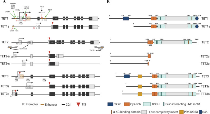
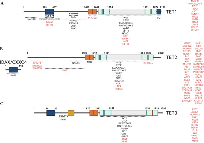
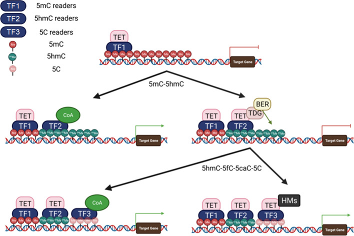
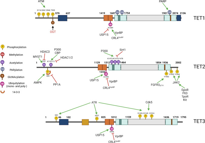
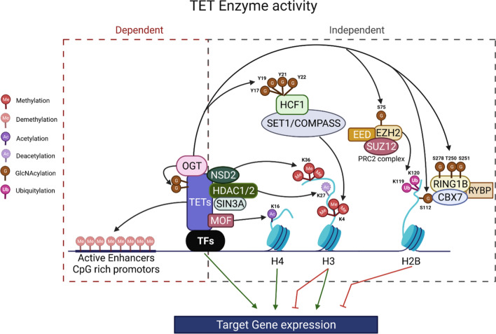
The ten-eleven translocation (TET) family of dioxygenases consists of three members, TET1, TET2, and TET3. All three TET enzymes have Fe+2 and α-ketoglutarate (α-KG)-dependent dioxygenase activities, catalyzing the 1st step of DNA demethylation by converting 5-methylcytosine (5mC) to 5-hydroxymethylcytosine (5hmC), and further oxidize 5hmC to 5-formylcytosine (5fC) and 5-carboxylcytosine (5caC). Gene knockout studies demonstrated that all three TET proteins are involved in the regulation of fetal organ generation during embryonic development and normal tissue generation postnatally. TET proteins play such roles by regulating the expression of key differentiation and fate-determining genes via (1) enzymatic activity-dependent DNA methylation of the promoters and enhancers of target genes; and (2) enzymatic activity-independent regulation of histone modification. Interacting partner proteins and post-translational regulatory mechanisms regulate the activities of TET proteins. Mutations and dysregulation of TET proteins are involved in the pathogenesis of human diseases, specifically cancers. Here, we summarize the research on the interaction partners and post-translational modifications of TET proteins. We also discuss the molecular mechanisms by which these partner proteins and modifications regulate TET functioning and target gene expression. Such information will help in the design of medications useful for targeted therapy of TET-mutant-related diseases.

Keywords: Gene expression; Interaction partners; Mutations; Post-translational modifications; TETs.


Conflict of interest statement

The authors declare that they have no competing financial or professional interests.

Figures

Fig. 1
Fig. 1
TET genes and TET proteins. A TET1, TET2, and TET3 have 2, 3, and 3 transcriptional products, respectively, due to the use of alternative promoters, which are regulated by the alternative activation of enhancers. The green arrow indicates induction of expression; the red cross depicts inhibition of expression. B. The corresponding protein isoforms of TET1, TET2, and TET3. The structural domains of the proteins are indicated
Fig. 2
Fig. 2
Interaction partner proteins of TET proteins. The interacting partner proteins of TET1, TET2, and TET3 are listed in a, b, and c, respectively. The partner proteins for which the interaction regions have been identified are listed under each TET at the corresponding regions. The partner proteins for which the interaction regions have not yet been defined are listed on the right side of each TET. The partners that are shared by all three TETs are listed in black font, while the partners that are specific for one or 2 TETs are listed in red font
Fig. 3
Fig. 3
Subgroups of TET-interacting TFs. The TET-interacting TFs can be divided into TF1, TF2, and TF3 based on their binding affinity for 5mC, 5hmC, and 5C, respectively. TF1 can bind 5mC DNA and recruits TET to initiate the first step of DNA demethylation by converting 5mC to 5hmC. TF2 can bind to 5hmC promoters/enhancers to turn on gene expression by recruiting co-activators (CoA) or to further complete the DNA demethylation elements by recruiting TET–TDG–BER complexes. TF3 binds 5C promoters/enhancers to promote gene expression by recruiting CoA or regulating gene expression by recruiting TET-histone modifiers (HMs)
Fig. 4
Fig. 4
Post-translational modifications of TET proteins. The post-translational modifications of TET1, TET2, and TET3 are listed in a, b, and c, respectively. The green arrow depicts the addition of modifications. The Red Cross indicates removal modifications
Fig. 5
Fig. 5
TETs regulate gene expression by both enzyme-dependent and -independent mechanisms. After binding to DNA regulatory regions, TETs regulate target gene expression by (1) enzymatic demethylation of 5mC; and (2) recruiting histone modifiers, including OGT, Sin3A/HDACs, or HATs.

References

    1. McKinney-Freeman S, et al. The transcriptional landscape of hematopoietic stem cell ontogeny. Cell Stem Cell. 2012;11:701–714. doi: 10.1016/j.stem.2012.07.018. - DOI - PMC - PubMed
    1. Long HK, Prescott SL, Wysocka J. Ever-changing landscapes: transcriptional enhancers in development and evolution. Cell. 2016;167:1170–1187. doi: 10.1016/j.cell.2016.09.018. - DOI - PMC - PubMed
    1. Kulis M, et al. Whole-genome fingerprint of the DNA methylome during human B cell differentiation. Nat Genet. 2015;47:746–756. doi: 10.1038/ng.3291. - DOI - PMC - PubMed
    1. Barwick BG, Scharer CD, Bally APR, Boss JM. Plasma cell differentiation is coupled to division-dependent DNA hypomethylation and gene regulation. Nat Immunol. 2016;17:1216–1225. doi: 10.1038/ni.3519. - DOI - PMC - PubMed
    1. Smith ZD, Meissner A. DNA methylation: roles in mammalian development. Nat Rev Genet. 2013;14:204–220. doi: 10.1038/nrg3354. - DOI - PubMed

MeSH terms

LinkOut - more resources