. 2022 Jun 15;19(1):152.
doi: 10.1186/s12974-022-02517-8.

FKBP51 mediates resilience to inflammation-induced anxiety through regulation of glutamic acid decarboxylase 65 expression in mouse hippocampus

Affiliations

FKBP51 mediates resilience to inflammation-induced anxiety through regulation of glutamic acid decarboxylase 65 expression in mouse hippocampus

Yu-Ling Gan et al. J Neuroinflammation. .

Abstract

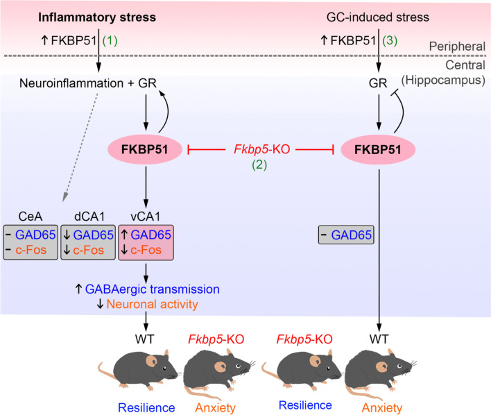
Background: Inflammation is a potential risk factor of mental disturbance. FKBP5 that encodes FK506-binding protein 51 (FKBP51), a negative cochaperone of glucocorticoid receptor (GR), is a stress-inducible gene and has been linked to psychiatric disorders. Yet, the role of FKBP51 in the inflammatory stress-associated mental disturbance remained unclear.

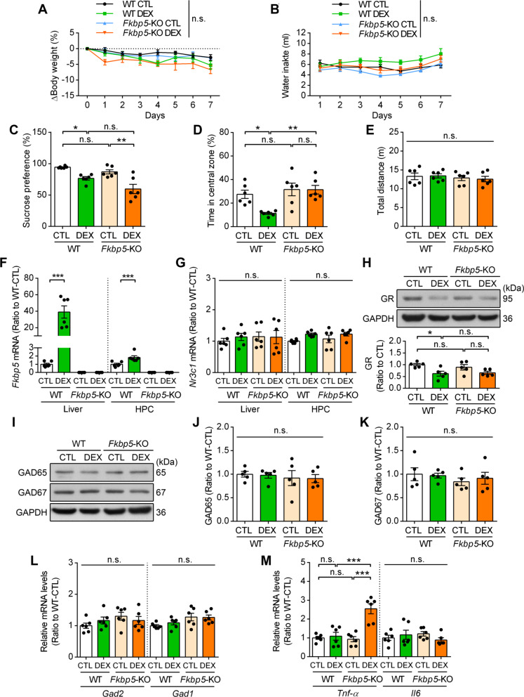
Methods: Fkbp5-deficient (Fkbp5-KO) mice were used to study inflammatory stress by a single intraperitoneal injection of lipopolysaccharide (LPS). The anxiety-like behaviors, neuroimaging, immunofluorescence staining, immunohistochemistry, protein and mRNA expression analysis of inflammation- and neurotransmission-related mediators were evaluated. A dexamethasone drinking model was also applied to examine the effect of Fkbp5-KO in glucocorticoid-induced stress.

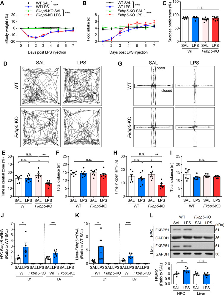
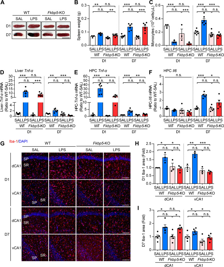
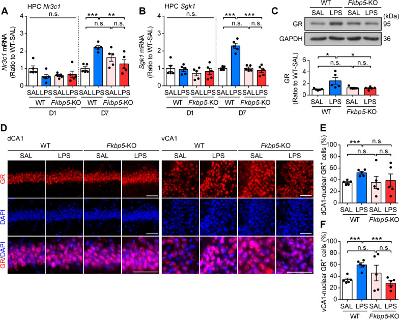
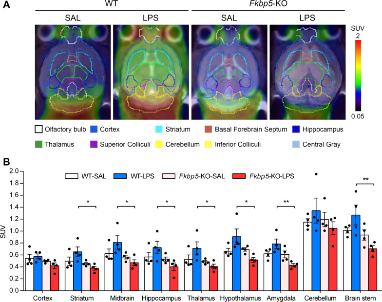
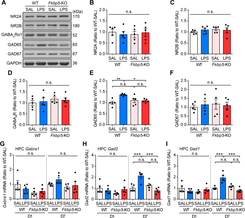
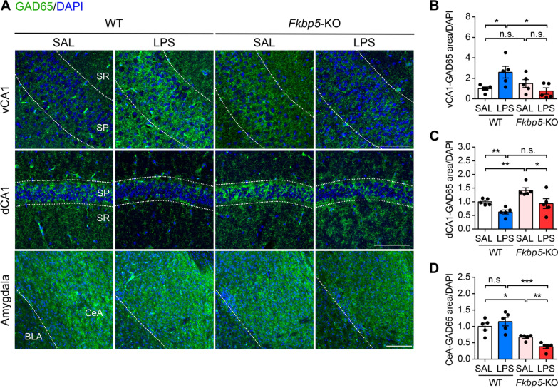
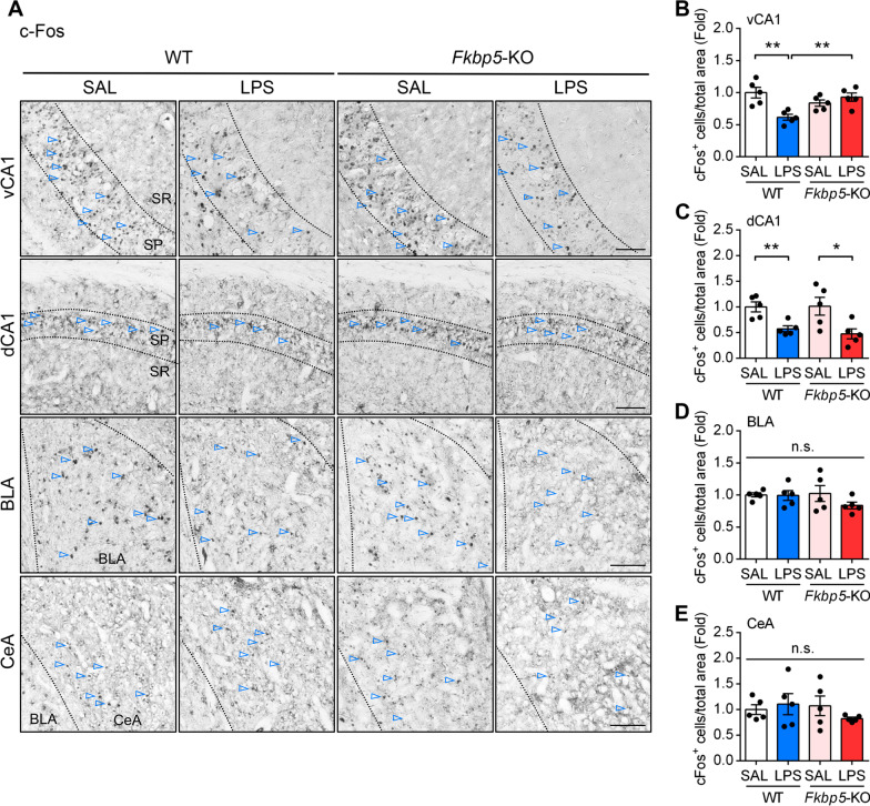
Results: LPS administration induced FKBP51 elevation in the liver and hippocampus accompanied with transient sickness. Notably, Fkbp5-KO but not wild-type (WT) mice showed anxiety-like behaviors 7 days after LPS injection (LPS-D7). LPS challenge rapidly increased peripheral and central immune responses and hippocampal microglial activation followed by a delayed GR upregulation on LPS-D7, and these effects were attenuated in Fkbp5-KO mice. Whole-brain [18F]-FEPPA neuroimaging, which target translocator protein (TSPO) to indicate neuroinflammation, showed that Fkbp5-KO reduced LPS-induced neuroinflammation in various brain regions including hippocampus. Interestingly, LPS elevated glutamic acid decarboxylase 65 (GAD65), the membrane-associated GABA-synthesizing enzyme, in the hippocampus of WT but not Fkbp5-KO mice on LPS-D7. This FKBP51-dependent GAD65 upregulation was observed in the ventral hippocampal CA1 accompanied by the reduction of c-Fos-indicated neuronal activity, whereas both GAD65 and neuronal activity were reduced in dorsal CA1 in a FKBP51-independent manner. GC-induced anxiety was also examined, which was attenuated in Fkbp5-KO and hippocampal GAD65 expression was unaffected.

Conclusions: These results suggest that FKBP51/FKBP5 is involved in the systemic inflammation-induced neuroinflammation and hippocampal GR activation, which may contribute to the enhancement of GAD65 expression for GABA synthesis in the ventral hippocampus, thereby facilitating resilience to inflammation-induced anxiety.

Keywords: Anxiety; FK506-binding protein 51 (FKBP51); GABA; Glucocorticoid receptor; Glutamic acid decarboxylase 65 (GAD65); Inflammation; Resilience; Ventral hippocampus.


Conflict of interest statement

The authors declare that they have no competing interests in this work.

Figures

Fig. 1
Fig. 1
Fkbp5 deletion increases sensitivity to LPS-induced anxiety responses. C57BL/6J (WT) and Fkbp5-KO mice were subjected to a single intraperitoneal injection of saline (SAL) or LPS (3 mg/kg of body weight). A, B The mean body weight (n = 7 mice per group) and food intake (n = 5 mice per group) of mice were tracked every day for 7 days to assess sickness behavior. Data are expressed as the mean ± SEM. ***p < 0.001 for the comparison between two groups in a two-way ANOVA. C The sucrose preference test was performed to assess the anhedonia-like phenotype. Representative traces (D) and the time spent in the central zone (E) of the open-field test (OFT). Spontaneous locomotor activity was expressed as the total distance traveled in the OFT (F). Representative traces (G), time spent in the open arms (H), and total distance traveled (I) in the elevated plus maze (EPM). n = 7 mice per group. J, K qRT-PCR analysis of both hippocampal (HPC) and liver Fkbp5 expression at days 1 and 7 after SAL or LPS injection (n = 5 mice per group). *p < 0.05, **p < 0.01, and ***p < 0.001 for the comparison between two groups in a two-way ANOVA followed by the Tukey post hoc test. L Western blotting indicated that LPS increased FKBP51 protein expression at day 7 in the HPC (n = 5 mice per group). The relative band intensities were normalized to GAPDH levels. *p < 0.05 for the comparison between two groups in an unpaired Student’s t test
Fig. 2
Fig. 2
Fkbp5 deletion attenuates LPS-induced hippocampal immune responses. A, B Spleens were dissected and weighed on day 1 and 7 post-injection of SAL or LPS (n = 6 mice per group). Scale bar: 1 cm. C Complete blood count analysis of peripheral blood samples collected from mice 1 or 7 days after saline or LPS injection. The monocyte counts are shown (D1, n = 3 mice per group; D7, n = 6 mice per group). D–F The liver and HPC of WT and Fkbp5-KO mice were analyzed by qRT-PCR for liver Tnf-α (D), HPC Tnf-α (E), and HPC Il-6 (F) mRNA. Note that Tnf-α expression was induced on day 1 in both the liver and HPC, whereas delayed induction of Il-6 in the HPC was observed on day 7. Data are indicated as the mean ± SEM. *p < 0.05, **p < 0.01, and ***p < 0.001 for the comparison between two groups in a two-way ANOVA followed by the Tukey post hoc test. G Representative immunofluorescence images of microglial marker Iba-1 (red) and DAPI (blue) in the mouse hippocampal dorsal and ventral CA1 (dCA1 and vCA1). Scale bar: 100 μm. H, I Quantification of the area expressing Iba-1 in the stratum pyramidale (SP) and stratum radiatum (SR) of the hippocampal CA1 region (D1, n = 3 mice per group; D7, n = 5 mice per group). Data are indicated as the mean ± SEM. *p < 0.05, **p < 0.01, and ***p < 0.001 for the comparison between two groups in an unpaired Student’s t test
Fig. 3
Fig. 3
Fkbp5 deletion attenuates LPS-induced GR activation. The HPC of WT and Fkbp5-KO mice were collected at days 1 and 7 after LPS injection, followed by mRNA and protein expression analysis. A, B qRT-PCR analysis of HPC Nr3c1 (encoding GR) and Sgk1 expression indicated that Fkbp5 deficiency reduced the LPS-induced delayed upregulation of Nr3c1 and Sgk1 expression at day 7 in the HPC. C Western blotting indicated an increase in GR on day 7 after LPS injection in the HPC of WT but not Fkbp5-KO mice. The relative band intensities were normalized to GAPDH levels. Data are indicated as the mean ± SEM. *p < 0.05, **p < 0.01, and ***p < 0.001 for the comparison between two groups in a two-way ANOVA followed by the Tukey post hoc test. D Immunostaining of the GR in the mouse hippocampal dorsal and ventral CA1 (dCA1 and vCA1) along with counterstaining of the cell nucleus with DAPI. GR-positive cells were predominantly located in CA1 pyramidal cell layers. E, F The nuclear localization of the GR, which indicated the activated state of the GR, was quantified by overlapping GR-DAPI signals in the total DAPI count. Note that LPS significantly increased the number of nuclear GR+ cells in both the dCA1 and vCA1 of WT but not Fkbp5-KO mice. ***p < 0.001 for the comparison between two groups in an unpaired Student’s t test. n = 5 mice per group. Scale bar: 50 μm
Fig. 4
Fig. 4
Neuroimaging of LPS-induced neuroinflammation in WT and Fkbp5-KO mice. A Representative small-animal 7-T PET/MRI of brain inflammation and TSPO radioligand [18F]-FEPPA imaging in WT versus Fkbp5-KO mice 7 days after LPS injection indicated that Fkbp5 deficiency reduced glial activation and neuroinflammation. B Quantitation of [18F]-FEPPA SUV (standard uptake value) in different brain regions. A marked decrease was observed in brain cells expressing TSPO in the mouse brain 7 days after the administration of LPS in Fkbp5-KO mice compared with WT mice. Data are expressed as the mean ± SEM (n = 4 mice per group). *p < 0.05, and **p < 0.01 for the comparison between two groups based on two-way ANOVA followed by the Tukey post hoc test
Fig. 5
Fig. 5
LPS increases GAD65 expression in the hippocampus of WT but not Fkbp5-KO mice. The hippocampal tissues of WT and Fkbp5-KO mice were collected 7 days after the injection of SAL or LPS. A–F Western blotting and quantification were performed to examine the expression of NMDA receptor subunits (NR2A, B; and NR2B, C), GABAARα1 (D), GAD65 (E), and GAD67 (F). The quantification of band densities was indicated by normalizing it to the GAPDH level. qRT-PCR analysis of Gabra1 (G), Gad1 (H), and Gad2 (I) expression. LPS increased both the protein and gene expression of GAD65 in the HPC of WT mice, and these effects were reversed by the knockout of Fkbp5. Data are expressed as the mean ± SEM (n = 5 mice per group). *p < 0.05, **p < 0.01, and ***p < 0.001 for the comparison between two groups based on two-way ANOVA followed by the Tukey post hoc test
Fig. 6
Fig. 6
Chronic GC intake induces FKBP51-dependent anxiety without upregulating hippocampal GAD65. Behavioral assessment and tissue collection were performed in mice with free drinking access to tap water (CTL) or dexamethasone (DEX, 0.016 mg/mL) for 7 days. A, B The body weight and water intake of mice during DEX challenge were tracked for 7 days (n = 6 mice per group). C–E Mice were assessed using the sucrose preference test (C) and open-field test (D, E) on day 7 after DEX intake (n = 6 mice per group). F, G qRT-PCR analysis of liver and hippocampal Fkbp5 and Nr3c1 mRNA levels (n = 6 mice per group). H–K Western blot analysis of GR (H), GAD65 (J), and GAD67 (K) expressions in the HPC (n = 5 mice per group). The band intensity was quantified after it was normalized to the GAPDH level. L, M qRT-PCR analysis of hippocampal Gad2, Gad1, Tnf-α and Il-6 mRNA levels (n = 6 mice per group). Data are expressed as the mean ± SEM. *p < 0.05, **p < 0.01, and ***p < 0.001 for the comparison between two groups in a two-way ANOVA followed by the Tukey post hoc test
Fig. 7
Fig. 7
Subregional alterations in GAD65 expression in the hippocampus and amygdala in LPS-injected mice. Mouse brains were harvested 7 days after the injection of SAL or LPS for immunohistochemical analysis. A Representative immunofluorescent image of GAD65 (green) and DAPI (blue) staining in the hippocampal subregion of the ventral CA1 (vCA1, upper panel) and dorsal CA1 (dCA1, middle panel) as well as the amygdala (lower panel). B–D The area expressing GAD65 was quantified by calculating the ratio of their immunoreactive area to total number of DAPI in the stratum pyramidal (SP) layer of vCA1 (B), dCA1 (C), or central nucleus of the amygdala (CeA, D). Data are indicated as the mean ± SEM (n = 5 mice per group). *p < 0.05, **p < 0.01, and ***p < 0.001 for the comparison between two groups in an unpaired Student’s t test. Scale bar: 100 μm. SR: stratum radiatum, BLA: basolateral amygdala
Fig. 8
Fig. 8
Fkbp5 deficiency LPS-reduced c-Fos expression in the hippocampus in ventral CA1. WT and Fkbp5-KO mouse brains were collected 7 days after the intraperitoneal injection of SAL or LPS. A Representative immunofluorescent images of the neuronal activity marker c-Fos (arrowheads) in the hippocampal subregion of the ventral CA1 and dorsal CA1 (vCA1 and dCA1, upper panel) as well as the amygdala (BLA and CeA, lower panel). B–E The c-Fos-labeled neurons were quantified by calculating the percentage of c-Fos-positive cells in the region of interest in the total area of the vCA1 (B), dCA1 (C), BLA (D), and CeA (E). Data are indicated as the mean ± SEM (n = 5 mice per group). *p < 0.05, and **p < 0.01 for the comparison between two groups in an unpaired Student’s t test. Scale bar: 100 μm
Fig. 9
Fig. 9
Schematic diagram showing that FKBP51 mediates inflammation-associated stress adaptation. The scheme illustrates the underlying mechanism of FKBP51-mediated stress adaptation after systemic inflammation. (1) In the inflammation-induced stress model, peripheral LPS stimulation causes activation of the GR signaling that upregulates FKBP51 as well as neuroinflammation, accompanied with upregulation of GAD65 for GABA synthesis, and suppression of c-Fos-indicated neuronal activity, in the ventral hippocampal CA1 subregion; whereas both GAD65 and neuronal activity were reduced in dorsal CA1. The mice under this condition appear to be resilient to inflammatory stress. (2) Although FKBP51 deficiency attenuates the LPS-induced GR expression and proinflammatory cytokines in the hippocampus, such deficiency results in the development of inflammation-induced anxiety, likely via blocking the GAD65 upregulation to de-suppress the neuronal activity in the ventral CA1. In contrast, FKBP51 deficiency does not block the LPS-induced suppression of GAD65 and neuronal activity in dorsal CA1. (3) GC-induced HPA axis hyperactivity does not increase GAD65 expression, thus resulting in the development of anxiety. Fkbp5 deficiency exerted an anxiolytic effect by regulating GR sensitivity in GC-induced stress. Taken together, FKBP51 modulates stress-induced anxiety in various stressors, and plays a critical role in stress adaptation and GABAergic neurotransmission after inflammation in the ventral hippocampus. (↑: increased, ↓: decreased, -: unchanged)

Similar articles

Cited by

References

    1. Wochnik GM, Ruegg J, Abel GA, Schmidt U, Holsboer F, Rein T. FK506-binding proteins 51 and 52 differentially regulate dynein interaction and nuclear translocation of the glucocorticoid receptor in mammalian cells. J Biol Chem. 2005;280:4609–4616. doi: 10.1074/jbc.M407498200. - DOI - PubMed
    1. Zannas AS, Wiechmann T, Gassen NC, Binder EB. Gene-stress-epigenetic regulation of FKBP5: clinical and translational implications. Neuropsychopharmacology. 2016;41:261–274. doi: 10.1038/npp.2015.235. - DOI - PMC - PubMed
    1. Binder EB. The role of FKBP5, a co-chaperone of the glucocorticoid receptor in the pathogenesis and therapy of affective and anxiety disorders. Psychoneuroendocrinology. 2009;34(Suppl 1):S186–195. doi: 10.1016/j.psyneuen.2009.05.021. - DOI - PubMed
    1. Menke A, Klengel T, Rubel J, Bruckl T, Pfister H, Lucae S, Uhr M, Holsboer F, Binder EB. Genetic variation in FKBP5 associated with the extent of stress hormone dysregulation in major depression. Genes Brain Behav. 2013;12:289–296. doi: 10.1111/gbb.12026. - DOI - PubMed
    1. Criado-Marrero M, Rein T, Binder EB, Porter JT, Koren J, 3rd, Blair LJ. Hsp90 and FKBP51: complex regulators of psychiatric diseases. Philos Trans R Soc Lond B Biol Sci. 2018;373:20160532. doi: 10.1098/rstb.2016.0532. - DOI - PMC - PubMed

MeSH terms