. 2022 Jun 15;19(1):148.
doi: 10.1186/s12974-022-02498-8.

Superior control of inflammatory pain by corticotropin-releasing factor receptor 1 via opioid peptides in distinct pain-relevant brain areas

Affiliations

Superior control of inflammatory pain by corticotropin-releasing factor receptor 1 via opioid peptides in distinct pain-relevant brain areas

Shaaban A Mousa et al. J Neuroinflammation. .

Abstract

Background: Under inflammatory conditions, the activation of corticotropin-releasing factor (CRF) receptor has been shown to inhibit pain through opioid peptide release from immune cells or neurons. CRF's effects on human and animal pain modulation depend, however, on the distribution of its receptor subtypes 1 and 2 (CRF-R1 and CRF-R2) along the neuraxis of pain transmission. The objective of this study is to investigate the respective role of each CRF receptor subtype on centrally administered CRF-induced antinociception during inflammatory pain.

Methods: The present study investigated the role of intracerebroventricular (i.c.v.) CRF receptor agonists on nociception and the contribution of cerebral CRF-R1 and/or CRF-R2 subtypes in an animal model of Freund's complete adjuvant (FCA)-induced hind paw inflammation. Methods used included behavioral experiments, immunofluorescence confocal analysis, and reverse transcriptase-polymerase chain reaction.

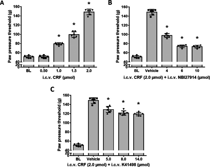
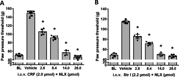
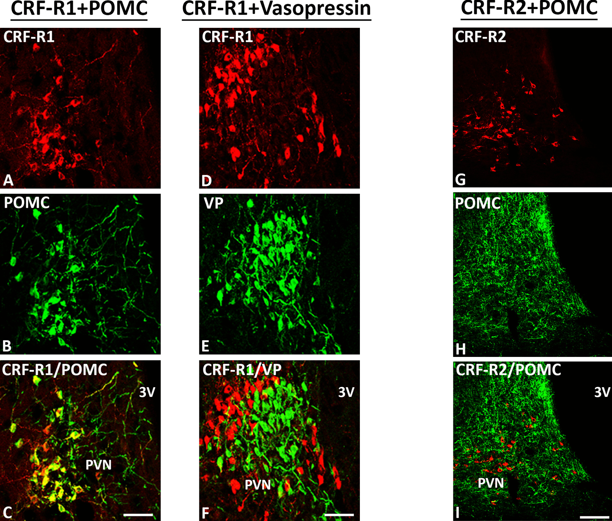
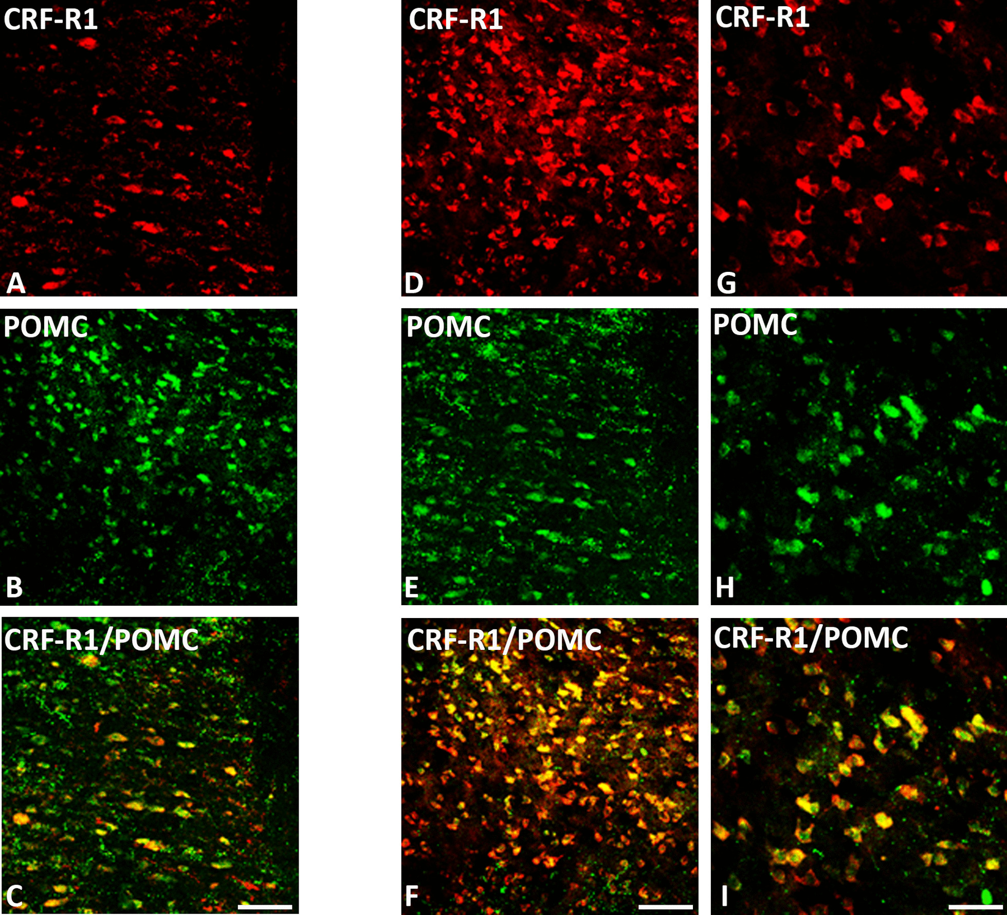
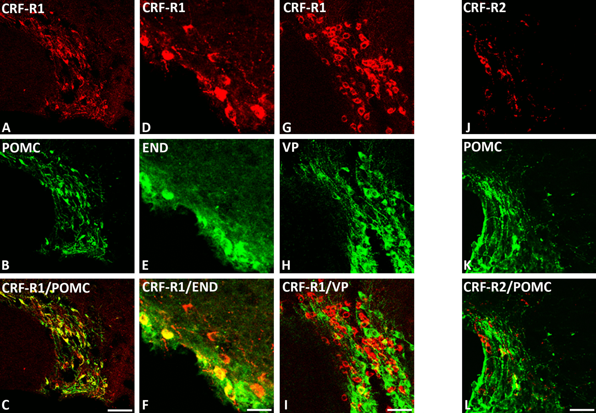
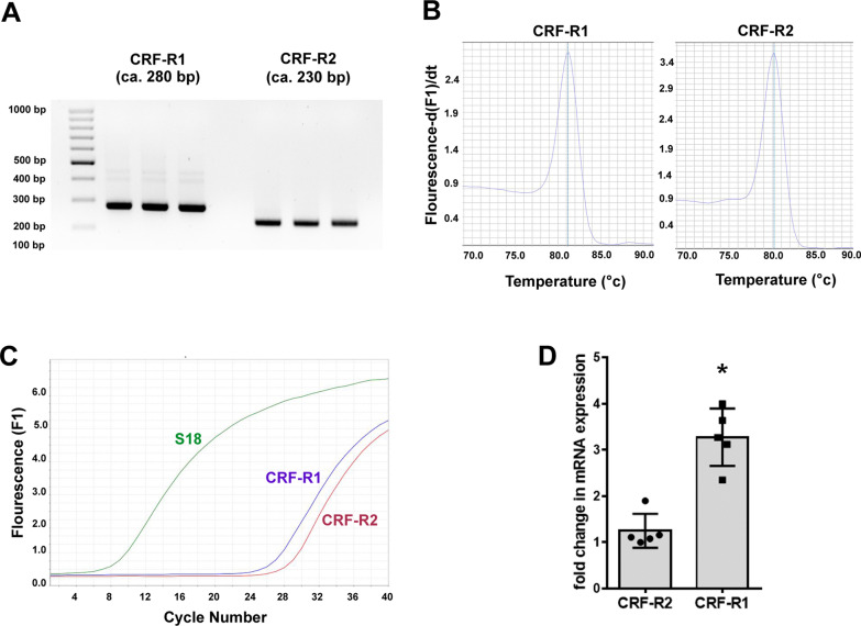
Results: Intracerebroventricular, but systemically inactive, doses of CRF elicited potent, dose-dependent antinociceptive effects in inflammatory pain which were significantly antagonized by i.c.v. CRF-R1-selective antagonist NBI 27914 (by approximately 60%) but less by CRF-R2-selective antagonist K41498 (by only 20%). In line with these findings, i.c.v. administration of CRF-R1 agonist stressin I produced superior control of inflammatory pain over CRF-R2 agonist urocortin-2. Intriguingly, i.c.v. opioid antagonist naloxone significantly reversed the CRF as well as CRF-R1 agonist-elicited pain inhibition. Consistent with existing evidence of high CRF concentrations in brain areas such as the thalamus, hypothalamus, locus coeruleus, and periaqueductal gray following its i.c.v. administration, double-immunofluorescence confocal microscopy demonstrated primarily CRF-R1-positive neurons that expressed opioid peptides in these pain-relevant brain areas. Finally, PCR analysis confirmed the predominant expression of the CRF-R1 over CRF-R2 in representative brain areas such as the hypothalamus.

Conclusion: Taken together, these findings suggest that CRF-R1 in opioid-peptide-containing brain areas plays an important role in the modulation of inflammatory pain and may be a useful therapeutic target for inflammatory pain control.

Keywords: Brain; Corticotropin-releasing factor; Immunofluorescence; Inflammatory pain; Opioid peptide.


Conflict of interest statement

The authors declare no potential conflicts of interest with respect to the research, authorship, and/or publication of this article.

Figures

Fig. 1
Fig. 1
Antagonism of antinociceptive effects of i.c.v. administered CRF by CRF-R1 antagonist NBI 27914 and CRF-R2 antagonist K41498. In Wistar rats with 4-day FCA-induced hind paw inflammation, effects of i.c.v. administered CRF on nociceptive paw pressure thresholds were measured by algesiometry. A i.c.v. application of CRF (0.5, 01.0, 1.5, 2.0 µmol) significantly increased nociceptive thresholds in a dose-dependent manner (F(4, 25) = 445; P < 0.001). B Dose-dependent antagonism of i.c.v. CRF’s antinociception by co-administered CRF-R1 antagonist NBI 27914 (F(4, 25) = 502.9; P < 0.001). C In contrast, increasing doses of the i.c.v. CRF-R2 antagonist K41498 only partially antagonized CRF’s antinociception (F(4, 25) = 315.7; P < 0.001). *indicates significant differences from vehicle treatment; data points (n = 6) represent means ± SD
Fig. 2
Fig. 2
Antinociceptive effects of the i.c.v. CRF-R1 agonist stressin I or CRF-R2 agonist Ucn-2 and their antagonism by the respective CRF-R1 (NBI 27914) or CRF-R2 (K41498) selective antagonists. The effects of i.c.v. CRF-R1 (stressin I) or CRF-R2 (Ucn-2) agonists on nociceptive paw pressure thresholds were measured by algesiometry. A i.c.v. administration of the CRF-R1 agonist stressin I significantly increased nociceptive thresholds in a dose-dependent manner (F(4, 25) = 552.4; P < 0.001). B i.c.v. administration of the CRF-R2 agonist Ucn-2 significantly increased nociceptive thresholds (F(4, 25) = 389.1; P < 0.001). C Dose-dependent antagonism of i.c.v. CRF-R1 agonist’s antinociception by co-administered CRF-R1 antagonist NBI 27914 (F(4, 25) = 73.9; P < 0.001, one-way ANOVA and Dunnett’s test). D) Dose-dependent antagonism of i.c.v. Ucn-2 antinociception by co-administered CRF-R2 antagonist K41498 was significant (F(5, 30) = 88.4; P < 0.001), *indicates significant differences from vehicle treatment; data points (n = 6) represent means ± SD
Fig. 3
Fig. 3
The antinociceptive effects of i.c.v. CRF and CRF-R1 agonist stressin I and their antagonism by the opioid receptor antagonist naloxone in rats with inflamed hindpaws. The effects of i.c.v. co-administration of the opioid receptor antagonist naloxone with CRF or CRF-R1 agonist stressin I on nociceptive thresholds were measured by algesiometer. A The i.c.v. CRF’s induced-antinociception was significantly reduced by co-administration of the opioid receptor antagonist naloxone (F(5, 30) = 384.8; P < 0.001). B Similarly, the antinociception resulting from i.c.v. CRF-R1 agonist stressin I was significantly attenuated by co-administered opioid receptor antagonist naloxone (F(5, 30) = 400.8; P < 0.001) *indicates significant differences from vehicle treatment); data points (n = 6) represent means ± SD
Fig. 4
Fig. 4
Double-immunofluorescence staining of CRF-R1 (red fluorescence) AF or CRF-R2 GI (red fluorescence) with proopiomelanocortin (POMC) (green fluorescence) B, H and vasopressin (green fluorescence) E in the paraventricular nucleus (PVN) of the rat hypothalamus. AC Double-immunofluorescence staining of coronal brain sections of the rat with hind paw inflammation showing that CRF-R1-immunoreactive neurons within PVN overlap with the opioid peptide precursor POMC (AC). Some neurons express CRF-R1 or POMC only. DF Show CRF-R1-immunoreactive neurons in the paraventricular nucleus (PVN) residing in close vicinity of vasopressin positive cells without any overlap of the two cell groups. GI Show only few scattered CRF-R2-immunoreactive neurons without POMC overlap in the same region. Bar = 20 μm
Fig. 5
Fig. 5
Double-immunofluorescence staining of CRF-R1 (red fluorescence) AF and POMC (green fluorescence) B, C or β-endorphin (END) (green fluorescence) E, F in the median eminence of the rat hypothalamus. AF Show that most of CRF-R1-immunoreactive fibers express POMC (C) or END (F) in coronal sections of the rat brain of Wistar rats, but few fibers contain CRF-R1, POMC or END only. Bar = 20 µm
Fig. 6
Fig. 6
Double-immunofluorescence staining of CRF-R1 (red fluorescence) A, D, G and POMC (green fluorescence) B, E, H (green fluorescence) E in the thalamus AC and periaqueductal grey DI of rat brain. AC Coronal sections of the rat brain with hindpaw inflammation show that most of CRF-R1-positive neurons in the thalamus region express POMC, but few fibers contain CRF-R1 or POMC only. DF Coronal sections of the rat brain with hindpaw inflammation show co-localization of CRF-R1 with POMC neurons in the periaqueductal grey, but few fibers contain CRF-R1 or POMC only. Bar = 20 µm, GI Higher magnification of DF. Bar = 40 µm
Fig. 7
Fig. 7
Double-immunofluorescence staining of CRF-R1 (red fluorescence) A, D, G or CRF-R2 Jl with proopiomelanocortin (POMC) (B, K), β-endorphin (END) E and vasopressin H (green fluorescence) in the supraoptic area (SOA) of the rat hypothalamus. AF Double-immunofluorescence staining of coronal sections of the rat brain with hind paw inflammation showing that CRF-R-immunoreactive neurons within SON overlap with the opioid peptide precursor POMC C or (F). Some neurons express CRF-R1 or POMC only. GI Show the majority of CRF-R1-immunoreactive neurons residing in close vicinity, and rarely overlap (as indicated by yellow fluorescence) with vasopressin positive cells. JL Show few scattered CRF-R2-immunoreactive neurons with only rare overlap with POMC-ir neurons. Bar = 20 μm
Fig. 8
Fig. 8
Double-immunofluorescence staining of CRF-R1 (red fluorescence) A, D with proopiomelanocortin (POMC) B or β-endorphin (END) E (green fluorescence) in the Locus coeruleus (LC) of the rat brain. AF Double-immunofluorescence staining of coronal sections of the rat brain with hind paw inflammation showing co-expression of CRF-R1 with POMC C or END F within LC. Some neurons express CRF-R1, POMC or END only. Bar = 20 µm for AC. Bar = 40 μm for (DF)
Fig. 9
Fig. 9
CRF-R1 and CRF-R2 mRNA expression in the hypothalamus region of the rat with 4 day FCA-induced hind paw inflammation. A RNA extraction from the rat hypothalamus, implementation of conventional PCR using specific primer pairs for CRF-R1 and CRF-R2, and subsequent visualization on a 2% agarose gel provided specific PCR products for the expression of CRF-R1 (280 bp) and CRF-R2 (230 bp) mRNA. B Shows the DNA melting profiles of the CRF-R1 (right) and CRF-R2 (left) specific primer pairs. C, D Quantification of CRF-R1 and CRF-R2 mRNA using Taqman® Real-Time PCR in the hypothalamus region of the rat brain. C The amplification profiles of the 18S- and CRF-R1- and CRF-R2-specific cDNA of rat hypothalamus. D The column graph representing % CRF-R1 mRNA expression relative to the expression of CRF-R2 mRNA. Note that CRF-R1 expression is more than threefold higher than that of CRF-R2 (experiments were done in duplicate from n = 5 rats, *P < 0.05, two-tailed independent Student t-test)

References

    1. Bale TL, Vale WW. CRF and CRF receptors: role in stress responsivity and other behaviors. Annu Rev Pharmacol Toxicol. 2004;44:525–557. doi: 10.1146/annurev.pharmtox.44.101802.121410. - DOI - PubMed
    1. Lariviere WR, Melzack R. The role of corticotropin-releasing factor in pain and analgesia. Pain. 2000;84:1–12. doi: 10.1016/S0304-3959(99)00193-1. - DOI - PubMed
    1. Andreoli M, Marketkar T, Dimitrov E. Contribution of amygdala CRF neurons to chronic pain. Exp Neurol. 2017;298:1–12. doi: 10.1016/j.expneurol.2017.08.010. - DOI - PMC - PubMed
    1. Mousa SA, Bopaiah CP, Richter JF, Yamdeu RS, Schäfer M. Inhibition of inflammatory pain by CRF at peripheral, spinal and supraspinal sites: involvement of areas coexpressing CRF receptors and opioid peptides. Neuropsychopharmacology. 2007;32:2530–2542. doi: 10.1038/sj.npp.1301393. - DOI - PubMed
    1. Taché Y. Corticotrophin-releasing factor 1 activation in the central amygdale and visceral hyperalgesia. Neurogastroenterol Motil. 2015;27:1–6. doi: 10.1111/nmo.12495. - DOI - PMC - PubMed

Substances

LinkOut - more resources