. 2022 Jun 15;29(1):42.
doi: 10.1186/s12929-022-00824-z.

MicroRNA-485-5p targets keratin 17 to regulate oral cancer stemness and chemoresistance via the integrin/FAK/Src/ERK/β-catenin pathway

Affiliations

MicroRNA-485-5p targets keratin 17 to regulate oral cancer stemness and chemoresistance via the integrin/FAK/Src/ERK/β-catenin pathway

Te-Hsuan Jang et al. J Biomed Sci. .

Abstract

Background: The development of drug resistance in oral squamous cell carcinoma (OSCC) that frequently leads to recurrence and metastasis after initial treatment remains an unresolved challenge. Presence of cancer stem cells (CSCs) has been increasingly reported to be a critical contributing factor in drug resistance, tumor recurrence and metastasis. Thus, unveiling of mechanisms regulating CSCs and potential targets for developing their inhibitors will be instrumental for improving OSCC therapy.

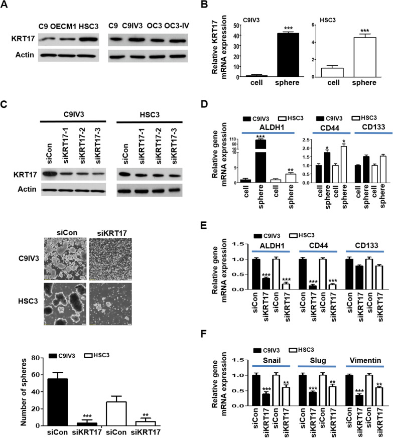
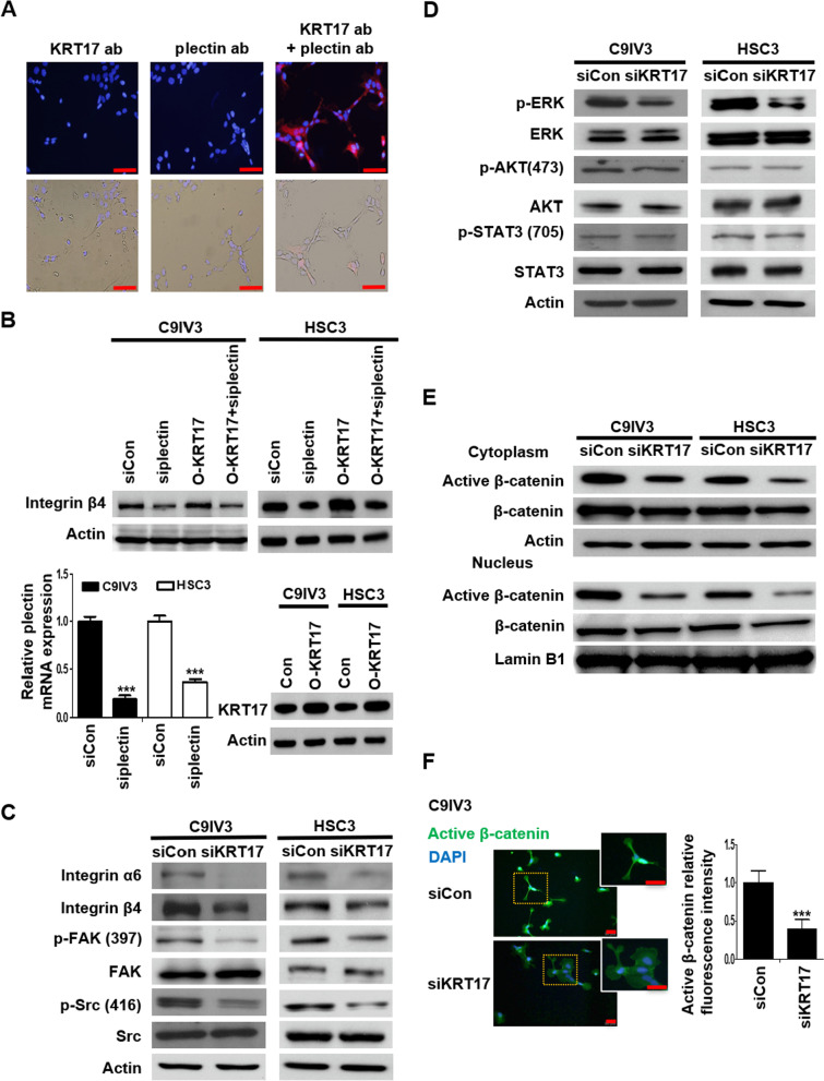
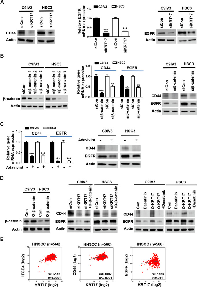
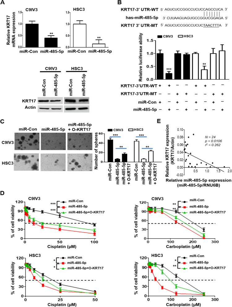
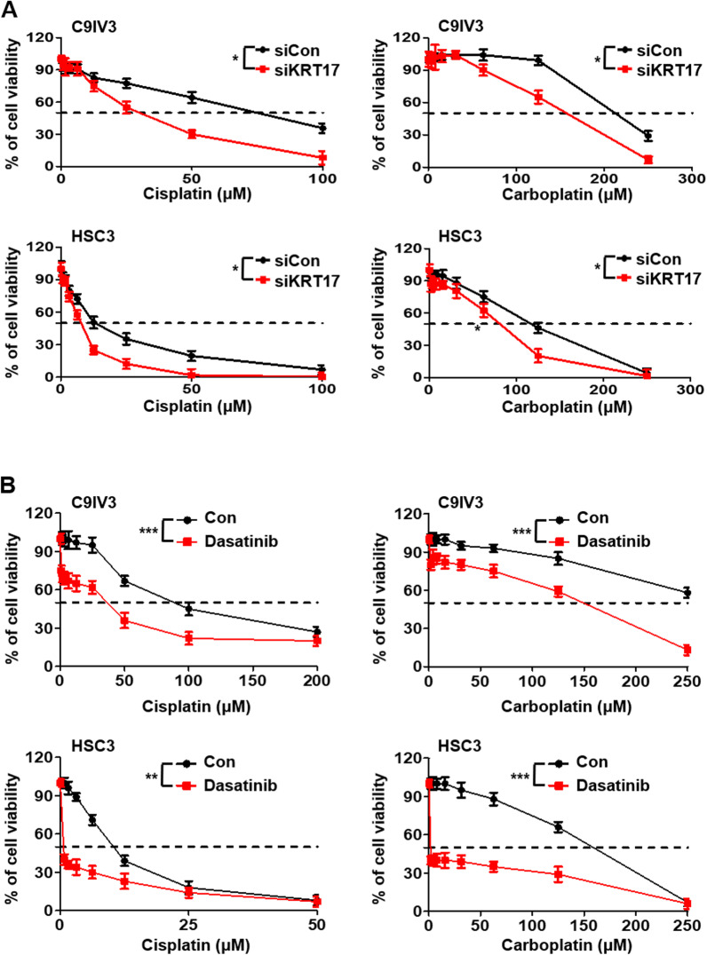
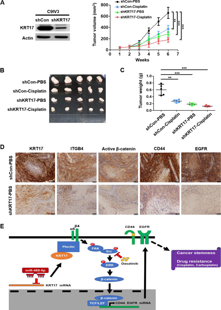
Methods: siRNA, shRNA and miRNA that specifically target keratin 17 (KRT17) were used for modulation of gene expression and functional analyses. Sphere-formation and invasion/migration assays were utilized to assess cancer cell stemness and epithelial mesenchymal transition (EMT) properties, respectively. Duolink proximity ligation assay (PLA) was used to examine molecular proximity between KRT17 and plectin, which is a large protein that binds cytoskeleton components. Cell proliferation assay was employed to evaluate growth rates and viability of oral cancer cells treated with cisplatin, carboplatin or dasatinib. Xenograft mouse tumor model was used to evaluate the effect of KRT17- knockdown in OSCC cells on tumor growth and drug sensitization.

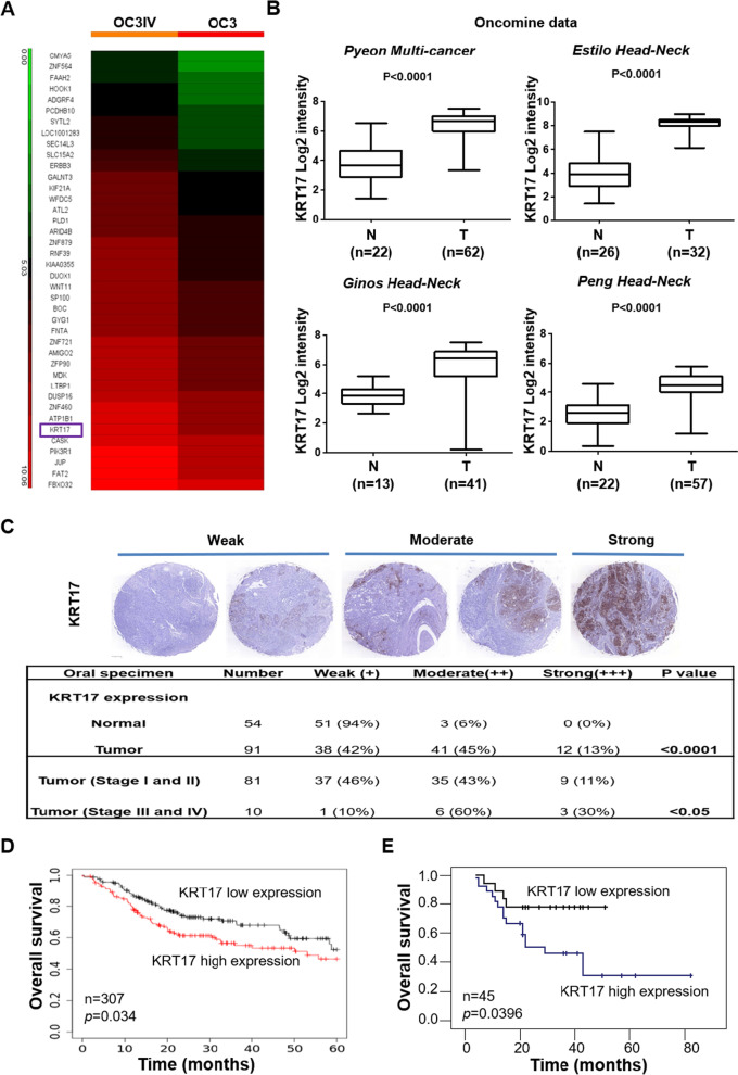
Results: Significantly elevated expression of KRT17 in highly invasive OSCC cell lines and advanced tumor specimens were observed and high KRT17 expression was correlated with poor overall survival. KRT17 gene silencing in OSCC cells attenuated their stemness properties including markedly reduced sphere forming ability and expression of stemness and EMT markers. We identified a novel signaling cascade orchestrated by KRT17 where its association with plectin resulted in activation of integrin β4/α6, increased phosphorylation of FAK, Src and ERK, as well as stabilization and nuclear translocation of β-catenin. The activation of this signaling cascade was correlated with enhanced OSCC cancer stemness and elevated expression of CD44 and epidermal growth factor receptor (EGFR). We identified and demonstrated KRT17 to be a direct target of miRNA-485-5p. Ectopic expression of miRNA-485-5p inhibited OSCC sphere formation and caused sensitization of cancer cells towards cisplatin and carboplatin, which could be significantly rescued by KRT17 overexpression. Dasatinib treatment that inhibited KRT17-mediated Src activation also resulted in OSCC drug sensitization. In OSCC xenograft mouse model, KRT17 knockdown significantly inhibited tumor growth, and combinatorial treatment with cisplatin elicited a greater tumor inhibitory effect. Consistently, markedly reduced levels of integrin β4, active β-catenin, CD44 and EGFR were observed in the tumors induced by KRT17 knockdown OSCC cells.

Conclusions: A novel miRNA-485-5p/KRT17/integrin/FAK/Src/ERK/β-catenin signaling pathway is unveiled to modulate OSCC cancer stemness and drug resistance to the common first-line chemotherapeutics. This provides a potential new therapeutic strategy to inhibit OSCC stem cells and counter chemoresistance.

Keywords: Cancer stemness; Chemoresistance; Dasatinib; Integrin β4; Keratin 17; Oral squamous cell carcinoma; miR-485-5p; β-catenin.


Conflict of interest statement

The authors declare that they have no competing interests.

Figures

Fig. 1
Fig. 1
The clinical importance of KRT17 as a prognostic marker in OSCC. A Heatmap analysis of differentially-expressed genes from cDNA microarrays data that were derived from OC3IV (left panel) and OC3 (right panel) OSCC cells. B Datamining analysis of KRT17 mRNA expression in the adjacent normal tissues (N) and OSCC tumor tissues (T) from Oncomine database. C Representative images of IHC staining for the KRT17 protein expression were shown using 3-level pathological ranking of weak, moderate and strong from a set of commercial tissue arrays containing a total of 145 normal and OSCC tissues (top panel). The bottom panel shows a table summarizing statistically the expression differences of KRT17 between normal and OSCC tissues with different pathological stages at I/II or III/IV. D KM plotter survival analysis was performed to examine KRT17 gene expression in a cohort of 307 Head-neck cancer patients (Oncomine database). E Correlation of KRT17 expression to overall survival rates was determined by performing cDNA microarray with 45 OSCC tumor specimens as described in “Methods”
Fig. 2
Fig. 2
Silencing of KRT17 downregulates cancer stemness in invasive OSCC. A KRT17 protein expressions in different OSCC cell lines were assessed by immunoblotting. B KRT17 mRNA expressions in the spheres formed from C9IV3 or HSC3 cells were analyzed by comparisons to their parental non-sphere C9IV3 or HSC3 cells, respectively, using qPCR. C Top, Protein expressions of KRT17 in C9IV3 and HSC3 cells transfected with Control-siRNAs (siCon) or three specific KRT17-siRNA plasmids (siKRT17-1, -2, -3) were assessed by immunoblotting. Middle, sphere-forming abilities of C9IV3 and HSC3 cells transfected with siCon or siKRT17 (siKRT17-3). Bottom, quantitative analysis of the number of spheres formed. D ALDH1, CD44 and CD133 expressions in spheres enriched from C9IV3 or HSC3 cells versus non-sphere cells were determined by qPCR. E ALDH1, CD44 and CD133 expressions in the C9IV3 or HSC3 spheres and non-sphere cells that had been transfected with siCon or siKRT17 were determined by qPCR. F Expressions of EMT markers Snail, Slug and Vimentin in the same spheres or non-sphere cells as in D, E. were determined by qPCR. Data are presented as mean ± SD (*p < 0.05, **p < 0.01 and ***p < 0.001)
Fig. 3
Fig. 3
KRT17 associates with the plectin-integrin β4 complex to activate downstream FAK/Src/ERK/β-catenin signaling pathway. A Molecular proximity between KRT17 and plectin in C9IV3 cells was analyzed by PLA as described in the Methods. Protein complexes under examination were visualized as red dots in fluorescence and bright field images; DAPI-stained cell nuclei are shown in blue. Scale bar shown is 100 μm. B ITGB4 protein expressions in C9IV3 and HSC3 cells that had been transfected with siCon, si-plectin, O-KRT17 (KRT17 overexpressing plasmid) or si-plectin + O-KRT17 were determined by immunoblotting (top). qPCR analysis was used to assess mRNA expression of plectin in C9IV3 and HSC3 cells transfected with siCon or si-plectin (bottom left); while immunoblotting was used to assess KRT17 protein expression in C9IV3 and HSC3 cells that had been transfected with empty control plasmid (Con) or O-KRT17 (bottom right). Data are presented as the mean ± SD (***p < 0.001). C–E Integrin α6, integrin β4, p-FAK, FAK, p-Src, Src, p-ERK, ERK, p-AKT, AKT, p-STAT3, STAT3, active β-catenin (unphosphorylated) and β-catenin protein expressions in C9IV3 and HSC3 cells transfected with siCon or siKRT17 were determined by immunoblotting with antibodies specifically recognizing these proteins. F. Immunofluorescent staining for active β-catenin (green) was conducted to examine the influence of KRT17-silencing on β-catenin in C9IV3 cells. Scale bar shown is 20 μm. Histograms show quantification of the fluorescence intensity for the expression of Active β-catenin. Data are presented as the mean ± SD (***p < 0.001)
Fig. 4
Fig. 4
KRT17 regulates CD44 and EGFR expression via activations of integrin/Src/β-catenin signaling cascade. A CD44 protein expression in C9IV3 and HSC3 cells transfected with siCon or siKRT17 was determined by western blotting (left panel), and EGFR expression was assessed by qPCR and western blotting (middle and right panels). B Left, Western blotting was used to assess the efficacy of three siRNAs (siβ-catenin-1, -2, -3) designed to specifically silence β-catenin in C9IV3 and HSC3 cells (left panel). Siβ-catenin-2 (siβ-catenin) that caused most effective β-catenin suppression was utilized to examine mRNA and protein expression of CD44 and EGFR in C9IV3 and HSC3 cells (middle and right). C The canonical Wnt/β-catenin inhibitor, adavivint (1 μM), was used to treat C9IV3 and HSC3 cells for 48 h before analyzing expressions of CD44 and EGFR at both mRNA and protein levels. D Expression efficiency of the β-catenin cDNA plasmid (O-β-catenin) was determined at protein level (left panel) prior to combinatorial treatment with either siKRT17 (middle panel) or dasatinib (right panel) for assessing their impacts on CD44 and EGFR expression in C9IV3 and HSC3 cells. Cells were transfected with the indicated siRNA or plasmids for 24 h, and then treated with 1 μM dasatinib for 48 h. E Correlation between the gene expression levels of KRT17 and ITGB4, CD44 or EGFR in clinical HNSCC specimens using TCGA database (n = 566)
Fig. 5
Fig. 5
miR-485-5p suppresses KRT17 expression and sphere formation and its effect on drug sensitivity of OSCC. A qPCR and immunoblotting were used to assess targeting effects of miR-485-5p on KRT17 mRNA (top panel) and protein (bottom panel) expression in C9IV3 and HSC3 cells in comparison with the miR-Con. B Sequence alignment of miR-485-5p binding site within the KRT17 3′-UTR (top panel). Luciferase activity assays from C9IV3 and HSC3 cells transfected with wild-type (WT) or mutant (MT) KRT17 3′-UTR reporter constructs in the presence of miRNA control (miR-Con) or miR-485-5p expression plasmids (bottom panel). C Sphere-formation assays using C9IV3 and HSC3 cells transfected with miR-Con, miR-485-5p or miR-485-5p + O-KRT17 (left panel). Quantitative analysis on the number of spheres formed is shown in the right panel. D Dose-dependent cellular growth assays using C9IV3 or HSC3 cells transfected with miR-Con, miR-485-5p or miR-485-5p + O-KRT17 plasmid in combination of treatment with cisplatin (0–100 μM) or carboplatin (0–250 μM). Data are presented as the mean ± SD (*p < 0.05, **p < 0.01 and ***p < 0.001). E Correlation analysis between miR-485-5p and KRT17 RNA levels in 24 OSCC tumor specimens from Kaohsiung Medical University, Taiwan
Fig. 6
Fig. 6
KRT17 silencing potentiates therapeutic efficacies of cisplatin, carboplatin and dasatinib in OSCC. A Dose-dependent cell viability assays were performed to examine the effect of KRT17 silencing on drug sensitivity of C9IV3 and HSC3 cells that were treated with cisplatin or carboplatin ranging from 0 to 100 μM. B Dose-dependent cell viability assays were performed to investigate whether combinatorial treatments of dasatinib with cisplatin or carboplatin could be therapeutically more effective against C9IV3 and HSC3 cells. Data are presented as the mean ± SD (*p < 0.05, **p < 0.01 and ***p < 0.001)
Fig. 7
Fig. 7
Knockdown of KRT17 inhibits tumor stemness and synergistically enhanced inhibitory effects of cisplatin on of OSCC tumor growth. A KRT17 knockdown (shKRT17) efficiency was determined via KRT17 protein expression by immunoblotting and comparison to the shRNA control (shCon) in C9IV3 cells (left panel). SCID mice were injected with 5 × 105 shCon or shKRT17 C9IV3 cells in right flank, and PBS or cisplatin was administered two times a week. Tumor volumes were measured weekly after transimplantation before tumor excision at day 42. Right panel shows the tumor growth curve of each treatment group. Data are presented as mean ± SD (n = 5, **p < 0.01 and ***p < 0.001). B Excised tumors from the xenograft model was shown at the end of 42 days. C Tumor weights of the excised tumors (**p < 0.01 and ***p < 0.001). D Immunohistochemical staining for protein expressions of KRT17, ITGB4, active β-catenin, CD44 and EGFR in representative tumor excised (Scale bar, 50 μm). E A schematic diagram that depicts a novel signaling mechanism mediated by miR-485-5p/KRT17/integrin/FAK/Src/ERK/β-catenin that contributes to cancer stemness and drug resistance in OSCC

References

    1. D'Souza S, Addepalli V. Preventive measures in oral cancer: an overview. Biomed Pharmacother. 2018;107:72–80. doi: 10.1016/j.biopha.2018.07.114. - DOI - PubMed
    1. Rahman QB, Iocca O, Kufta K, Shanti RM. Global burden of head and neck cancer. Oral Maxillofac Surg Clin North Am. 2020;32(3):367–375. doi: 10.1016/j.coms.2020.04.002. - DOI - PubMed
    1. Huang SH, O'Sullivan B. Overview of the 8th edition tnm classification for head and neck cancer. Curr Treat Options Oncol. 2017;18(7):40. doi: 10.1007/s11864-017-0484-y. - DOI - PubMed
    1. Naruse T, Yanamoto S, Matsushita Y, Sakamoto Y, Morishita K, Ohba S, et al. Cetuximab for the treatment of locally advanced and recurrent/metastatic oral cancer: an investigation of distant metastasis. Mol Clin Oncol. 2016;5(2):246–252. doi: 10.3892/mco.2016.928. - DOI - PMC - PubMed
    1. Mohan SP, Bhaskaran MK, George AL, Thirutheri A, Somasundaran M, Pavithran A. Immunotherapy in oral cancer. J Pharm Bioallied Sci. 2019;11(Suppl 2):S107–S111. doi: 10.4103/JPBS.JPBS_31_19. - DOI - PMC - PubMed

MeSH terms