. 2022 Jun 15;19(1):147.
doi: 10.1186/s12974-022-02486-y.

Microglial amyloid beta clearance is driven by PIEZO1 channels

Affiliations

Microglial amyloid beta clearance is driven by PIEZO1 channels

Henna Jäntti et al. J Neuroinflammation. .

Abstract

Background: Microglia are the endogenous immune cells of the brain and act as sensors of pathology to maintain brain homeostasis and eliminate potential threats. In Alzheimer's disease (AD), toxic amyloid beta (Aβ) accumulates in the brain and forms stiff plaques. In late-onset AD accounting for 95% of all cases, this is thought to be due to reduced clearance of Aβ. Human genome-wide association studies and animal models suggest that reduced clearance results from aberrant function of microglia. While the impact of neurochemical pathways on microglia had been broadly studied, mechanical receptors regulating microglial functions remain largely unexplored.

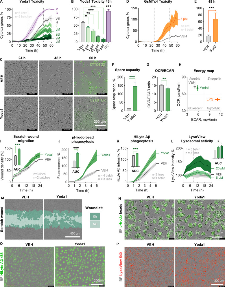
Methods: Here we showed that a mechanotransduction ion channel, PIEZO1, is expressed and functional in human and mouse microglia. We used a small molecule agonist, Yoda1, to study how activation of PIEZO1 affects AD-related functions in human induced pluripotent stem cell (iPSC)-derived microglia-like cells (iMGL) under controlled laboratory experiments. Cell survival, metabolism, phagocytosis and lysosomal activity were assessed using real-time functional assays. To evaluate the effect of activation of PIEZO1 in vivo, 5-month-old 5xFAD male mice were infused daily with Yoda1 for two weeks through intracranial cannulas. Microglial Iba1 expression and Aβ pathology were quantified with immunohistochemistry and confocal microscopy. Published human and mouse AD datasets were used for in-depth analysis of PIEZO1 gene expression and related pathways in microglial subpopulations.

Results: We show that PIEZO1 orchestrates Aβ clearance by enhancing microglial survival, phagocytosis, and lysosomal activity. Aβ inhibited PIEZO1-mediated calcium transients, whereas activation of PIEZO1 with a selective agonist, Yoda1, improved microglial phagocytosis resulting in Aβ clearance both in human and mouse models of AD. Moreover, PIEZO1 expression was associated with a unique microglial transcriptional phenotype in AD as indicated by assessment of cellular metabolism, and human and mouse single-cell datasets.

Conclusion: These results indicate that the compromised function of microglia in AD could be improved by controlled activation of PIEZO1 channels resulting in alleviated Aβ burden. Pharmacological regulation of these mechanoreceptors in microglia could represent a novel therapeutic paradigm for AD.

Keywords: Alzheimer’s disease; Amyloid; Mechanoreceptor; Microglia; Piezo1; iMGL; iPSC-derived microglia.


Conflict of interest statement

Malm and Giniatullin are inventors of PCT patent application related to this topic.

Figures

Fig. 1
Fig. 1
Human and mouse microglia sense mechanical forces through PIEZO1 receptor. A Piezo1 gene expression in murine trigeminal neurons, astrocytes, microglia (MG) and microglial cell line (BV2); and in human microglial cell line (SV40) and iPSC-derived microglia (iMGL) analyzed by RT-qPCR (N = 3–4). B PIEZO1 and PIEZO2 gene expression in human iMGLs, fetal and adult MG, iPSCs, induced hematopoietic progenitor cells (iHPCs), CD14 + and CD16 + monocytes (CD14M, CD16M), and dendritic cells (DC) obtained from human RNA-seq datasets [26, 37]. N = 3–6. Immunostaining of PIEZO1 (green) and nuclei DAPI (blue) in C iMGLs and D MGs. E Schematic for PIEZO1 activation by mechanical fluid puff, small molecule agonist Yoda1, and hypo-osmotic solution (HOS). Ca2+ transients of single F iMGLs and G MGs evoked by 2-min applications of HOS. H Ca2+ transients in iMGLs induced by mechanical fluid puffs (500 ms, 30 psi) followed with chemical activation by 0.1 µM Yoda1. n = 7 coverslips. Dose–response curves for Yoda1 as I fold change (fc) to maximum Ca2+ amplitudes normalized to baseline (F/F0), J and as a percentage of responsive cells. Dashed line, maximum response; dotted line, 5 µM. n = 2–14 with N = 2–5. K Ca2+ transients of 0.3 µM and 5 µM Yoda1 in single iMGLs. L Maximum Ca2+ amplitudes as % compared to vehicle control (VEH) after 1 h preincubation with 5 µM GsMTtx4 inhibitor followed by 1-min Yoda1 application (n = 4). Dose–response curves for M normalized maximum amplitudes and N percentage of responsive mouse MG (N = 6). O Ca2+ transients of 1 µM and 5 µM Yoda1 in single mouse MGs. Unpaired t-test, ***p < 0.001, **p < 0.01, *p < 0.05; data repeated in n = experiments with N biological replicates. Data as mean ± SEM. See also Additional file 1: Fig. S1 and Table 1
Fig. 2
Fig. 2
PIEZO1 orchestrates a unique immune response in human iMGLs. A Toxicity as count of Cytotox Green cells per confluence for iMGLs treated with 2–20 µM Yoda1 and live-imaged for 60 h. Normalized to positive control (PC) of 200 µM 1-Methyl-4-phenylpyridinium iodide. N = 3 in n = 3. B Quantification at 48 h. C Representative images of confluency and labeling for fluorescent Cytotox Green reagent. DO The cells were treated with 5 µM Yoda1 24 h before starting the assays. D Respective toxicity for iMGLs treated with 5.0 µM GsMTx4 and E quantification at 48 h. n = 6 wells. F Spare respiratory capacity calculated from the oxygen consumption rate (OCR) profiles in mitostress test. n = 7 with N = 5. G Ratio of OCR to extracellular acidification (ECAR). H A mitostress energy map depicting OCR and ECAR compared to 20 ng/ml LPS. I Scratch wound density normalized to vehicle. Areas under the curves (AUC) presented as bars. N = 3 in n = 2. Green fluorescence intensity of phagocytosed. J pHrodo beads (n = 4 with N = 3) and K 0.5 µM green HiLyte Aβ 1–42 (N = 3 in n = 1) per confluence over 5 h with AUC normalized to vehicle. L Lysosomal activity over time as intensity of pH-sensitive fluorescent red LysoView 540 reagent per confluency. n = 5 with N = 3. Representative images of M scratch wounds at 24 h overlayed with masks for the original scratches at 0 h and at 24 h, N cells with internalized green fluorescent pHrodo beads that show as black dots outside the cells at 3 h; O cells with internalized Hilyte Aβ42 488 at 5 h; and, P cells treated with 20 µM Yoda1 and fluorescent red LysoView 540 reagent. Two-way ANOVA with Sidak’s multiple comparisons or unpaired t-test. Significance ***p < 0.001, **p < 0.01, *p < 0.05. Data as mean ± SEM. All data repeated in n = experiments/batches with N biological replicates. See also Additional file 1: Fig. S2
Fig. 3
Fig. 3
Aβ inhibits PIEZO1 activation in human iMGLs. A Average representative Ca2+ transients in iMGLs after 30 min preincubation with soluble 1 µM Aβ followed by 1-min 0.3 µM Yoda1 application from one experiment. B Corresponding maximum Ca2+ amplitudes as the % compared to VEH control. n = 4 cell batches. Unpaired t-test. Significance ***p < 0.001, *p < 0.05. Data as mean ± SEM
Fig. 4
Fig. 4
Activation of PIEZO1 mediates microglial clearance of Aβ plaques in 5xFAD mice. A A Schematic for in vivo treatments of 5-month-old 5xFAD mice with 1% DMSO in saline (VEH) or 0.25 µg Yoda1 with total of ten daily 5 µl infusions over 12-day period through intracranial ventricular cannula. Created with BioRender.com. Representative immunofluorescence images of B microglia (IBA1, green) and C Aβ plaques (WO2, magenta). Scale bar 200 µm. DE Corresponding quantifications of immunoreactive areas in hippocampus and cortex. Representative maximum intensity projections of confocal z-stack images of double-immunostaining for F microglia (IBA1, green) and G Aβ plaques (WO2, magenta), with H merged images showing colocalization (white). Scale bar 20 µm. I Quantification of overlap of IBA1 and WO2 in small, medium, and large Aβ plaques. N = 5 VEH, N = 6 Yoda1 mice. Statistical outliers removed. Unpaired t-test. Significance ***p < 0.001, *p < 0.05. Data as mean ± SEM. See also Additional file 1: Fig. S4
Fig. 5
Fig. 5
PIEZO1 expression is altered in AD-specific subpopulations of human and murine microglia. Piezo1 Log2 gene expression overlaid onto the Uniform Manifold Approximation and Projection (UMAP) plots of the clusters in A 5xFAD scRNA [41], B Trem2−/− 5xFAD snRNA [42], and C human AD patient snRNA [40] dataset. Corresponding consensus scores summarizing the ranks of Piezo1 frequency and expression in respect to the other genes in all annotated clusters (see Additional file 1: Fig. S5): D for A data, E for B data and F for C data. Respective Piezo1 expression level in microglial subclusters of G WT and 5xFAD mice in (A), H all Trem2−/− 5xFAD subclusters in (B), and I in human microglial subtypes m1–m5 in (C). Subtypes m1 and m2 are only present in AD patients PIEZO1. Unpaired t-test, one-way ANOVA with Dunnett’s multiple comparisons, Kruskal–Wallis p.v. Significance *p < 0.05. Data as mean ± min and max. See also Additional file 1: Fig. S5

References

    1. Hammond TR, Robinton D, Stevens B. Microglia and the brain: complementary partners in development and disease. Annu Rev Cell Dev Biol. 2018;34:523–544. doi: 10.1146/annurev-cellbio-100616-060509. - DOI - PubMed
    1. Malm TM, Jay TR, Landreth GE. The evolving biology of microglia in Alzheimer’s disease. Neurotherapeutics. 2015;12:81–93. doi: 10.1007/s13311-014-0316-8. - DOI - PMC - PubMed
    1. Mawuenyega KG, Sigurdson W, Ovod V, Munsell L, Kasten T, Morris JC, et al. Decreased clearance of CNS β-amyloid in Alzheimer’s disease. Science. 2010;330:1774. doi: 10.1126/science.1197623. - DOI - PMC - PubMed
    1. Wildsmith KR, Holley M, Savage JC, Skerrett R, Landreth GE. Evidence for impaired amyloid β clearance in Alzheimer’s disease. Alzheimer’s Res Therapy. 2013;5:33. doi: 10.1186/alzrt187. - DOI - PMC - PubMed
    1. Hickman SE, Allison EK, El Khoury J. Microglial dysfunction and defective β-amyloid clearance pathways in aging Alzheimer’s disease mice. J Neurosci. 2008;28:8354–8360. doi: 10.1523/JNEUROSCI.0616-08.2008. - DOI - PMC - PubMed