. 2022 Jul;26(14):3977-3994.
doi: 10.1111/jcmm.17434. Epub 2022 Jun 15.

Analysis and pharmacological modulation of senescence in human epithelial stem cells

Affiliations

Analysis and pharmacological modulation of senescence in human epithelial stem cells

Vanessa Barbaro et al. J Cell Mol Med. 2022 Jul.

Abstract

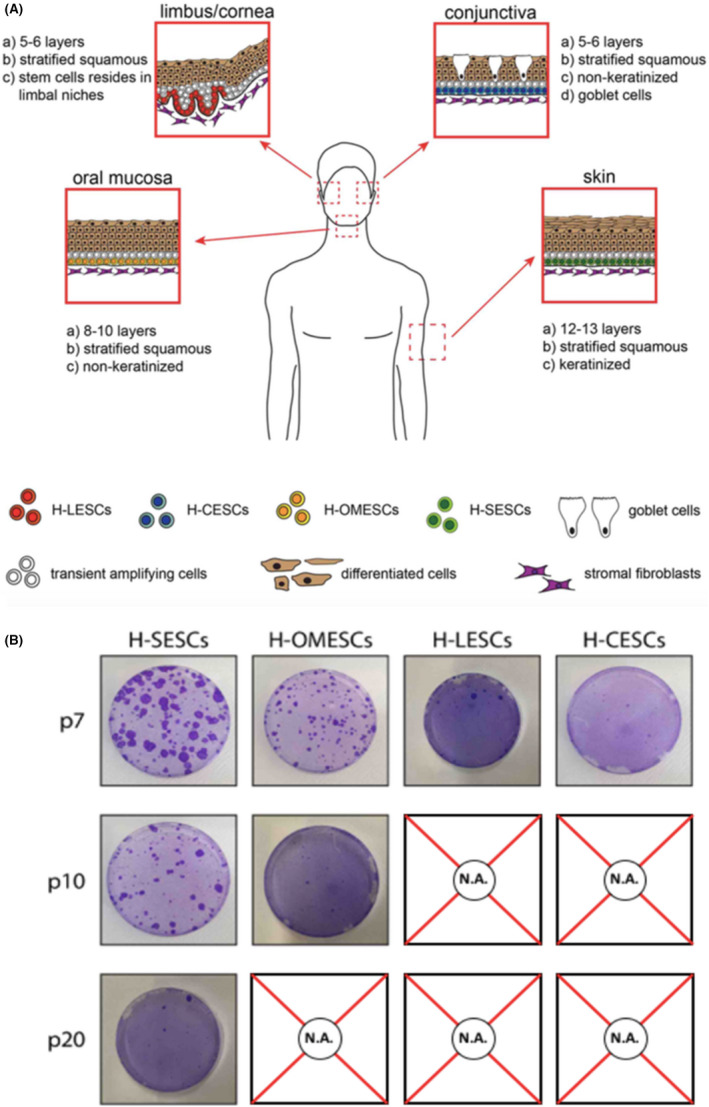
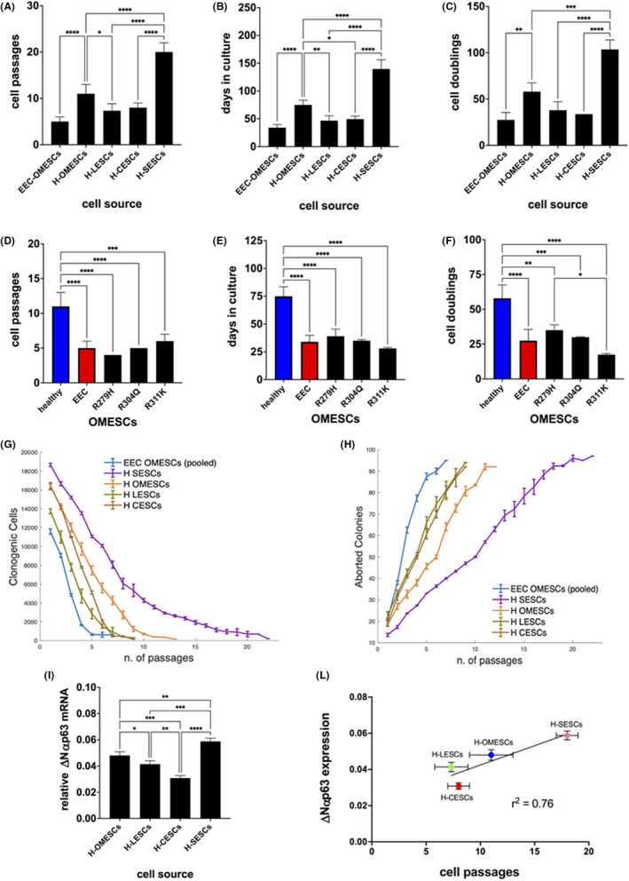
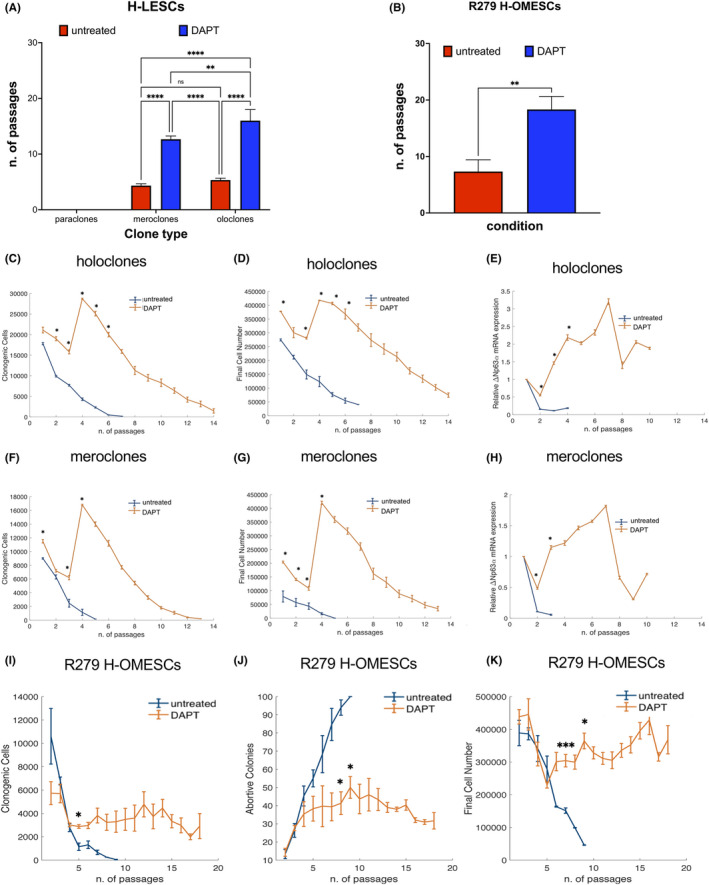
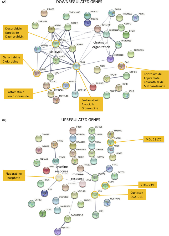
Human epithelial stem cells (ESCs) are characterized by long-term regenerative properties, much dependent on the tissue of origin and varying during their lifespan. We analysed such variables in cultures of ESCs isolated from the skin, conjunctiva, limbus and oral mucosa of healthy donors and patients affected by ectrodactyly-ectodermal dysplasia-clefting syndrome, a rare genetic disorder caused by mutations in the p63 gene. We cultured cells until exhaustion in the presence or in the absence of DAPT (γ-secretase inhibitor; N-[N-(3, 5-difluorophenacetyl)-L-alanyl]-S-phenylglycine T-butyl ester). All cells were able to differentiate in vitro but exhibited variable self-renewal potential. In particular, cells carrying p63 mutations stopped prematurely, compared with controls. Importantly, administration of DAPT significantly extended the replicative properties of all stem cells under examination. RNA sequencing analysis revealed that distinct sets of genes were up- or down-regulated during their lifetime, thus allowing to identify druggable gene networks and off-the-shelf compounds potentially dealing with epithelial stem cell senescence. These data will expand our knowledge on the genetic bases of senescence and potentially pave the way to the pharmacological modulation of ageing in epithelial stem cells.

Keywords: DAPT (γ-secretase inhibitor; N-[N-(3,5-difluorophenacetyl)-L-alanyl]-S-phenylglycine T-butyl ester); EEC syndrome; RNAseq; lifespan; p63; self-renewal; stem cells.


Conflict of interest statement

The authors confirm that there are no conflicts of interest.

Figures

FIGURE 1
FIGURE 1
Comparative features of stem cells obtained from four different epithelial tissues. (A) Model of the four epithelia investigated in this study: skin, oral mucosa, cornea and conjunctiva and their organization at tissue level. (B) Colony forming efficiency of H‐SESCs, H‐OMESCs, H‐LESCs and H‐CESCs evaluated at the last passage in culture. H‐SESCs, human skin epithelia stem cells; H‐OMESCs, human oral mucosa epithelial stem cells; H‐LESCs, human limbal epithelia stem cells; H‐CESCs, human conjunctival epithelial stem cells
FIGURE 2
FIGURE 2
Proliferative potential of epithelial stem cells from different sources in vitro. (A) Cell passages, (B) days in culture and (C) cell doublings of EEC‐OMESCs, H‐OMESCs, H‐LESCs, H‐CESCs and H‐SESCs. (D) Cell passages, (E) days in culture and (F) cell doublings of healthy (blue) and EEC (red) OMESCs. EEC stands for the average of the values found for the three mutants R279H, R304Q and R311K OMESCs. (G) Clonogenic cell number and (H) percentage of aborted colonies relative to ESCs obtained from the indicated sources. For statistical analysis calculated by ONE way anova, please refer to Figure S1A,B. (I) Real‐time quantitative analysis of ∆Np63α expression in H‐OMESCs, H‐SESCs, H‐LESCs and H‐CESCs. Results are normalized against GAPDH. Data shown are mean + standard deviation of the mean along with the results of statistical significance as calculated by ordinary One‐Way anova with multiple comparisons (*p < .05; **p < .005; ***p < .0005; ****p < .0001). (L) The ∆Np63α expression levels of each individual ESC are plotted against the number of passages in culture. Mean values ± standard error of the means are shown, along with the chi‐squared value calculated for the linear regression. SESCs, skin epithelial stem cells; OMESCs, oral mucosa epithelial stem cells; LESCs, limbal epithelial stem cells; CESCs, conjunctival epithelial stem cells; GAPDH, glyceraldehyde 3‐phosphate dehydrogenase
FIGURE 3
FIGURE 3
DAPT administration extends the lifespan of stem cells. Cell cultures derived from holoclones and meroclones obtained from donor limbus and EEC‐OMESCs were treated with DAPT. The number of passages in the absence (red columns) or presence (blue columns) of DAPT (A). Significant increase in the number of passages after DAPT treatment of EEC‐OMESCs (B). Clonogenic cells, final cell number and ∆Np63α expression quantified by qPCR from holoclones (C–E), meroclones (F–H) and R279 H‐OMESCs (I–K) either untreated or treated with DAPT. Data are shown as mean ± standard deviation along with the results of statistical significance as calculated by ordinary one‐way anova with multiple comparisons (*p < .05; **p < .005; ***p < .0005; ****p < .0001). EEC‐OMESCs: ectrodactyly‐ectodermal dysplasia‐clefting oral mucosa epithelial stem cells; DAPT: N‐[N‐(3, 5‐difluorophenacetyl)‐l‐alanyl]‐S‐phenylglycine t‐butyl ester
FIGURE 4
FIGURE 4
Changes in cell morphology following DAPT discontinuation. Cells cultured in medium containing DAPT show a stem‐like morphology (A). DAPT removal from culturing medium leads to morphological changes compatible with senescence (B). Scale bar: 100 μm. Abbreviations: DAPT: N‐[N‐(3, 5‐difluorophenacetyl)‐l‐alanyl]‐S‐phenylglycine t‐butyl ester
FIGURE 5
FIGURE 5
DAPT induces telomere elongation without altering the karyotype. Representative pictures displaying EEC OMESCs karyotype. (A) EEC‐OMESCs karyotype at passage I, (B) EEC‐OMESCs + DAPT karyotype at passage IV, (C) EEC‐OMESCs + DAPT karyotype at passage VII, (D) EEC‐OMESCs + DAPT at passage XVII. In (E), bar plot comparing the relative telomere length in EEC‐OMESCs pI, EEC‐OMESCs + DAPT pIV, EEC‐OMESCs + DAPT pVII, EEC‐OMESCs + DAPT pXVII. EEC‐OMESCs: ectrodactyly‐ectodermal dysplasia‐clefting oral mucosa epithelial stem cells; DAPT: N‐[N‐(3, 5‐difluorophenacetyl)‐l‐alanyl]‐S‐phenylglycine t‐butyl ester
FIGURE 6
FIGURE 6
Comparative epithelial stem cells transcriptomics analysis. Heatmaps depicting mean of ex vivo gene expression data sets, representing (A) genes and (B) isoforms negatively correlated with lifespan and up‐regulated during senescence and vice versa (red: high values, blue: low values)
FIGURE 7
FIGURE 7
Gene ontology (GO) enrichment analysis of deregulated genes in epithelial SCs. Genes either down‐regulated (A) or up‐regulated (B) during SC lifespans were used to perform GO enrichment analysis as described in the materials and methods section. All significantly enriched biological processes are shown on the left: the size of each slice is proportional to the enrichment of each biological process with respect to the reference list, and the p value is indicated. All other relevant information is reported in the right panels
FIGURE 8
FIGURE 8
Expression profiles of senescence markers. The expression profiles of a panel of genes involved in the maintenance of telomeric integrity and/or associated with differentiation and senescence in keratinocyte stem cells were obtained from Limbal Holoclone cultures either untreated (LH) or treated with DAPT (LH‐DAPT) by means of RNAseq analysis. Normalized expression of the indicated markers is plotted against the culture passage. The time series relative to LH and LH‐DAPT is compared using an analysis of covariance test (p < .05). LH, limbal holoclones
FIGURE 9
FIGURE 9
Identification of druggable targets involved in SCs ageing and their connections. Genes that are either down‐regulated (A) or up‐regulated (B) were used to reconstruct their potential connections using String software and drugs targeting them were retrieved from the Genecards database. Line thickness is proportional to the levels of confidence, druggable gene products are circled in yellow and potential drugs targeting such genes are listed in the yellow boxes

Similar articles

Cited by

References

    1. Das D, Fletcher RB, Ngai J. Cellular mechanisms of epithelial stem cell self‐renewal and differentiation during homeostasis and repair. Wiley Interdisciplinary Rev Dev Biol. 2020;9:e361. - PubMed
    1. Montagnani S, Rueger MA, Hosoda T, Nurzynska D. Adult stem cells in tissue maintenance and regeneration. Stem Cells Int. 2016;2016:7362879. - PMC - PubMed
    1. Blanpain C, Fuchs E. Plasticity of epithelial stem cells in tissue regeneration. Science. 2014;344:1242281. - PMC - PubMed
    1. Rowat JS, Squier CA. Rates of epithelial cell proliferation in the oral mucosa and skin of the tamarin monkey (Saguinus fuscicollis). J Dent Res. 1986;65:1326‐1331. - PubMed
    1. Gibbs S, Ponec M. Intrinsic regulation of differentiation markers in human epidermis, hard palate and buccal mucosa. Arch Oral Biol. 2000;45:149‐158. - PubMed

Publication types

Substances