

A Systematic Review Comparing Digital Subtraction Angiogram With Magnetic Resonance Angiogram Studies in Demonstrating the Angioarchitecture of Cerebral Arteriovenous Malformations
- PMID: 35706438
- PMCID: PMC9187205
- DOI: 10.7759/cureus.25803
A Systematic Review Comparing Digital Subtraction Angiogram With Magnetic Resonance Angiogram Studies in Demonstrating the Angioarchitecture of Cerebral Arteriovenous Malformations
Abstract
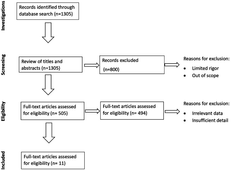
In brain arteriovenous malformations (AVMs), there is mismatched communication between arteries and veins, causing a nidal bed between them. This systematic review explores whether a magnetic resonance angiogram (MRA) can be used as a diagnostic imaging tool instead of a digital subtraction angiogram (DSA). Utilizing PubMed, Cochrane, and Google Scholar, as well as the Preferred Reporting Items for Systematic Reviews and Meta-Analyses (PRISMA) guidelines for article selection, a literature search was conducted over the past five years. Eleven studies were included, with a majority of the articles suggesting a potential for consideration. Arterial spin labeling (ASL) versus time-of-flight (TOF) scans was a comparison study, in addition to the study on pseudo-continuous arterial spin labeling (pc-ASL), which proved its high sensitivity in comparison with DSA scans. Other studies included quantitative magnetic resonance angiogram (Q-MRA) measuring the blood flow and susceptibility weighted imaging (SWI) modality. Although promising, digital subtraction angiogram (DSA) scans have diagnostic superiority. In addition, articles discussed follow-up magnetic resonance angiogram (MRA) scans after surgery. Overall, digital subtraction angiogram remains the gold standard due to its superior spatial resolution and hemodynamic properties; these are the key limitations of magnetic resonance studies. MRA has demonstrated its ability to reproduce high-quality diagnostic images for arteriovenous malformation (AVM) angioarchitecture; however, coupled with their limitations, not many studies with large sample sizes over longer periods have been conducted, and we urge more research into it.
Keywords: 4d dsa; asl-mra; avm; brain avm; dsa; mra; swi; tof-mra.
Copyright © 2022, Raman et al.
Conflict of interest statement
The authors have declared that no competing interests exist.
Figures
References
-
- Angioarchitectural features amongst patients with unruptured brain arteriovenous malformations presenting with headache: findings from a single center retrospective review of 76 patients. Africk BN, Heiferman DM, Wozniak AW, et al. http://pubmed.ncbi.nlm.nih.gov/34627140/ J Headache Pain. 2021;22:122. - PMC - PubMed
-
- Radiosurgery for cerebral arteriovenous malformation (AVM): current treatment strategy and radiosurgical technique for large cerebral AVM. Byun J, Kwon DH, Lee DH, Park W, Park JC, Ahn JS. https://pubmed.ncbi.nlm.nih.gov/32423182/ J Korean Neurosurg Soc. 2020;63:415–426. - PMC - PubMed
-
- Is four-dimensional CT angiography as effective as digital subtraction angiography in the detection of the underlying causes of intracerebral haemorrhage: a systematic review. Denby CE, Chatterjee K, Pullicino R, Lane S, Radon MR, Das KV. https://pubmed.ncbi.nlm.nih.gov/31901972/ Neuroradiology. 2020;62:273–281. - PMC - PubMed
-
- The efficacy of surgical treatment of cerebral arteriovenous malformations in a single academic institution: a case series. Smrcka M, Navratil O, Hovorka E, Duris K. https://pubmed.ncbi.nlm.nih.gov/34472738/ Croat Med J. 2021;62:353–359. - PMC - PubMed
-
- 4D-DSA: development and current neurovascular applications. Ruedinger KL, Schafer S, Speidel MA, Strother CM. http://pubmed.ncbi.nlm.nih.gov/33243899/ AJNR Am J Neuroradiol. 2021;42:214–220. - PMC - PubMed
Publication types
LinkOut - more resources
Full Text Sources
Miscellaneous