. 2022 May 27;4(3):fcac140.
doi: 10.1093/braincomms/fcac140. eCollection 2022.

A connectivity model of the anatomic substrates underlying Gerstmann syndrome

Affiliations

A connectivity model of the anatomic substrates underlying Gerstmann syndrome

Qazi S Shahab et al. Brain Commun. .

Abstract

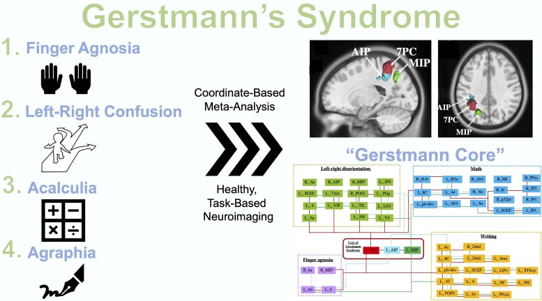
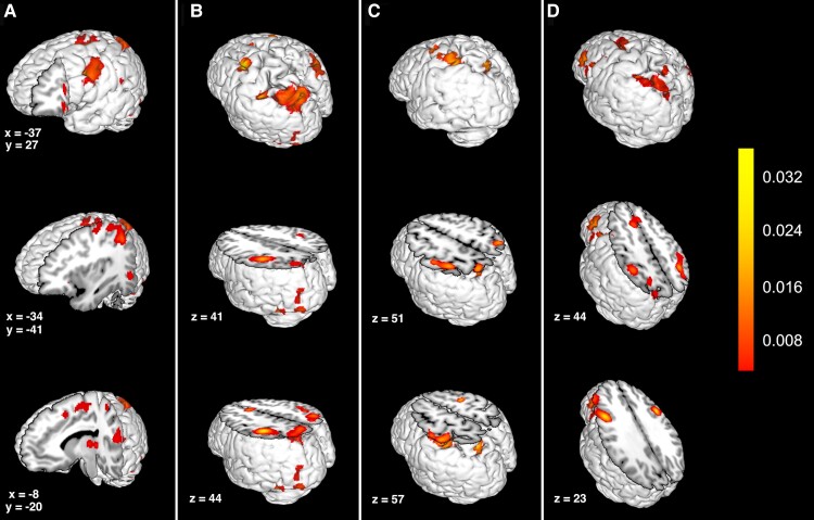
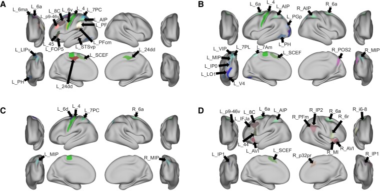
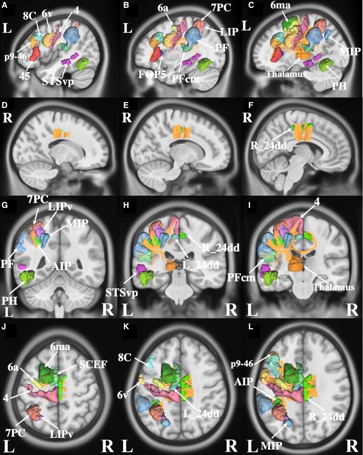
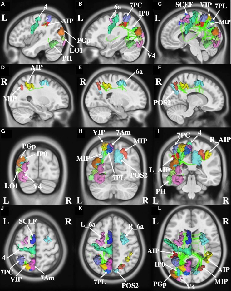
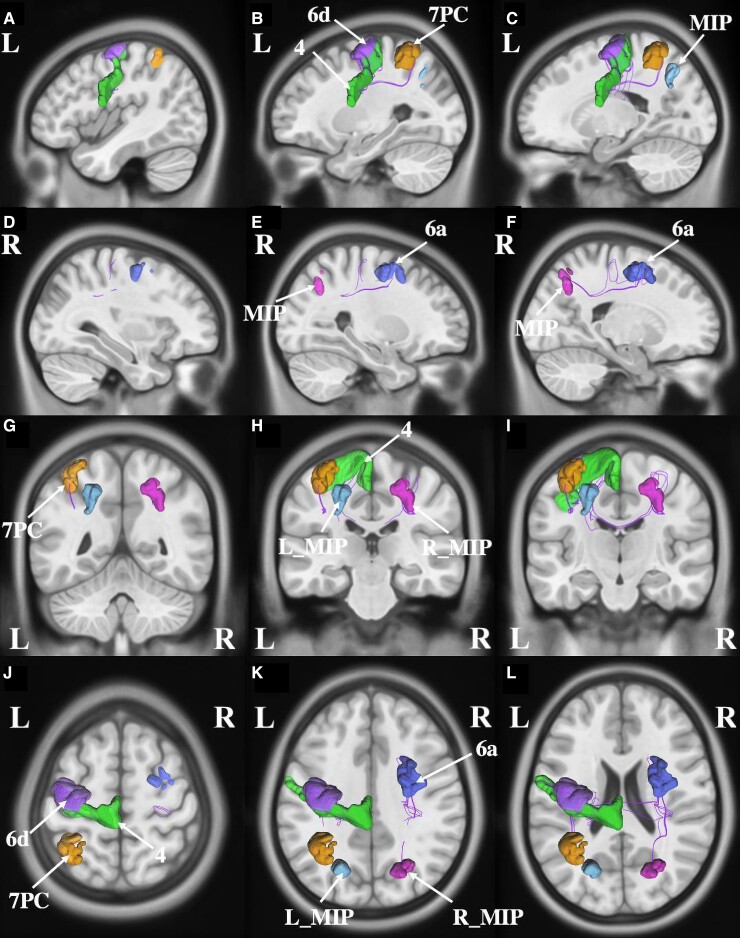
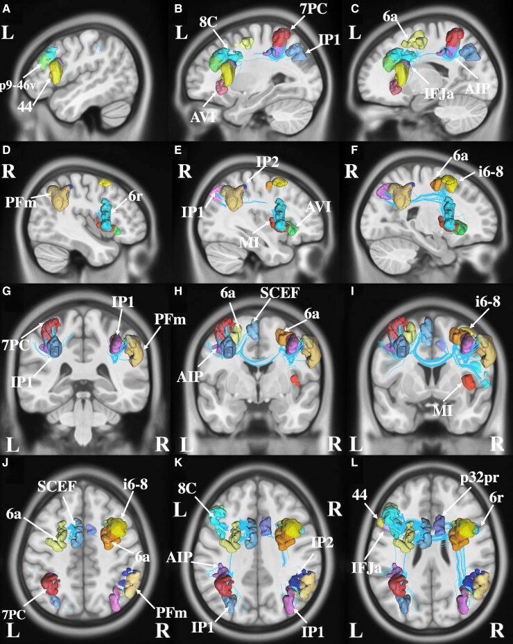
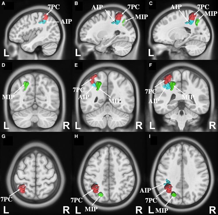
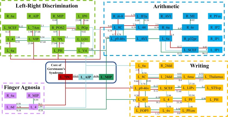
The Gerstmann syndrome is a constellation of neurological deficits that include agraphia, acalculia, left-right discrimination and finger agnosia. Despite a growing interest in this clinical phenomenon, there remains controversy regarding the specific neuroanatomic substrates involved. Advancements in data-driven, computational modelling provides an opportunity to create a unified cortical model with greater anatomic precision based on underlying structural and functional connectivity across complex cognitive domains. A literature search was conducted for healthy task-based functional MRI and PET studies for the four cognitive domains underlying Gerstmann's tetrad using the electronic databases PubMed, Medline, and BrainMap Sleuth (2.4). Coordinate-based, meta-analytic software was utilized to gather relevant regions of interest from included studies to create an activation likelihood estimation (ALE) map for each cognitive domain. Machine-learning was used to match activated regions of the ALE to the corresponding parcel from the cortical parcellation scheme previously published under the Human Connectome Project (HCP). Diffusion spectrum imaging-based tractography was performed to determine the structural connectivity between relevant parcels in each domain on 51 healthy subjects from the HCP database. Ultimately 102 functional MRI studies met our inclusion criteria. A frontoparietal network was found to be involved in the four cognitive domains: calculation, writing, finger gnosis, and left-right orientation. There were three parcels in the left hemisphere, where the ALE of at least three cognitive domains were found to be overlapping, specifically the anterior intraparietal area, area 7 postcentral (7PC) and the medial intraparietal sulcus. These parcels surround the anteromedial portion of the intraparietal sulcus. Area 7PC was found to be involved in all four domains. These regions were extensively connected in the intraparietal sulcus, as well as with a number of surrounding large-scale brain networks involved in higher-order functions. We present a tractographic model of the four neural networks involved in the functions which are impaired in Gerstmann syndrome. We identified a 'Gerstmann Core' of extensively connected functional regions where at least three of the four networks overlap. These results provide clinically actionable and precise anatomic information which may help guide clinical translation in this region, such as during resective brain surgery in or near the intraparietal sulcus, and provides an empiric basis for future study.

Keywords: Gerstmann’s syndrome; connectivity; connectome; network.


Figures

Graphical abstract
Graphical abstract
Figure 1
Figure 1
The activation likelihood estimation for the neurocognitive domains involved in Gerstmann syndrome. The activation likelihood estimation clusters of the task-based fMRI experiments that were included in our meta-analysis for (A) writing, (B) left–right discrimination, (C) finger agnosia and (D) arithmetic. The colour bar represents the ALE statistic, which increases in significance from bottom (red) to top (yellow).
Figure 2
Figure 2
The parcels with the largest percentage overlap with each activation likelihood estimation cluster. Largest percentage overlap parcel for (A) writing, (B) left–right discrimination, (C) finger agnosia and (D) arithmetic. 24dd, Dorsal Area 24d; 6a, Area 6 Anterior; 6d, Dorsal Area 6; 6ma, Area 6m Anterior; 6r, Rostral Area 6; 6v, Ventral Area 6; 7Am, Medial Area 7A; 7PC, Area 7 postcentral; 7PL, Lateral Area 7P; 8C, Area 8C; AIP, Anterior Intraparietal Area; AVI, Anterior Ventral Insular Area; FOP5, Area Frontal Opercular 5; i6-8, Inferior 6-8 Transitional Area; IFJa, Area IFJa; IP0, Area Intraparietal 0; IP1, Area Intraparietal 1; IP2, Area Intraparietal 2; LIPv, Area Lateral Intraparietal Ventral; LO1, Area Lateral Occipital 1; MI, Middle Insular Area; MIP, medial Intraparietal Area; p32pr, Area p32 Prime; p9-46v, Area Posterior 9-46v; PF, Area PF Complex; PFcm, Area PFcm; PFm, Area PFm Complex; PGp, Area PGp; PH, area PH; POS2, Parieto-Occipital Sulcus Area 2; SCEF, Supplementary and Cingulate Eye Field; STSvp, superior temporal sulcus ventral posterior; VIP, Ventral Intraparietal Complex.
Figure 3
Figure 3
Connectivity among parcels overlapping with the activation likelihood estimation for writing. (A–C) sagittal sections, from lateral to medial, demonstrating the parcels in the left hemisphere that are involved in writing. (D–F) sagittal sections, from lateral to medial, demonstrating the parcels in the right hemisphere that are involved in writing. (G–I) coronal sections, from the posterior to anterior, highlighting the left and right-sided parcels and their tractography patterns. (J–L) axial sections, from superior to inferior, demonstrating the tractography and the anatomical relationships between parcels that contribute to writing.
Figure 4
Figure 4
Connectivity among parcels overlapping with the activation likelihood estimation for left–right discrimination. (A–C) Sagittal sections, from lateral to medial, demonstrating the parcels and their tractography in the left hemisphere that are involved in the left–right discrimination. (D–F) Sagittal sections, from lateral to medial, demonstrating the parcels and their tractography in the right hemisphere that are involved in the left–right discrimination. (G–I) Coronal sections, from the posterior to anterior, highlighting the left and right-sided parcels and their tractography patterns. (J–L) Axial sections, from superior to inferior, demonstrating the tractography and the anatomical relationships between parcels that contributes to left–right discrimination.
Figure 5
Figure 5
Connectivity among parcels overlapping with the activation likelihood estimation for finger agnosia. (A–C) Sagittal sections, from lateral to medial, demonstrating the parcels and their tractography in the left hemisphere that are involved in finger agnosia. (D–F) Sagittal sections, from lateral to medial, demonstrating the parcels and their tractography in the right hemisphere that are involved in finger agnosia. (G–I) Coronal sections, from the posterior, to anterior highlighting the left and right-sided parcels and their tractography patterns. (J–L) Axial sections, from superior to inferior, demonstrating the tractography and the anatomical relationships between parcels that contributes to finger agnosia.
Figure 6
Figure 6
Connectivity among parcels overlap with the activation likelihood estimation for arithmetic. (A–C) sagittal sections, from lateral to medial, demonstrating the parcels in the left hemisphere that are involved in math. (D–F) sagittal sections, from lateral to medial, demonstrating the parcels in the right hemisphere that are involved in math. (G–I) coronal sections, from the posterior to anterior, highlighting the left and right-sided parcels and their tractography patterns. (J–L) axial sections, from superior to inferior, demonstrating the tractography and the anatomical relationships between parcels that contributes to math function.
Figure 7
Figure 7
Parcels that represent the core of the Gerstmann’s syndrome. 7PC (red), MIP (green) and AIP (blue) in the left hemisphere. (A–C) Sagittal sections, from lateral to medial (D–F) coronal sections, from the posterior to anterior. (G–I) Axial sections, from superior to inferior.
Figure 8
Figure 8
Simplified schematic of the white matter connections identified between individual parcels and the core of the Gerstmann’s syndrome. L, left. R, right. Resultant tract volumes are listed for each parcel which is connected directly on the diagram. Of note is the connecting lines do not show a relationship between all parcels they connect, but rather the three parcels in the Gerstmann Core with all other parcels, and a true connection is only there if there is an associated number. The number associated with each connection represents the average number of streamlines between given two regions on tractography across all subjects.

Similar articles

Cited by

References

    1. Wingard EM, Barrett AM, Crucian GP, Doty L, Heilman KM. The Gerstmann syndrome in Alzheimer's disease. J Neurol Neurosurg Psychiatry. 2002;72:403–405. - PMC - PubMed
    1. Gerstmann J. Fingeragnosie-eine umschriebene storung der orientierung am eigenen Korper. Wiener Klinische Wochenschrift. 1924;37:1010–1012.
    1. Roux FE, Boetto S, Sacko O, Chollet F, Trémoulet M. Writing, calculating, and finger recognition in the region of the angular gyrus: a cortical stimulation study of Gerstmann syndrome. J Neurosurg. 2003;99:716–727. - PubMed
    1. Rusconi E. Gerstmann syndrome: historic and current perspectives. Handb Clin Neurol. 2018;151:395–411. - PubMed
    1. Rusconi E, Pinel P, Dehaene S, Kleinschmidt A. The enigma of Gerstmann’s syndrome revisited: a telling tale of the vicissitudes of neuropsychology. Brain. 2009;133:320–332. - PubMed

LinkOut - more resources