Review
. 2022 Jun 6:2022:5164970.
doi: 10.1155/2022/5164970. eCollection 2022.

Modern Diagnostic Imaging Technique Applications and Risk Factors in the Medical Field: A Review

Affiliations
Review

Modern Diagnostic Imaging Technique Applications and Risk Factors in the Medical Field: A Review

Shah Hussain et al. Biomed Res Int. .

Abstract

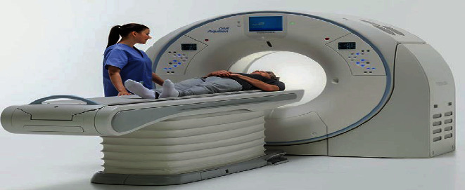
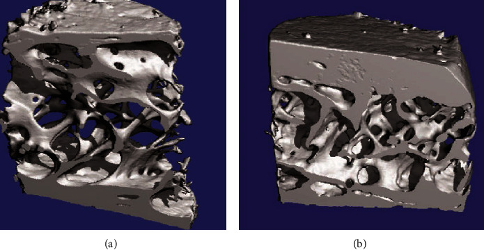
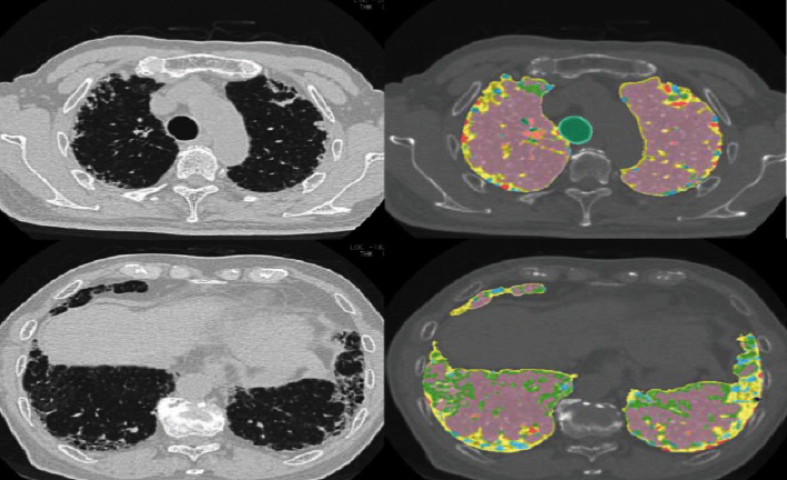
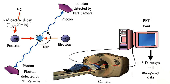
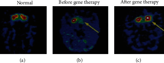
Medical imaging is the process of visual representation of different tissues and organs of the human body to monitor the normal and abnormal anatomy and physiology of the body. There are many medical imaging techniques used for this purpose such as X-ray, computed tomography (CT), positron emission tomography (PET), magnetic resonance imaging (MRI), single-photon emission computed tomography (SPECT), digital mammography, and diagnostic sonography. These advanced medical imaging techniques have many applications in the diagnosis of myocardial diseases, cancer of different tissues, neurological disorders, congenital heart disease, abdominal illnesses, complex bone fractures, and other serious medical conditions. There are benefits as well as some risks to every imaging technique. There are some steps for minimizing the radiation exposure risks from imaging techniques. Advance medical imaging modalities such as PET/CT hybrid, three-dimensional ultrasound computed tomography (3D USCT), and simultaneous PET/MRI give high resolution, better reliability, and safety to diagnose, treat, and manage complex patient abnormalities. These techniques ensure the production of new accurate imaging tools with improving resolution, sensitivity, and specificity. In the future, with mounting innovations and advancements in technology systems, the medical diagnostic field will become a field of regular measurement of various complex diseases and will provide healthcare solutions.


Conflict of interest statement

The authors declare no conflicts of interest.

Figures

Figure 1
Figure 1
CT scanner.
Figure 2
Figure 2
Distraction injury scanned by CT scan (showing damage occurrence at the 11th and 12th thoracic vertebrae).
Figure 3
Figure 3
An axial scan of a spinal injury by computed tomography (CT) at the 2nd and 3rd lumbar vertebrae.
Figure 4
Figure 4
(a) Femora undergo vQCT to determine BMD and macrostructure. (b) Ultradistal forearm undergoes CT to measure the structure of the trabecular complex network and its texture.
Figure 5
Figure 5
Microstructure of transiliac bone biopsies is determined to undergo 3D micro-CT of two postmenopausal women who have accomplished strontium ranelate therapy for 36 months: (a) strontium ranelate therapy; (b) placebo.
Figure 6
Figure 6
Images derived using a system known as GHNC (Gaussian Histogram Normalized Correlation). Lung images of a 73-year-old male IPF patient have been taken by computed tomography (CT). Light blue and yellow color, fibrosis; dark blue color, emphysema; pink color, normal; and light green color is indicated as ground-glass opacity.
Figure 7
Figure 7
Basic principle of PET scan: (1) a positive electron (positron) is emitted by radioactive decay of radioisotope, for example, carbon-11; (2) this positron hits an electron present in the tissue to be analyzed and emits two photons having low energy; (3) scintillation crystals are present in the PET camera to absorb this emitted photon with low energy; (4) the light is produced that is converted into another signal such as electrical signals used by the computer system to produce 3D images.
Figure 8
Figure 8
PET images of gene therapy in unilateral MPTP monkey as Parkinson's model: (a) image of normal dopamine production; (b) the image is representing autonomous dopamine MPTP-induced shortage (before gene therapy); (c) image of the restoration of dopamine production in the caudate and putamen (after gene therapy) [56].
Figure 9
Figure 9
Abnormal cerebral asymmetry in schizophrenia patients compared with control is shown by functional-MRI imaging technique.
Figure 10
Figure 10
Breast adenocarcinoma mouse model formed for the detection of lung metastases. (a) High-resolution hyperpolarized 3He MRI (HP 3He MRI) images were taken from the control mouse. Screening as normal ventilation forms of lungs. (b) After injection of LHRH-SPIONs, images were produced from human breast adenocarcinoma abnormal mouse (model), showing defects in the right lobe (under circles).
Figure 11
Figure 11
Dual-headed single-photon emission computed tomography (SPECT) system.
Figure 12
Figure 12
(a) The SPECT/CT scan of the knee joint shows clear articular cartilage. (b) The SPECT/CT arthrography image of the knee joint showing enhance the value of SPECT/CT screening.
Figure 13
Figure 13
(a) A healthy breast tissue mammogram recorded by digital mammogram; (b) the same breast tissue mammogram recorded by film mammography. White spots shown in the above images are deposits of calcium which can consider the mark of cancer when they form clusters.
Figure 14
Figure 14
(a) Hyphemia is due to injury; blood diffuses the anterior chamber of the eye. (b) Hyphemia scan image by Sonomed UBM.
Figure 15
Figure 15
Images of mouse fibroblast cell (a) normal smooth form and (b) abnormal cell in its rough shape. Due to the diagnostic ultrasound effect, fingerlike projections are produced on the smooth surface of the fibroblast cell.

References

    1. Laal M. Innovation process in medical imaging. Procedia-Social and Behavioral Sciences . 2013;81:60–64. doi: 10.1016/j.sbspro.2013.06.388. - DOI
    1. Kasban H., El-Bendary M. A. M., Salama D. H. A comparative study of medical imaging techniques. International Journal of Information Science and Intelligent System . 2015;4(2):37–58.
    1. National Research Council. Mathematics and Physics of Emerging Biomedical Imaging . National Academies Press; 1996. - PubMed
    1. Flower M. A. Webb's Physics of Medical Imaging . CRC Press; 2012.
    1. Roobottom C. A., Mitchell G., Morgan-Hughes G. Radiation-reduction strategies in cardiac computed tomographic angiography. Clinical radiology . 2010;65(11):859–867. doi: 10.1016/j.crad.2010.04.021. - DOI - PubMed

MeSH terms

LinkOut - more resources