. 2022 May 30:13:864330.
doi: 10.3389/fpls.2022.864330. eCollection 2022.

Molecular and Biochemical Analysis of Duplicated Cytosolic CuZn Superoxide Dismutases of Rice and in silico Analysis in Plants

Affiliations

Molecular and Biochemical Analysis of Duplicated Cytosolic CuZn Superoxide Dismutases of Rice and in silico Analysis in Plants

Ravi Prakash Sanyal et al. Front Plant Sci. .

Abstract

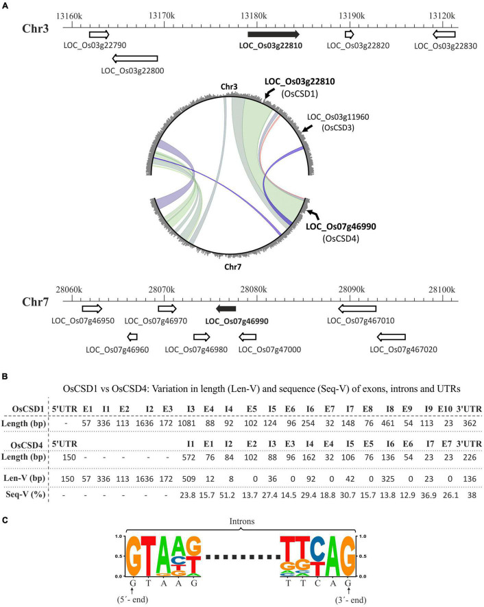
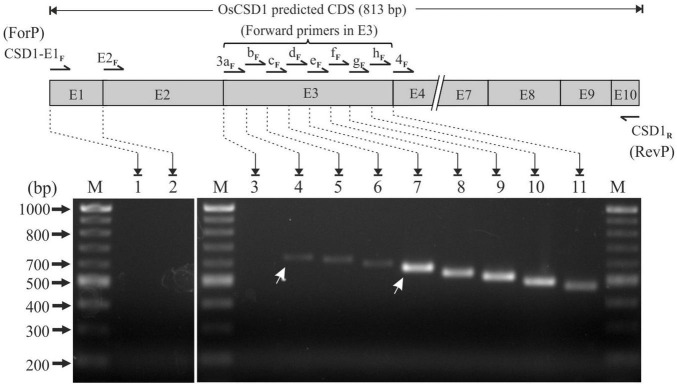
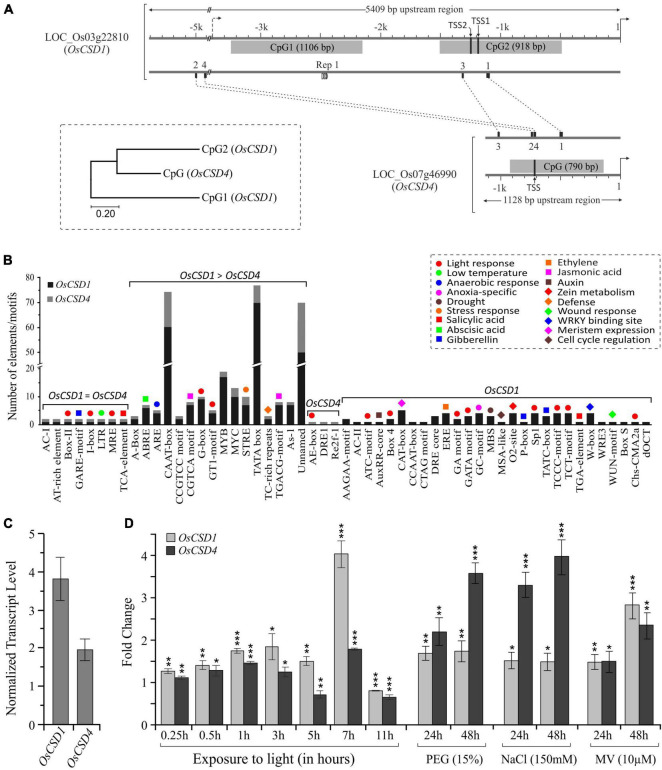
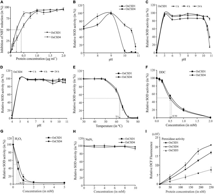
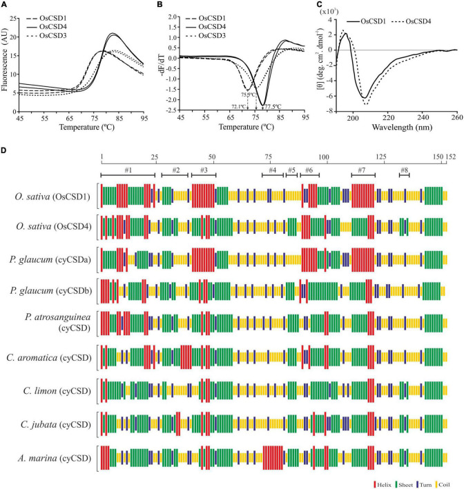
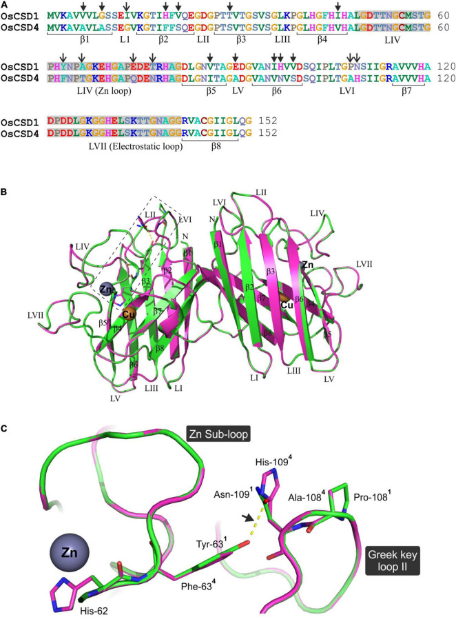
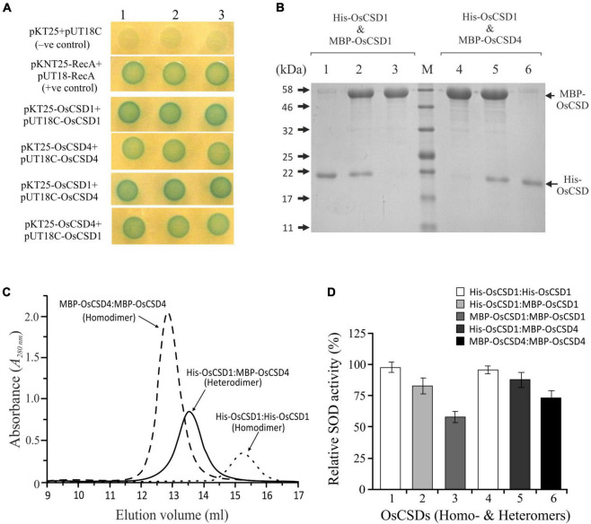
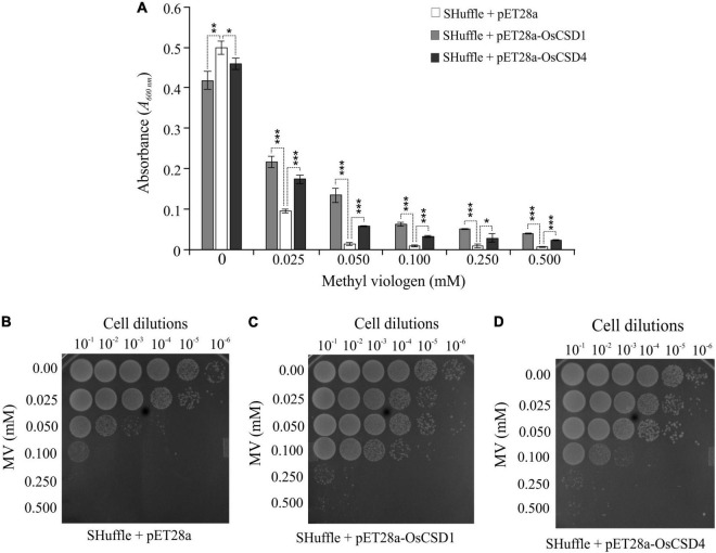
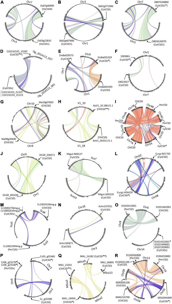
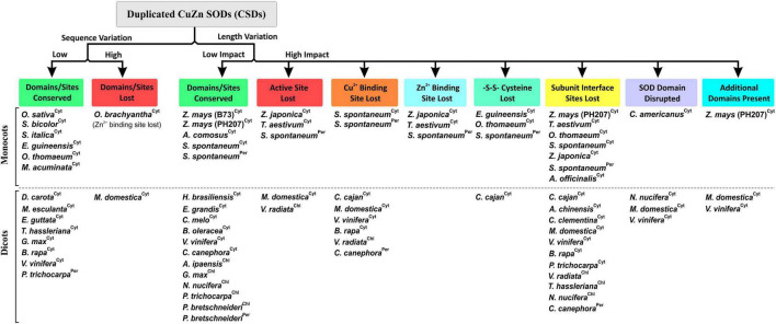
Superoxide dismutases (SODs, EC 1.15.1.1) are ubiquitous antioxidant metalloenzymes important for oxidative stress tolerance and cellular redox environment. Multiple factors have contributed toward the origin and diversity of SOD isoforms among different organisms. In plants, the genome duplication events, responsible for the generation of multiple gene copies/gene families, have also contributed toward the SOD diversity. However, the importance of such molecular events on the characteristics of SODs has not been studied well. This study investigated the effects of divergence on important characteristics of two block-duplicated rice cytosolic CuZn SODs (OsCSD1, OsCSD4), along with in silico assessment of similar events in other plants. The analysis revealed heterogeneity in gene length, regulatory regions, untranslated regions (UTRs), and coding regions of two OsCSDs. An inconsistency in the database-predicted OsCSD1 gene structure was also identified and validated experimentally. Transcript analysis showed differences in the basal levels and stress responsiveness of OsCSD1 and OsCSD4, and indicated the presence of two transcription start sites in the OsCSD1. At the amino acid level, the two OsCSDs showed differences at 18 sites; however, both exist as a homodimer, displaying typical CuZn SOD characteristics, and enhancing the oxidative stress tolerance of Escherichia coli cells. However, OsCSD4 showed higher specific activity as well as stability. The comparison of the two OsCSDs with reported thermostable CSDs from other plants identified regions likely to be associated with stability, while the homology modeling and superposition highlighted structural differences. The two OsCSDs displayed heteromeric interaction capability and forms an enzymatically active heterodimer (OsCSD1:OsCSD4) on co-expression, which may have significance as both are cytosolic. In silico analysis of 74 plant genomes revealed the prevalence of block duplications for multiple CSD copies (mostly cytosolic). The divergence and clustering analysis of CSDs suggested the possibility of an ancestral duplication event in monocots. Conserved SOD features indicating retention of SOD function among CSD duplicates were evident in few monocots and dicots. In most other species, the CSD copies lacked critical features and may not harbor SOD function; however, other feature-associated functions or novel functions might be present. These aspects of divergent CSD copies encoding co-localized CSDs may have implications in plant SOD functions in the cytosol and other organelles.

Keywords: Oryza sativa; block duplication; cytosolic CuZn superoxide dismutase; heteromeric interaction; homology modeling; in silico analysis; oxidative stress; thermostability.


Conflict of interest statement

The authors declare that the research was conducted in the absence of any commercial or financial relationships that could be construed as a potential conflict of interest.

Figures

FIGURE 1
FIGURE 1
(A) Schematic representation of chromosomal locations encoding two rice cytosolic CuZn SODs, OsCSD1 (LOC_Os03g22810, chromosome 3, Chr3) and OsCSD4 (LOC_Os07g46990, chromosome 7, Chr7) as per RGAP database. The neighboring genes are shown with arrowheads indicating the direction of the transcription, while the scale indicates the chromosomal coordinates. The circle plot generated at the Monocot PLAZA 4.5 web server shows segmental duplications between chromosomes 3 and 7 along with positions of region-harboring rice cytosolic CSDs (LOC_Os03g22810, LOC_Os07g46990) and peroxisomal CSD (LOC_Os03g11960). (B) Overview of variation in length (Len-V) and sequence (Seq-V) between the UTRs, exons, and introns of OsCSD1 and OsCSD4 genes. (C) Sequence logo generated by aligning the 5′- and 3′-ends of the all introns of OsCSD1 and OsCSD4 genes.
FIGURE 2
FIGURE 2
RT-PCR-based analysis of transcription status of OsCSD1 exon1, exon2, and fine mapping of exon3 (E3 box) using different forward and common reverse primers (CSD1R). Lane 1, CSD1-E1F + CSD1R; lane 2, CSD1-E2F + CSD1R; lane 3, CSD1-E3aF + CSD1R; lane 4, CSD1-E3bF + CSD1R; lane 5, CSD1-E3cF + CSD1R; lane 6, CSD1-E3dF + CSD1R; lane 7, CSD1-E3eF + CSD1R; lane 8, CSD1-E3fF + CSD1R; lane 9, CSD1-E3gF + CSD1R; lane 10, CSD1-E3hF + CSD1R; lane 11, CSD1-E4F + CSD1R. White arrows indicate the two differentially abundant OsCSD1 transcripts. Lane M, 100 bp DNA ladder.
FIGURE 3
FIGURE 3
Promoter region differences and expression pattern of OsCSD1 and OsCSD4 genes. (A) The length and location of CpG islands (gray boxes), positions of conserved motifs (1, 2, 3, 4), repetitive motif (Rep1), and two potential TSS sites in OsCSD1-CpG2 and one in OsCSD4-CPG are indicated. The scale on the top indicates the length of the upstream region. The dendrogram shows the divergence between the OsCSD1 and OsCSD4 CpG islands. (B) The bar plot shows the abundance of different categories of cis-elements (indicated by color-coded symbols) in the promoter of two OsCSDs. Single copy, differentially abundant, and gene-specific cis-elements are grouped separately. (C) Reverse transcription-quantitative PCR (RT-qPCR)-based analysis of relative abundance of OsCSD1 and OsCSD4 transcripts in rice shoot tissue. (D) Expression pattern of OsCSD1 and OsCSD4 transcripts in light (0–11 h) and in response to polyethylene glycol (PEG, 15%), sodium chloride (NaCl, 150 mM), and methyl viologen (MV, 10 μM) at 24 and 48 h time points after stress treatment. The transcript levels were estimated using actin as a reference gene. The experiment was repeated three times and data are represented as mean value ± SD. Statistical significance is indicated by *(p < 0.05), **(p < 0.01), ***(p < 0.001).
FIGURE 4
FIGURE 4
Overexpression and purification of recombinant rice cytosolic OsCSDs. (A) OsCSD1: lane 1, pET28a(+) uninduced; lane 2, pET28a(+) induced; lane 3, pET28(+)-OsCSD1 uninduced; lane 4, pET(+)-OsCSD1 induced; lane 5, pET(+)-OsCSD1-induced supernatant fraction; lane 6, Ni-NTA affinity-purified OsCSD1; lane M, Protein molecular weight standard. (B) OsCSD4: lane 1, pET28a(+) uninduced; lane 2, pET28a(+) induced; lane 3, pET28(+)-OsCSD4 uninduced; lane 4, pET(+)-OsCSD4 induced; lane 5, pET(+)-OsCSD4-induced supernatant fraction; lane 6, Ni-NTA column-purified OsCSD4; lane M, Protein molecular weight standard. (C) Gel-filtration chromatography elution profiles of OsCSD1 and OsCSD4. The standard protein markers used were (a) bovine serum albumin (66.5 kDa), (b) chicken egg albumin (45 kDa), (c) carbonic anhydrase (29 kDa), and (d) cytochrome C (12.4 kDa). The gel photograph shows relative mobility and SOD activities of native OsCSD1 (1) and OsCSD4 (2) in a non-denaturing polyacrylamide gel. “SC” indicates net surface charge on the proteins.
FIGURE 5
FIGURE 5
Specific activity, effect of pH, and temperature. (A) Estimation of specific activity: increasing protein amount (0–2 μg ml–1) was used for SOD assay, and % inhibition of NBT reduction was measured to determine the SOD activity (1 U of SOD activity—amount of protein required to inhibit the NBT reduction by 50%). (B) Optimum pH: SOD activity of OsCSDs was assayed at different pH (range: 7.0–10.8) and relative SOD activity was estimated by considering the maximum activity as 100%. (C,D) Effect of pH on stability: purified OsCSDs were incubated at different pH (range: 4.0–10.8), aliquots were removed at different time points (1–24 h) and assayed for SOD activity (activity before pre-incubation was considered as 100%). (E) Effect of temperature: purified OsCSDs were pre-incubated at different temperatures (30–80°C) for 1 h, and assayed for SOD activity (activity before pre-incubation was considered as 100%), and plotted as a function of temperature. The vertical dotted lines indicate T1/2 value (temperature at 50% of SOD activity is lost). (F–H) Effect of SOD inhibitors on enzyme activity, diethyldithiocarbamate (DDC, 0–2.0 mM), hydrogen peroxide (H2O2, 0–5.0 mM), and sodium azide (NaN3, 0–10.0 mM). Relative SOD activity was estimated by considering the enzyme activity before pre-incubation with inhibitor as 100%. The arrows indicate IC50 value (concentration that inhibits 50% of SOD activity). (I) For analysis of bicarbonate-dependent peroxidase activity of OsCSDs, dichlorofluorescein (DCF) formation was monitored with increasing amount of protein (0–250 nM). For the SOD activity-based assays, 1U equivalent of purified protein was used, and data are represented as mean ± SD of three independent replicates.
FIGURE 6
FIGURE 6
Differential scanning fluorimetry (DSF) and secondary structure analysis. (A) DSF profiles of OsCSD1, OsCSD4, and OsCSD3 generated using a Roche LightCycler LC480 II real-time PCR system. (B) First derivative curve (-dF/dT vs. Temperature) generated using the Tm calling software function for estimation of melting temperature (Tm) of respective OsCSDs, as indicated by arrows. (C) Circular dichroism spectra of OsCSD1 and OsCSD4 (parameters, path length: 0.5 cm, Acq duration: 0.2 s, and band width: 2.0 nm). (D) Analysis of plant cytosolic CSDs (cyCSDs) of Oryza sativa (OsCSD1 and OsCSD4, this study), P. glaucum (cyCSDa, ABP65325, Mahanty et al., 2012), P. glaucum (cyCSDb, this study), P. atrosanguinea (EU532614, Kumar et al., 2012), C. aromatica (FJ5896638, Kumar et al., 2014), C. limon (AF318938, Lin et al., 2002), C. jubata (EF530044, Kumar et al., 2016b), and A. marina (ACA50531.1, Fesharaki-Esfahani et al., 2021) by the CFSSP online tool. The scale on the top indicates the position of amino acids while #1-#8 indicate the regions affected with substitutions.
FIGURE 7
FIGURE 7
Comparative in silico structural analysis of two rice cytosolic CSDs. (A) Pair-wise alignment of OsCSD1 and OsCSD4 amino acid sequences. The arrows show the positions of 18 amino acid variations affecting the beta strands (β) and loop (L) regions. (B) Dimeric superposed 3D homology models of OsCSD1 (green) and OsCSD4 (magenta) generated using the SWISS MODEL workspace using S. lycopersicum CuZn SOD crystal structure (PDB ID: 3PU7) as template. Beta strands (β1- β8), loops (LI-LVII), and positions of Cu and Zn metal co-factors are indicated. (C) Enlarged view of the rectangular region (marked in B) to show the effect of amino acid substitutions on structural variations in Zn sub-loop and Greek key loop II regions of OsCSD1 and OsCSD4. The superscripts indicate the residues in two proteins (1: OsCSD1 and 2: OsCSD4), while an arrow shows H-bond between Asn-109 and Tyr-63 (OsCSD1).
FIGURE 8
FIGURE 8
Analysis of heteromeric interaction between rice OsCSD1 and OsCSD4 subunits by BACTH system. (A) E. coli (BTH101) cells co-transformed with different combinations of pKT25 and pUT18C plasmids expressing OsCSD1 and OsCSD4 fusion proteins were analyzed by spot test assay. Cells containing empty plasmids served as negative control, while the positive control contained plasmids pKNT25-RecA + pUT18-RecA. (B) SDS-PAGE analysis of heterodimer and homodimer forms separated after Ni-NTA/amylose affinity purification of co-expressed OsCSDs, lane 1, His-OsCSD1:His-OsCSD1 (homodimer); lane 2, His-OsCSD1:MBP-OsCSD1 (heterodimer); lane 3, MBP-OsCSD1:MBP-OsCSD1 (homodimer); lane 4, MBP-OsCSD4:MBP-OsCSD4 (homodimer); lane 5, His-OsCSD1:MBP-OsCSD4 (heterodimer); lane 6, His-OsCSD1:His-OsCSD1 (homodimer); lane M, protein molecular weight standards. (C) Gel-filtration profiles of two different homodimers (MBP-OsCSD4:MBP-OsCSD4 and His-OsCSD1:His-OsCSD1) and heterodimer (His-OsCSD1:MBP-OsCSD4) forms. (D) Analysis of SOD activity of OsCSD homodimer and heterodimer forms by NBT reduction method.
FIGURE 9
FIGURE 9
Analysis of oxidative stress tolerance of E. coli SHuffle T7 Express cells overexpressing OsCSD1 and OsCSD4. (A) Comparative analysis of growth of E. coli cells (absorbance at 600 nm) containing pET28a (empty vector), pET28a-OsCSD1, and pET28a-OsCSD4 plasmids. Cells induced with IPTG were treated with increasing methyl viologen concentration (0–0.500 mM) and monitored spectrophotometrically. The experiment was repeated three times and data are represented as mean value ± SD. Statistical significance is indicated by *p < 0.05, **p < 0.01, ***p < 0.001. (B–D) Spot test assay for evaluation of oxidative stress tolerance of E. coli SHuffle T7 Express cells containing empty pET28a (B), pET28a-OsCSD1, (C) and pET28a-OsCSD4 (D). Cells were induced with IPTG, treated with methyl viologen (0.0–0.500 mM), and analyzed by spot test. The numbers on the top indicates the serial dilution of the E. coli cells.
FIGURE 10
FIGURE 10
Circle plots generated at PLAZA database showing tandem/block duplications of cytosolic (CytCSDs; OsCSD1, OsCSD4 in rice), chloroplastic (ChlCSD), and peroxisomal (PerCSDs) CSD isoforms among some monocots [(A) O. sativa, (B) S. bicolor, (C) O. brachyantha, (D) O. thomaeum, (E) Z. mays (PH207), (F) C. americanus, (G) M. acuminata, (H) A. officinalis, and (I) T. aestivum] and dicots [(J) D. carota, (K) E. guttata, (L) E. grandis, (M) C. clementina, (N) A. chinensis, (O) V. vinifera, (P) C. canephora, (Q) N. nucifera, and (R) P. trichocarpa]. In general, the chromosomes involved in block/tandem are shown with designations (Chr, chromosome; LG, linkage group; Sca, scaffold, etc.) and gene ids as per the PLAZA database. The arrows indicate the location of CSDs, “*” indicate copies with variations in SOD domain/important features, and “^” indicate copies containing variation due to indels.
FIGURE 11
FIGURE 11
Phylogenetic analysis of cytosolic, chloroplastic, and peroxisomal CSDs (including duplicate copies) among monocots (A) and dicots (B) using neighbor-joining algorithm (pairwise-deletion option) in the MEGA software. Clusters specific to cytosolic (I), chloroplastic (II), and peroxisomal (III) CSD isoforms are indicated, and the numbers at the nodes represent bootstrap values (in%) for a 500-replicate analysis. Taxa designation includes the species name followed by chromosomal designation and duplication type (BD: block duplication and TD: tandem duplication) in parenthesis and symbols indicating extent of variation (“*” variation in SOD domain/important features; “^” variation due to indels). Conservation of amino acid residues at three important positions (63 in Loop IV; 108 and 109 in Loop VI) are shown with different color codes for cytosolic CSD copies in monocots and dicots.
FIGURE 12
FIGURE 12
Simplified grouping of divergence-mediated effects on the important features of duplicated copies of CuZn SOD isoforms (Cyt, cytosolic; Per, peroxisomal; Chl, chloroplastic) among monocots and dicots.

Similar articles

Cited by

References

    1. Alscher R. G., Erturk N., Heath L. S. (2002). Role of superoxide dismutases (SODs) in controlling oxidative stress in plants. J. Exp. Bot. 53 1331–1341. 10.1093/jexbot/53.372.1331 - DOI - PubMed
    1. Bannister W. H., Bannister J. V., Barra D., Bond J., Bossa F. (1991). Evolutionary aspects of superoxide dismutase: the copper/zinc enzyme. Free Radic. Res. 12 349–361. 10.3109/10715769109145804 - DOI - PubMed
    1. Barker M. S., Baute G. J., Liu S. L. (2012). “Duplications and turnover in plant genomes,” in Plant Genome Diversity, Vol. 1 eds Wendel J., Greilhuber J., Doleze J., Leitch I. (Vienna: Springer; ), 155–169. 10.1007/978-3-7091-1130-7_11 - DOI
    1. Battesti A., Bouveret E. (2012). The bacterial two-hybrid system based on adenylate cyclase reconstitution in Escherichia coli. Methods 58 325–334. 10.1016/j.ymeth.2012.07.018 - DOI - PubMed
    1. Beauclair L., Yu A., Bouché N. (2010). MicroRNA-directed cleavage and translational repression of the copper chaperone for superoxide dismutase mRNA in Arabidopsis. Plant J. 62 454–462. 10.1111/j.1365-313X.2010.04162.x - DOI - PubMed

LinkOut - more resources