

Opportunistic diagnosis of osteoporotic vertebral fractures on standard imaging performed for alternative indications
- PMID: 35707752
- PMCID: PMC9185849
- DOI: 10.1259/bjro.20210053
Opportunistic diagnosis of osteoporotic vertebral fractures on standard imaging performed for alternative indications
Abstract
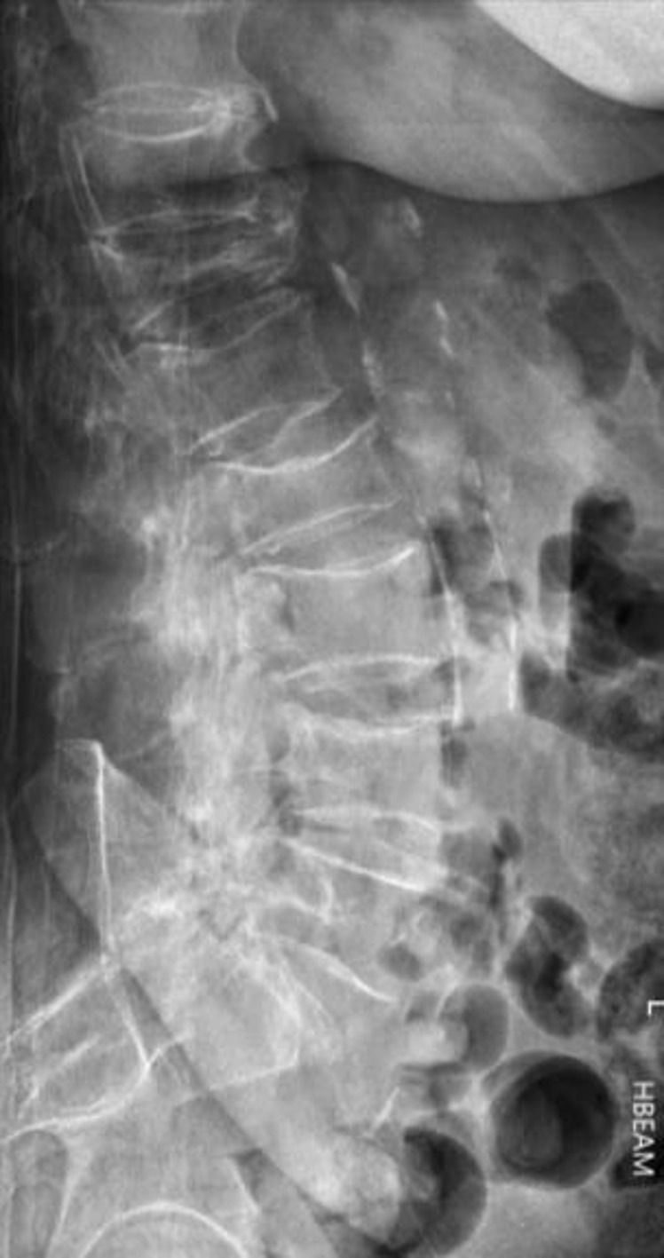
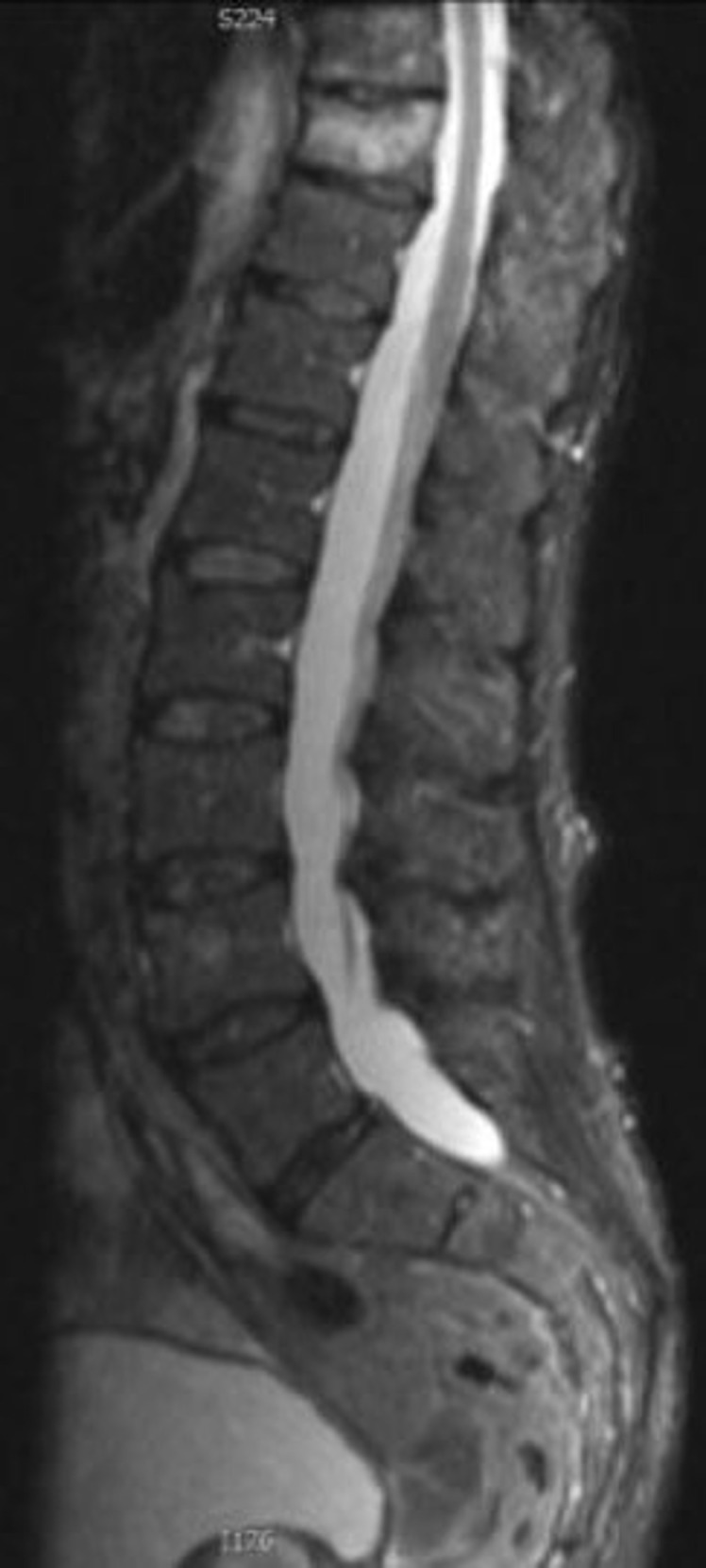
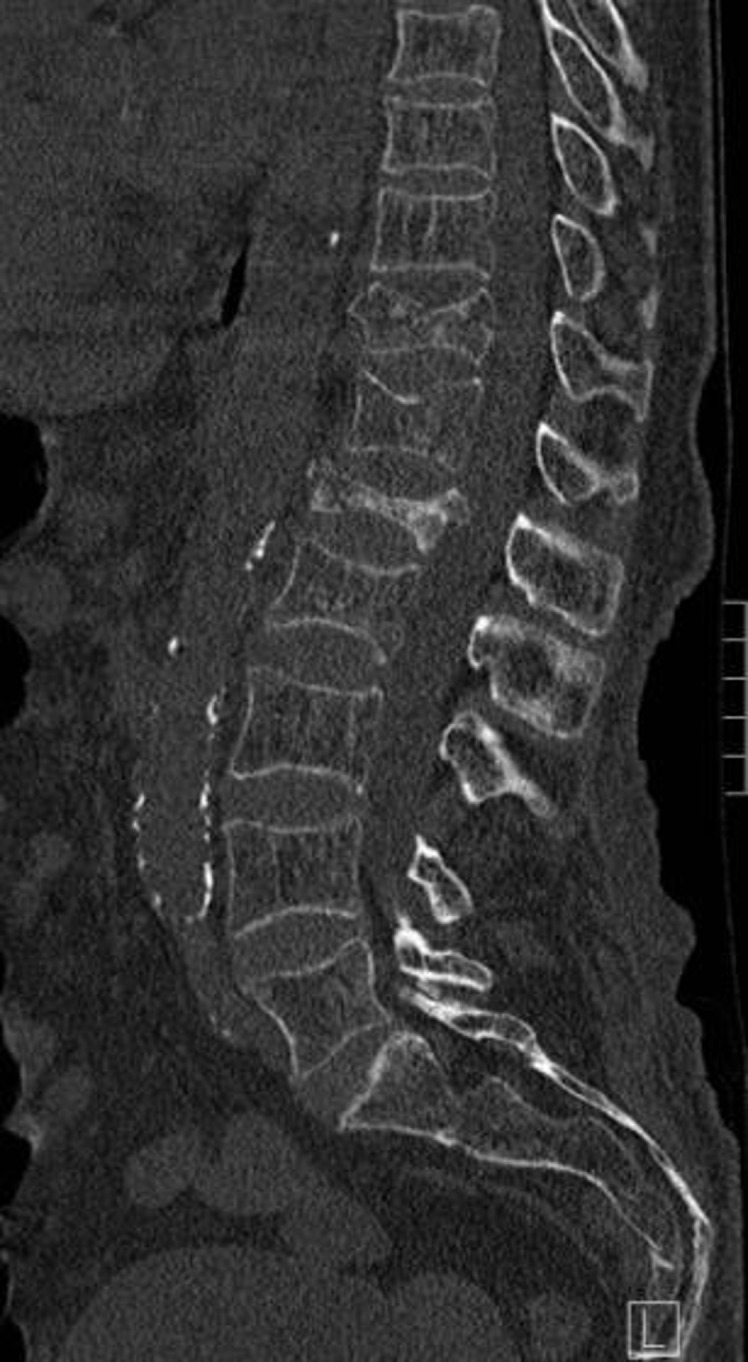
Osteoporotic vertebral fractures (VFs) are the most common type of osteoporotic fracture. Patients with VF are at increased risk of hip fractures or additional VFs, both of which contribute to patient morbidity and mortality. Early diagnosis of VFs is essential so patients can be prescribed appropriate medical therapy. Most patients with clinical suspicion for VF have an X-ray of the spine. Many VFs are invisible on X-ray and require further imaging. CT can provide excellent bony detail but uses high doses of ionising radiation. MRI provides excellent soft tissue detail and can distinguish old from new fractures in addition to differentiating osteoporotic VFs from other causes of back pain. Bone scans have a limited role due to poor specificity. The literature suggests that radiologists frequently miss or do not report VFs when imaging is requested for an alternative clinical indication and when there is no clinical suspicion of VF. Common examples include failure to identify VFs on lateral chest X-rays, sagittal reformats of CT thorax and abdomen, lateral localizers on MRI and scout views on CT. Failure to diagnose a VF is a missed opportunity to improve management of osteoporosis and reduce risk of further fractures. This article discusses the role of radiographs, CT, MRI and bone scintigraphy in the assessment and recognition of osteoporotic fractures. This article focuses on opportunistic diagnosis of VFs on imaging studies that are performed for other clinical indications. It does not discuss use of dual energy X-ray absorptiometry which is a specific imaging modality for osteoporosis.
© 2021 The Authors. Published by the British Institute of Radiology.
Figures


References
-
- (N.d.). Foundation no.National Osteoporosis Foundation Website. . Available from: https://www.nof.org/preventing-fractures/general-facts/2019
Publication types
LinkOut - more resources
Full Text Sources
