. 2022 Jun 2:13:15-21.
doi: 10.1016/j.ibneur.2022.05.008. eCollection 2022 Dec.

Endogenous dopamine transmission is crucial for motor skill recovery after stroke

Affiliations

Endogenous dopamine transmission is crucial for motor skill recovery after stroke

Clément Vitrac et al. IBRO Neurosci Rep. .

Abstract

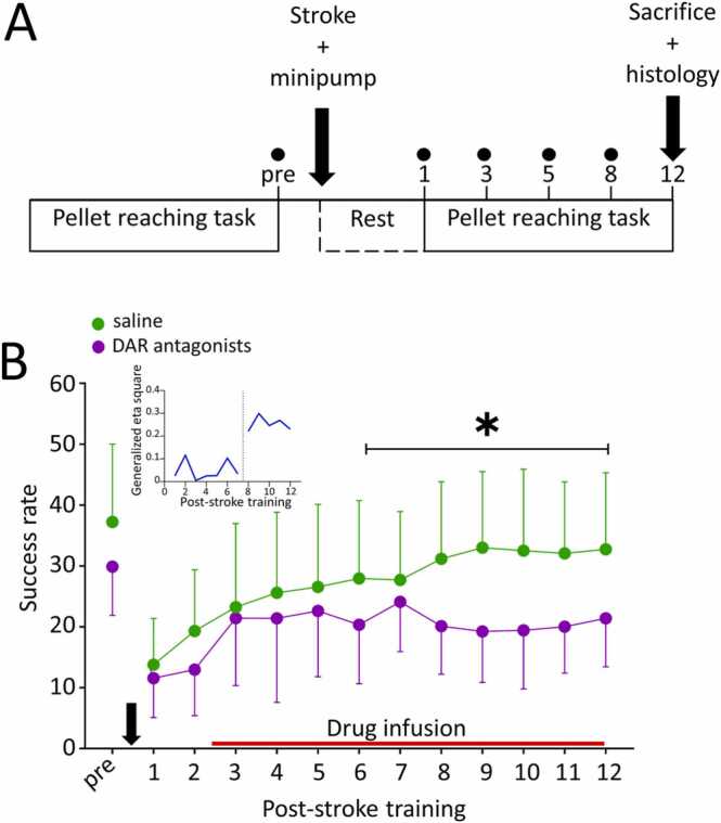
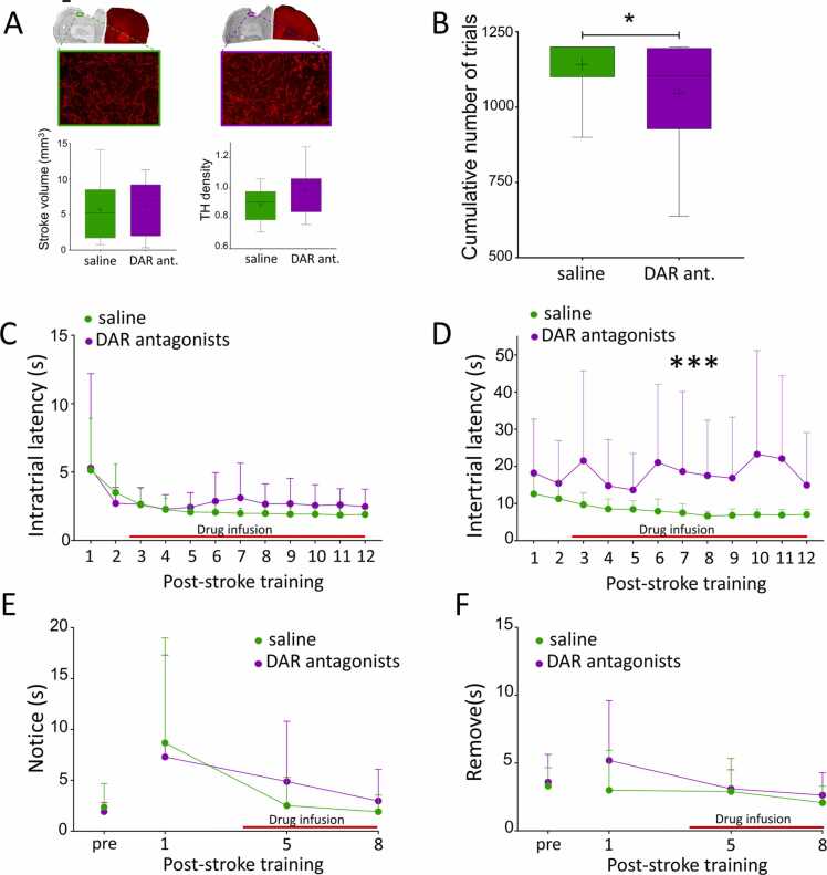
Ischemic stroke frequently causes motor impairments. Despite exercise can improve motor outcomes, many stroke survivors remain life-long disabled. Understanding the mechanisms associated with motor recovery after a stroke is necessary to develop treatments. Here, we show that endogenous DA transmission is required for optimal motor skill recovery following photothrombotic stroke in rats. Blockade of dopamine D1 and D2 receptors impaired the recovery of a forelimb reaching task and decreased the rats' motivation to complete full training sessions. Our data indicate that dopamine transmission is important to drive motor rehabilitation after stroke through motivational aspects and ultimately suggest that augmented motivation and reward feedback could be an interesting strategy to increase the effectiveness or rehabilitation.

Keywords: Dopamine; Motivation; Motor skill learning; Rehabilitation; Stroke.


Figures

Fig. 1
Fig. 1
Dopamine transmission is necessary for optimal motor recovery from stroke. A: Schematic representation of the experimental protocol. Rats were trained in the pellet reaching task to plateau before receiving a stroke and a minipump infusing either 0.9 % saline (control) or a mixture of D1R and D2R antagonists. Training was resumed 3 days post-stroke and continued for 12 days. Dots represent the days of tape removal test. B: Post-stroke learning curves of DAR antagonist treated (n = 16, purple) and saline treated rats (n = 18, green). Blocking DAR significantly impaired motor recovery starting on training day 8 compared to saline. * : p < 0.05. The red line indicates drug delivery. The black arrow pictures the stroke induction and minipump implantation. The Inset shows that the effect of drug treatment was negligible during the first 8 days of post-stroke training and jumped to much larger values during the second week of training indicating a stronger association between drug and success rate. (For interpretation of the references to colour in this figure, the reader is referred to the web version of this article.)
Fig. 2
Fig. 2
DA antagonists alter motivation after stroke. A. Anatomical characterization of the stroke volume and TH innervation of the peri-infarct area. Analysis of stroke volume and TH innervation of the peri-infarct area (green: saline n = 10, purple: DAR antagonists n = 7) revealed no difference. Scale bar= 1 mm for upper panel, 1 µm for lower panel. B. Effects of DAR antagonists on training intensity. Rats treated with DAR antagonists (purple, n = 16) performed significantly fewer trials during the 12 days of post-stroke training than saline treated rats (green, n = 18). * : p < 0.05. C. Effects of DAR antagonists on intratrial latency. DAR blockade did not impair the speed of execution of a trial compared to saline. D. DAR antagonists increased intertrial latencies. DAR antagonist-treated rats took significantly longer to initiate a new trial than saline treated rats. *** : p < 0.001. E-F. Effect of DAR antagonists on somatosensory function. No difference was detected between saline (green, n = 16) and DAR antagonists (purple, n = 12) on the time to notice (E) or to remove (F) a piece of tape placed on the lesioned forelimb. A, B: Boxes: median, 25th and 75th percentile. Whiskers: 5th and 95th percentile. + : sample mean. C-F: Red line: timecourse of drug infusion. (For interpretation of the references to colour in this figure, the reader is referred to the web version of this article.)

References

    1. Akiyama Y., Ito A., Koshimura K., Ohue T., Yamagata S., Miwa S., Kikuchi H. Effects of transient forebrain ischemia and reperfusion on function of dopaminergic neurons and dopamine reuptake in vivo in rat striatum. Brain Res. 1991;561:120–127. 〈https://www.sciencedirect.com/science/article/pii/000689939190756L〉 (Available at) - PubMed
    1. Alling K., Poling A. The effects of differing response-force requirements on fixed-ratio responding of rats. J. Exp. Anal. Behav. 1995;63:331–346. Available at: 〈https://www.ncbi.nlm.nih.gov/pmc/articles/PMC1334449/〉. - PMC - PubMed
    1. Biernaskie J., Chernenko G., Corbett D. Efficacy of rehabilitative experience declines with time after focal ischemic brain injury. J. Neurosci. 2004;24:1245–1254. - PMC - PubMed
    1. Buitrago M.M., Ringer T., Schulz J.B., Dichgans J., Luft A.R. Characterization of motor skill and instrumental learning time scales in a skilled reaching task in rat. Behav. Brain Res. 2004;155:249–256. - PubMed
    1. Carmichael S.T. Cellular and molecular mechanisms of neural repair after stroke: making waves. Ann. Neurol. 2006;59:735–742. - PubMed

LinkOut - more resources