. 2022 Jul;29(7):103313.
doi: 10.1016/j.sjbs.2022.103313. Epub 2022 May 22.

Melatonin relieves diabetic complications and regenerates pancreatic beta cells by the reduction in NF-kB expression in streptozotocin induced diabetic rats

Affiliations

Melatonin relieves diabetic complications and regenerates pancreatic beta cells by the reduction in NF-kB expression in streptozotocin induced diabetic rats

Alyaa Farid et al. Saudi J Biol Sci. 2022 Jul.

Abstract

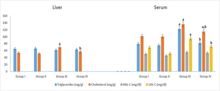
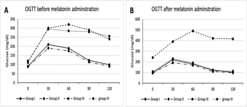
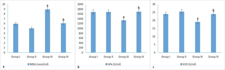
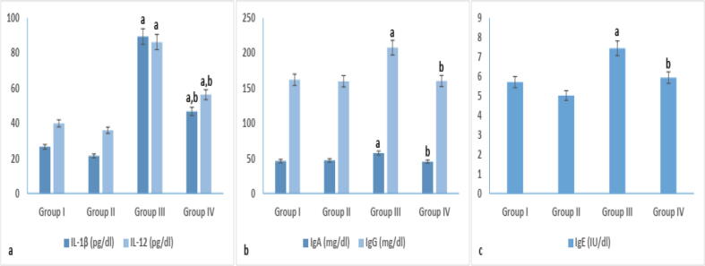
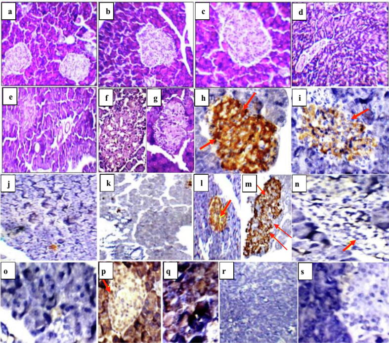
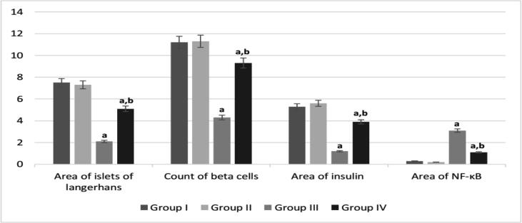
Melatonin, a pleiotropic hormone, has many regulatory effects on the circadian and seasonal rhythms, sleep and body immune system. It is used in the treatment of blind circadian rhythm sleep disorders, delayed sleep phase and insomnia. It is a potent antioxidant, anti-inflammatory, free radical scavenger, helpful in fighting infectious disease and cancer treatment. Decreased level of circulating melatonin was associated with an increased blood glucose level, losing the anti-oxidant protection and anti-inflammatory responses. We aimed to evaluate the effect of melatonin administration, in streptozotocin (STZ) induced diabetic rats, on blood glucose level and pancreatic beta (β) cells. Diabetes mellitus was induced in Sprague dawley male rats by the intravenous (i.v) injection of 65 mg/kg of STZ. Diabetic rats received melatonin at a dose of 10 mg/kg daily for 8 weeks by oral routes. The results showed, after 8 weeks of melatonin administration, a reduction in: 1- fasting blood glucose (FBG) and fructosamine (FTA) levels, 2- kidney and liver function parameters, 3- levels of serum triglycerides, cholesterol and LDL-C, 4- malondialdehyde (MDA), 5- NF-κB expression in treated group, 6- pro-inflammatory cytokines (IL-1β and IL-12) and immunoglobulins (IgA, IgE and IgG). Furthermore, an elevation in insulin secretion was noticed in melatonin treated group that indicated β cells regeneration. Therefore, melatonin administration, in STZ induced diabetic rats; reduced hyperglycemia, hyperlipidemia and oxidative stress. Melatonin acted as an anti-inflammatory agent that reduced pro-inflammatory cytokines (IL-1β and IL-12) and oxidative stress biomarkers (MDA). Melatonin succeeded in protecting β cells under severe inflammatory situations, which was apparent by the regeneration of islets of Langerhans in treated diabetic rats. Moreover, these results can open a gate for diabetes management and treatment.

Keywords: Diabetes mellitus; IL-12; IL-1β; Inflammation; β-cells.


Conflict of interest statement

The authors declare that they have no known competing financial interests or personal relationships that could have appeared to influence the work reported in this paper.

Figures

Fig. 1
Fig. 1
Serum and liver lipid profile in different experimental groups. arepresents significance compared with the corresponding healthy control group I and brepresents significance compared with the corresponding diabetic group III (P < 0.05). Group I: healthy control rats, group II: melatonin administrated control rats, group III: non-treated STZ diabetic rats and group IV: melatonin treated STZ diabetic rats.
Fig. 2
Fig. 2
Oral glucose tolerance test (OGTT): (A) before; and (B) after 8-week of melatonin administration. Group I: healthy control rats, group II: melatonin administrated control rats, group III: non-treated STZ diabetic rats and group IV: melatonin treated STZ diabetic rats.
Fig. 3
Fig. 3
Oxidative stress in different experimental groups. arepresents significance compared with the corresponding healthy control group I and brepresents significance compared with the corresponding diabetic group III (P < 0.05). Group I: healthy control rats, group II: melatonin administrated control rats, group III: non-treated STZ diabetic rats and group IV: melatonin treated STZ diabetic rats.
Fig. 4
Fig. 4
Immunological parameters (cytokines and immunoglobulin) in different experimental groups. arepresents significance compared with the corresponding healthy control group I and brepresents significance compared with the corresponding diabetic group III (P < 0.05). Group I: healthy control rats, group II: melatonin administrated control rats, group III: non-treated STZ diabetic rats and group IV: melatonin treated STZ diabetic rats.
Fig. 5
Fig. 5
Pancreatic sections of rat showing a], b] and c] normal sized stained islets of Langerhans in healthy control group I and melatonin administrated control group II (H&E; x200&400, respectively), d] and e] small-sized hypocellular pale-staining islets of Langerhans in non-treated diabetic group III (H&E; x200&400, respectively), f] and g] restored islets of Langerhans in melatonin treated diabetic group IV (H&E; x400), h] and i] positive anti-insulin antibody reaction as indicated by brown colour (group I and II; x400), j] and k] negative anti-insulin antibody reaction in non-treated diabetic group III (x200), l] and m] positive anti-insulin antibody reaction in melatonin treated diabetic group IV (x200&x400, respectively), n] and o] no NF- κB expression in group I and II (x400), p] and q] significant elevation of NF- κB expression as brown colour indicates positive immunstaining in non treated diabetic group III, r] and s] no NF- κB expression in melatonin treated diabetic group IV (x200&400, respectively).
Fig. 6
Fig. 6
Quantitative analysis of histopathological and immunohistochemial results. arepresents significance compared with the corresponding healthy control group I and brepresents significance compared with the corresponding diabetic group III (P < 0.05). Group I: healthy control rats, group II: melatonin administrated control rats, group III: non-treated STZ diabetic rats and group IV: melatonin treated STZ diabetic rats.
Fig. 7
Fig. 7
a] and b] liver sections of normal control group I and melatonin administrated group II showing average central veins with average hepatocytes arranged in single cell cords and average intervening blood sinusoids (H&E; x400), c] and d] liver sections of diabetic untreated group III showing mildly dilated congested central veins with marked micro-vesicular steatosis of hepatocytes (arrows) (H&E; x400), e] and f] liver sections of melatonin treated diabetic group IV showing average central veins (arrow) with average hepatocytes (H&E; x200& 400, respectively), g] and h] Kidney sections of normal control group I and melatonin administrated group II showing average renal capsule (arrow), average glomeruli with average proximal tubules and preserved brush borders (H&E, x200), i] and j] kidney sections of diabetic untreated group III showing small-sized glomeruli with widened Bowman’s spaces, proximal tubules with mildly vacuolated epithelial lining and partial loss of brush borders (H&E, x400), k] and l] kidney sections of melatonin treated diabetic group IV showing average renal capsule (head arrow), average glomeruli with average proximal tubules, normal epithelial lining and preserved brush borders (arrow) (H&E, x400). CV: central vein and CT: collecting tubules.

References

    1. Abdel-Monaem G., Farid A., Rabia I., El-Amir A. Evaluation of Echinostoma liei worm, metacercaria and redia antigens for schistosomiasis control. Experimental Parasitology. 2015;157:23–29. - PubMed
    1. Adorini L. Interleukin 12 and autoimmune diabetes. Nature Genet. 2001;27(2):131–132. - PubMed
    1. Agil A., Rosado I., Ruiz R., Figueroa A., Zen N., Fernández-Vázquez G. Melatonin improves glucose homeostasis in young zucker diabetic fatty rats. J. Pineal Res. 2012;52(2):203–210. - PubMed
    1. Ahmad Haja Y., Rai S., Basheer M., Ghosh H., Singh S. Protective role of melatonin in streptozotocin induced pancreatic damages in diabetic wistar rat. Pak J. Biol. Sci. 2018;21(9):423–431. - PubMed
    1. Ahmed O., Abdel-Halim M., Farid A., Elamir A. Taurine loaded chitosan-pectin nanoparticle shows curative effect against acetic acid-induced colitis in rats. Chemico-Biological Interactions. 2022;351:109715. - PubMed

LinkOut - more resources