. 2022 Aug 30;13(4):e0142022.
doi: 10.1128/mbio.01420-22. Epub 2022 Jun 16.

S. cerevisiae Cells Can Grow without the Pds5 Cohesin Subunit

Affiliations

S. cerevisiae Cells Can Grow without the Pds5 Cohesin Subunit

Karan Choudhary et al. mBio. .

Abstract

During DNA replication, the newly created sister chromatids are held together until their separation at anaphase. The cohesin complex is in charge of creating and maintaining sister chromatid cohesion (SCC) in all eukaryotes. In Saccharomyces cerevisiae cells, cohesin is composed of two elongated proteins, Smc1 and Smc3, bridged by the kleisin Mcd1/Scc1. The latter also acts as a scaffold for three additional proteins, Scc3/Irr1, Wpl1/Rad61, and Pds5. Although the HEAT-repeat protein Pds5 is essential for cohesion, its precise function is still debated. Deletion of the ELG1 gene, encoding a PCNA unloader, can partially suppress the temperature-sensitive pds5-1 allele, but not a complete deletion of PDS5. We carried out a genetic screen for high-copy-number suppressors and another for spontaneously arising mutants, allowing the survival of a pds5Δ elg1Δ strain. Our results show that cells remain viable in the absence of Pds5 provided that there is both an elevation in the level of Mcd1 (which can be due to mutations in the CLN2 gene, encoding a G1 cyclin), and an increase in the level of SUMO-modified PCNA on chromatin (caused by lack of PCNA unloading in elg1Δ mutants). The elevated SUMO-PCNA levels increase the recruitment of the Srs2 helicase, which evicts Rad51 molecules from the moving fork, creating single-stranded DNA (ssDNA) regions that serve as sites for increased cohesin loading and SCC establishment. Thus, our results delineate a double role for Pds5 in protecting the cohesin ring and interacting with the DNA replication machinery. IMPORTANCE Sister chromatid cohesion is vital for faithful chromosome segregation, chromosome folding into loops, and gene expression. A multisubunit protein complex known as cohesin holds the sister chromatids from S phase until the anaphase stage. In this study, we explore the function of the essential cohesin subunit Pds5 in the regulation of sister chromatid cohesion. We performed two independent genetic screens to bypass the function of the Pds5 protein. We observe that Pds5 protein is a cohesin stabilizer, and elevating the levels of Mcd1 protein along with SUMO-PCNA accumulation on chromatin can compensate for the loss of the PDS5 gene. In addition, Pds5 plays a role in coordinating the DNA replication and sister chromatid cohesion establishment. This work elucidates the function of cohesin subunit Pds5, the G1 cyclin Cln2, and replication factors PCNA, Elg1, and Srs2 in the proper regulation of sister chromatid cohesion.

Keywords: Cln2; DNA replication; ELG1; Elg1; Mcd1; PCNA and PDS5; Pds5; Rad51; SUMO; Srs2; cohesin; sister chromatid cohesion; yeast.


Conflict of interest statement

The authors declare no conflict of interest.

Figures

FIG 1
FIG 1
Screen for suppressors of the pds5Δ elg1Δ double mutant. (A) Illustration of the experimental scheme for the high-copy-number suppressor screen; (B) 5-fold serial dilutions of cells harboring either empty vector or high-copy-number vectors overexpressing MCD1 or PDS5 in addition to the covering plasmid (carrying the URA3 and PDS5 genes); (C, D) spot assay with 5-fold serial dilutions of cells harboring either empty vector or high-copy-number plasmids overexpressing MCD1 with different mutations at specified residues in addition to the covering plasmid (carrying the URA3 and PDS5 genes). (E) Experimental regimen of a screen looking for the spontaneous suppressor mutants able to grow in the complete absence of PDS5 and ELG1; (F) spot assay with 5-fold serial dilutions of the pds5Δ background strains carrying specified gene deletions on Ura and 5-FOA plates. All mentioned strains carry a Pds5 covering plasmid (carrying the URA3 selection marker).
FIG 2
FIG 2
Deletion of ELG1 and CLN2 restores the Mcd1 protein level in the absence of Pds5. (A) Western blot showing the Mcd1 protein level in different AID-PDS strains. Cells were harvested after arresting them in the G2/M phase by treatment with nocodazole (15 μg/mL) for 2 h, followed by the treatment with auxin (IAA [300 μM]). The experimental scheme is represented below the Western blot panel. Mcd1 was probed with an anti-Mcd1 antibody, Pds5 was detected using anti-V5, and tubulin was used as a loading control. (B) Mcd1 protein levels normalized to those of tubulin (mean ± standard deviation [SD]; n = 3). **, P ≤ 0.01 by t test. (C to E) Western blot for the auxin chase experiment. The cells of the indicated strains were grown until the log phase (time zero) and then treated with auxin (300 μM). Samples were taken every 20 min until completion of a 2-h experiment. (F) Relative levels of Mcd1 protein normalized to those of tubulin used as a loading control (mean ± SD percentage; n = 3).
FIG 3
FIG 3
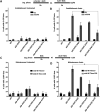
Mcd1 is overexpressed in elg1Δ cln2Δ double mutants. (A) GFP-RFP plasmid with a short-lived GFP gene under the control of the Mcd1 promoter and internal control mCherry under the control of ADH1 promoter; (B) mean fluorescent intensity of the GFP/mCherry ratio from flow cytometry for different strains treated with auxin (IAA [300 μM]) for 2 h (right) and without auxin (left). Results represent 20,000 events (n = 3). ***, P ≤ 0.001 by one-way analysis of variance (ANOVA). (C) Western blot (anti-GFP) monitoring the GFP fused to CL1 degron protein levels in different strains expressed from a 2μ plasmid. Actin was used as a loading control. (D) Western blot quantification of GFP levels normalized to the loading control actin (mean ± SD; n = 3). ***, P ≤ 0.001 by t test. (E) Western blot (anti-GFP) monitoring the GFP-CL1 fusion protein levels expressed from a construct carrying a mentioned deletion in the MCB box in Mcd1 promoter; (F) Western blot quantification of GFP levels normalized to the loading control actin (mean ± SD; n = 3). ***, P ≤ 0.001 by one-way ANOVA.
FIG 4
FIG 4
Deletion of ELG1 and CLN2 restores the sister chromatid cohesion defects in the absence of Pds5. Results from cohesion establishment analysis are shown in panels A and B. The experimental scheme for the cohesion establishment assay is shown above panels A and B. (A) Percentage of cells with 2 dots in mid-M phase without auxin treatment (mean ± SD; n = 3 with >200 cells per strain and experiment). (B) Establishment assay for auxin-treated cells. α-Factor was used at 50 ng/mL, nocodazole (NOC) at 15 μg/mL, and pronase E (PRON) at 0.1 mg/mL. Results from cohesion maintenance analysis are shown in panels C and D. The experimental scheme for the cohesion maintenance assay with auxin (IAA [300 μM]) is shown above panels C and D. The untreated experimental process was the same as for cohesion establishment, but without auxin. (C) Percentage of cells with 2 dots for every strain without auxin treatment (mean ± SD; n = 3, with 200 cells per strain and experiment). (D) Maintenance assay for auxin-treated cells in different strains. Nocodazole was used at 15 μg/mL.
FIG 5
FIG 5
PCNA accumulation on chromatin promotes sister chromatid cohesion in the absence of Pds5. (A) Spot assay with 5-fold serial dilution of the pds5Δ cln2Δ mutant plus the CEN PDS5 URA background strain carrying different mutations of Elg1 at the ELG1 locus in the genome. The assay was performed on 5-FOA medium and plates containing the DNA-damaging agent MMS at the mentioned concentrations. (B) Spot assay with 5-fold serial dilution of the pds5Δ cln2Δ elg1Δ mutant plus the CEN PDS5 URA background strain harboring disassembly-prone PCNA mutations in the genomic copy of the POL30 gene. The assay was performed on 5-FOA plates. (C) Chromatin fractionation experiment showing the EcoI-3HA levels on chromatin in untreated and auxin-treated (2 h) samples. Histone H3 was used as a chromatin marker and loading control; Rps6 was used as a cytoplasmic marker. (D) The graph represents the Western blot quantification of the relative abundance of EcoI protein on chromatin (mean ± SD; n = 3). ns, not significant by Student’s t test.
FIG 6
FIG 6
SUMO-PCNA accumulation on chromatin and Srs2 promote sister chromatid cohesion in the absence of Pds5. (A) Spot assay with 5-fold serial dilution of the pds5Δ cln2Δ elg1Δ mutant plus the CEN PDS5 URA background strain harboring point mutations at the key lysine residue in the genomic copy of the POL30 gene. The assay was performed on 5-FOA plates. (B) Spot assay with 5-fold serial dilution of the pds5Δ cln2Δ elg1Δ mutant plus the CEN PDS5 URA background strain carrying deletion of genes involved in PCNA ubiquitination (Rad5 and Rad18) or PCNA SUMOylation pathways (Siz1) and the SUMO-PCNA interactor Srs2. The assay was performed on 5-FOA plates. (C) Five-fold serial dilution of pds5Δ cln2Δ elg1Δ srs2Δ rad51Δ mutant plus the CEN PDS5 URA background strain and control strains on 5-FOA plates.
FIG 7
FIG 7
Model for the bypass of Pds5 function by elg1Δ cln2Δ. (A) The WT cells properly establish cohesion during the S phase and maintain it throughout the following cell cycle to allow faithful chromosome segregation. (B) The deletion of Pds5 results in hyper-SUMOylation of the Mcd1 cohesin subunit, leading to its premature degradation, followed by loss of cohesion and cell death. (C) The deletion of the G1 cyclin Cln2 results in overproduction of Mcd1; however, it cannot produce sufficient cohesion to sustain the high cohesin turnover associated with the loss of Pds5 protein. As a result, the pds5Δ cln2Δ strain is inviable and shows cohesion defects. (D) The deletion of PCNA unloader Elg1 results in accumulation of SUMO-PCNA on chromatin, which might allow a wider window for cohesin establishment. However, the pds5Δ elg1Δ strain is inviable due to the insufficient levels of Mcd1 protein available during cohesion establishment. (E) The deletion of PCNA unloader Elg1 along with G1 cyclin Cln2 (or with Mcd1 overexpression) results in stable cohesion in the absence of the Pds5 cohesin subunit, rendering yeast cells viable. In other words, the high cohesin turnover associated with pds5Δ might be compensated by overestablishing functional cohesion during DNA replication in this scenario. The SUMO-PCNA accumulation recruits Srs2 to remove Rad51 protein from ssDNA, which might allow the increased establishment of cohesion during DNA replication. Estb., establishment.

Similar articles

Cited by

References

    1. Onn I, Heidinger-Pauli JM, Guacci V, Ünal E, Koshland DE. 2008. Sister chromatid cohesion: a simple concept with a complex reality. Annu Rev Cell Dev Biol 24:105–129. doi:10.1146/annurev.cellbio.24.110707.175350. - DOI - PubMed
    1. Barrington C, Finn R, Hadjur S. 2017. Cohesin biology meets the loop extrusion model. Chromosome Res 25:51–60. doi:10.1007/s10577-017-9550-3. - DOI - PMC - PubMed
    1. Matityahu A, Onn I. 2021. Hit the brakes—a new perspective on the loop extrusion mechanism of cohesin and other SMC complexes. J Cell Sci 134:jcs247577. doi:10.1242/jcs.247577. - DOI - PubMed
    1. Peters JM, Nishiyama T. 2012. Sister chromatid cohesion. Cold Spring Harb Perspect Biol 4:a011130. doi:10.1101/cshperspect.a011130. - DOI - PMC - PubMed
    1. Uhlmann F. 2016. SMC complexes: from DNA to chromosomes. Nat Rev Mol Cell Biol 17:399–412. doi:10.1038/nrm.2016.30. - DOI - PubMed

Publication types

MeSH terms

LinkOut - more resources