. 2022 Aug 30;13(4):e0051922.
doi: 10.1128/mbio.00519-22. Epub 2022 Jun 16.

Metalloproteinase-Dependent and TMPRSS2-Independent Cell Surface Entry Pathway of SARS-CoV-2 Requires the Furin Cleavage Site and the S2 Domain of Spike Protein

Affiliations

Metalloproteinase-Dependent and TMPRSS2-Independent Cell Surface Entry Pathway of SARS-CoV-2 Requires the Furin Cleavage Site and the S2 Domain of Spike Protein

Mizuki Yamamoto et al. mBio. .

Abstract

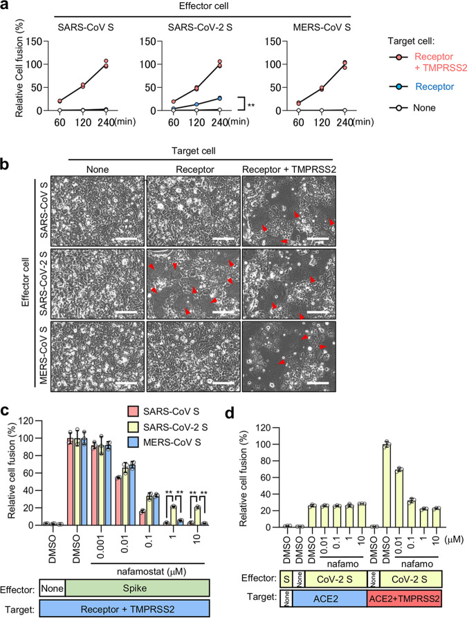
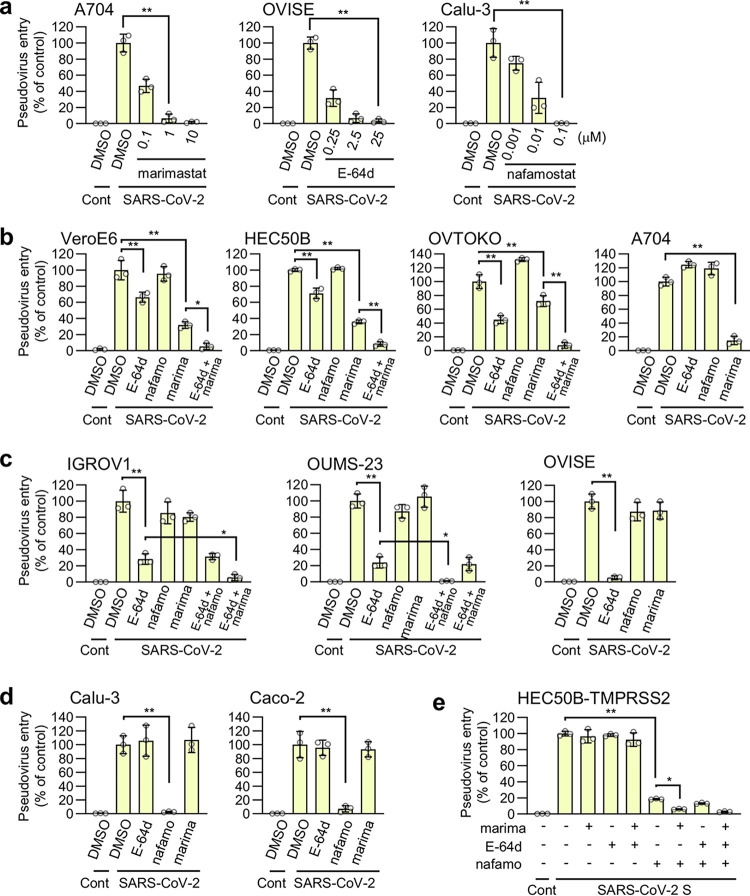
The ongoing global vaccination program to prevent SARS-CoV-2 infection, the causative agent of COVID-19, has had significant success. However, recently, virus variants that can evade the immunity in a host achieved through vaccination have emerged. Consequently, new therapeutic agents that can efficiently prevent infection from these new variants, and hence COVID-19 spread, are urgently required. To achieve this, extensive characterization of virus-host cell interactions to identify effective therapeutic targets is warranted. Here, we report a cell surface entry pathway of SARS-CoV-2 that exists in a cell type-dependent manner and is TMPRSS2 independent but sensitive to various broad-spectrum metalloproteinase inhibitors such as marimastat and prinomastat. Experiments with selective metalloproteinase inhibitors and gene-specific small interfering RNAS (siRNAs) revealed that a disintegrin and metalloproteinase 10 (ADAM10) is partially involved in the metalloproteinase pathway. Consistent with our finding that the pathway is unique to SARS-CoV-2 among highly pathogenic human coronaviruses, both the furin cleavage motif in the S1/S2 boundary and the S2 domain of SARS-CoV-2 spike protein are essential for metalloproteinase-dependent entry. In contrast, the two elements of SARS-CoV-2 independently contributed to TMPRSS2-dependent S2 priming. The metalloproteinase pathway is involved in SARS-CoV-2-induced syncytium formation and cytopathicity, leading us to theorize that it is also involved in the rapid spread of SARS-CoV-2 and the pathogenesis of COVID-19. Thus, targeting the metalloproteinase pathway in addition to the TMPRSS2 and endosomal pathways could be an effective strategy by which to cure COVID-19 in the future. IMPORTANCE To develop effective therapeutics against COVID-19, it is necessary to elucidate in detail the infection mechanism of the causative agent, SARS-CoV-2. SARS-CoV-2 binds to the cell surface receptor ACE2 via the spike protein, and then the spike protein is cleaved by host proteases to enable entry. Here, we found that the metalloproteinase-mediated pathway is important for SARS-CoV-2 infection in addition to the TMPRSS2-mediated pathway and the endosomal pathway. The metalloproteinase-mediated pathway requires both the prior cleavage of spike into two domains and a specific sequence in the second domain, S2, conditions met by SARS-CoV-2 but lacking in the related human coronavirus SARS-CoV. Besides the contribution of metalloproteinases to SARS-CoV-2 infection, inhibition of metalloproteinases was important in preventing cell death, which may cause organ damage. Our study provides new insights into the complex pathogenesis unique to COVID-19 and relevant to the development of effective therapies.

Keywords: SARS-CoV-2; furin; membrane fusion; metalloproteinase; virus entry.


Conflict of interest statement

The authors declare no conflict of interest.

Figures

FIG 1
FIG 1
ACE2-dependent but TMPRSS2-independent membrane fusion activity of the SARS-CoV-2 S protein. (a) Cell fusion kinetics induced by the S proteins from SARS-CoV, SARS-CoV-2, and MERS-CoV were determined using the DSP assay. Target cells expressing ACE2 alone or together with TMPRSS2 were used for coculturing with effector cells expressing SARS-CoV S and SARS-CoV-2 S, and cells expressing CD26 alone or together with TMPRSS2 were used for coculturing with effector cells expressing MERS-CoV S. Relative cell fusion values were calculated by normalizing the RL activity of each coculture to that of the coculture with cells expressing both receptor and TMPRSS2 at 240 min, which was set to 100%. Values are means ± standard deviations (SD) (n =3/group). **, P < 0.01. (b) Phase-contrast images of S protein-mediated cell fusion 16 h after coculture. Red arrowheads indicate syncytium formation. Scale bars, 100 μm. (c) Effect of nafamostat on TMPRSS2-dependent cell fusion. Target cells expressing ACE2 with TMPRSS2 were used for coculturing with effector cells expressing SARS-CoV S and SARS-CoV-2 S, and cells expressing CD26 with TMPRSS2 were used for coculturing with effector cells expressing MERS-CoV S. Relative cell fusion values were calculated by normalizing the RL activity for each coculture to that of the coculture with cells expressing both receptor and TMPRSS2 in the presence of DMSO, which was set to 100%. Values are means ± SD (n =3/group). **, P < 0.01. (d) Effects of nafamostat on TMPRSS2-independent or -dependent cell fusion. Target cells expressing ACE2 alone or together with TMPRSS2 were used for coculturing with effector cells expressing SARS-CoV-2 S. Relative cell fusion values were calculated by normalizing the RL activity for each coculture to that of the coculture with cells expressing both ACE2 and TMPRSS2 in the presence of DMSO, which was set to 100%. Values are means ± SD (n =3/group). nafamo, nafamostat.
FIG 2
FIG 2
TMPRSS2-independent membrane fusion induced by the S protein of SARS-CoV-2 is blocked by various metalloproteinase inhibitors. (a) High-throughput screening of the Validated Compound Library (1,630 clinically approved compounds and 1,885 pharmacologically active compounds) obtained from the Drug Discovery Initiative (The University of Tokyo) by the DSP assay using the SARS-CoV-2 S protein. The x axis shows the relative cell fusion value using cells expressing both TMPRSS2 and ACE2 in the presence of each compound (1 μM in DMSO) (n = 1). The y axis shows the relative cell fusion value using cells expressing ACE2 alone in the presence of each compound (1 μM in DMSO) (n = 1). The relative cell fusion value was calculated by normalizing the RL activity for each compound to that of the control assay (DMSO alone; set to 100%). Each dot represents an individual compound. Dots in the red dashed box represent compounds that preferentially inhibit TMPRSS2-independent membrane fusion (<30% inhibition of the relative cell fusion value using the target cells expressing both TMPRSS2 and ACE2 and >40% inhibition of the relative cell fusion value using the target cells expressing ACE2 alone). The compound names for the candidates are indicated. (b) Effects of the metalloproteinase inhibitors on cell fusion in the cocultures of cells expressing SARS-CoV-2 S protein with those expressing ACE2 alone or in combination with TMPRSS2. Relative cell fusion values were calculated by normalizing the RL activity for each coculture to that of the coculture with cells expressing both ACE2 and TMPRSS2 in the presence of DMSO, which was set to 100%. Values are means ± SD (n =3/group). (c) Expression of ACE2 in target cells (top). Tubulin was used as a loading control (bottom). ACE2-WT, wild-type ACE2; ACE2-NN, enzymatically inactive ACE2 with H374N and H378N mutations. (d) Effects of the metalloproteinase inhibitor on cell fusion in the cocultures of cells expressing SARS-CoV-2 S protein with those expressing wild-type ACE2 (ACE2-WT) or enzymatically inactive ACE2 (ACE2-NN). Relative cell fusion values were calculated by normalizing the RL activity for each coculture to that of the coculture with cells expressing wild-type ACE2 in the presence of DMSO, which was set to 100%. Values are means ± SD (n =3/group). marima, 1 μM marimastat. **, P < 0.01.
FIG 3
FIG 3
The metalloproteinase-dependent viral entry pathway is cell type dependent. The effects of drugs on the entry of SARS-CoV-2 S-bearing vesicular stomatitis virus (VSV) pseudotype virus produced by 293T cells are shown. The relative pseudovirus entry was calculated by normalizing the FL activity for each condition to the FL activity of cells infected with SARS-CoV-2 S-bearing pseudovirus in the presence of DMSO alone, which was set to 100%. Values are means ± SD (n =3/group). *, P < 0.05; **, P < 0.01. Cont, control (cells infected with pseudovirus without S protein); SARS-CoV-2, cells infected with SARS-CoV-2 S-bearing pseudovirus; E-64d, 25 μM E-64d; nafamo, 10 μM nafamostat; marima, 1 μM marimastat. (a) Effects of marimastat, E-64d, and nafamostat on pseudovirus entry in A704, OVISE, and Calu-3 cells, respectively. (b to e) Effects of a single drug treatment or a combination treatment on pseudovirus entry in VeroE6, HEC50B, OVTOKO, and A704 cells (b), IGROV1, OUMS-23, and OVISE cells (c), Calu-3 and Caco-2 cells (d), and HEC50B-TMPRSS2 cells (e).
FIG 4
FIG 4
The metalloproteinase-dependent entry pathway requires both the furin cleavage site and S2 region of the SARS-CoV-2 S protein. The effects of drugs on the entry of S protein-bearing vesicular stomatitis virus (VSV) pseudotype virus produced by 293T cells are shown. The relative pseudovirus entry was calculated by normalizing the FL activity for each condition to the FL activity of cells infected with pseudovirus in the presence of DMSO alone, which was set to 100%. Values are means ± SD (n =3/group). **, P < 0.01. Cont, control (cells infected with pseudovirus without S protein); E-64d, 25 μM E-64d; marima, 1 μM marimastat; nafamo, 10 μM nafamostat. (a) Effects of E-64d and marimastat on the entry of pseudoviruses bearing SARS-CoV S, SARS-CoV-2 S, MERS-CoV S, or VSV G in HEC50B cells. (b) Effects of E-64d and marimastat on the entry of HCoV-NL63 S and WIV1-CoV S pseudovirus in HEC50B cells. (c) Schematic illustration of C-terminally Flag-tagged chimeric S proteins in which the S1, S1/S2 boundary, and S2 domain from SARS-CoV S (red) and SARS-CoV-2 S (yellow) are indicated (top). Amino acid sequences of the residues around the S1/S2 boundary of the coronaviruses (bottom). Numbers refer to the amino acid residues. F, Flag tag. Arginine residues in the S1/S2 cleavage site and furin cleavage motif are highlighted in red. (d) Expression of chimeric S protein in pseudoviruses. S proteins were detected using an anti-Flag tag antibody that binds to a Flag tag on the C terminus of the S proteins (top). Detection of the vesicular stomatitis virus matrix protein (VSV M) served as the control (bottom). Culture supernatants of 293T cells containing the pseudoviruses were centrifuged at 109,000 × g for 35 min at 4°C using a TLA100.3 rotor with an Optima TLX ultracentrifuge (Beckman Coulter, CA, USA), and the pellet was then lysed for Western blotting. S0, uncleaved S protein; S2, cleaved S2 domain of the S protein. (e and f) Effects of E-64d and marimastat on the entry of pseudoviruses bearing chimeric S proteins in HEC50B cells. (g) Effects of E-64d and nafamostat on the entry of pseudoviruses bearing SARS-CoV S, SARS-CoV-2 S, MERS-CoV S, or VSV G in HEC50B-TMPRSS2 cells in the presence of marimastat. (h and i) Effects of E-64d and nafamostat on the entry of pseudoviruses bearing chimeric S proteins in HEC50B-TMPRSS2 cells in the presence of marimastat.
FIG 5
FIG 5
Possible involvement of ADAM-10 in the metalloproteinase-dependent entry of SARS-CoV-2. (a) Effects of metalloproteinase inhibitors on the entry of pseudoviruses bearing SARS-CoV-2 S or VSV G in VeroE6 and HEC50B cells in the presence of E-64d and in A704 cells in the absence of E-64d. The relative pseudovirus entry was calculated by normalizing the FL activity for each condition to the FL activity of cells infected with pseudovirus in the presence of DMSO alone, which was set to 100%. Values are means ± SD (n =3/group). Data were compared with those obtained from cells infected with pseudoviruses bearing SARS-CoV-2 S in the presence of E-64d for HEC50B and VeroE6 cells and in the presence of DMSO alone for A704 cells. *, P < 0.05; **, P < 0.01. Cont, control (cells infected with pseudovirus without S protein); marima, marimastat; prinoma, prinomastat; iloma, ilomastat; CTS, CTS-1027; UK, UK370106; GW, GW280264X; GI, GI254023X; MLN, MLN-4760; BK, BK-1361; MMP2/9i, MMP2/9 inhibitor I; MMP9i, MMP9 inhibitor I. (b) Effects of the ADAM10 knockdown on ACE2 (top), ADAM10 (middle), and tubulin (bottom) expression. HEC50B cells were transfected with two distinct control siRNAs or three distinct siRNAs against ADAM10 for 48 h. (c) Effect of the ADAM10 knockdown on the entry of pseudoviruses bearing SARS-CoV-2 S, SARS-CoV S, MERS-CoV S, or VSV G. HEC50B cells were transfected with siRNAs for 48 h and then infected with pseudoviruses. Relative pseudovirus entry was calculated by normalizing the FL activity for each condition to the FL activity of cells infected with pseudovirus in the absence of siRNA (mock), which was set to 100%. Values are means ± SD (n =3/group). *, P < 0.05; **, P < 0.01. (d and e) Effect of ADAM10 knockdown on the patterns of the entry pathways for SARS-CoV-2 S pseudovirus in HEC50B cells. HEC50B cells were transfected with siRNAs for 48 h and then infected with pseudoviruses in the presence of drugs. Values are means ± SD (n =3/group). **, P < 0.01. E-64d, 25 μM E-64d; marima, 1 μM marimastat. Data are displayed as the conditions of siRNA treatment (d) and drug treatment (e).
FIG 6
FIG 6
The metalloproteinase-dependent entry pathway of authentic SARS-CoV-2 is involved in syncytium formation and cytopathicity. The effects of the drugs on the cytoplasmic viral RNA after SARS-CoV-2 infection are shown. Cells were treated with inhibitors for 1 h and added with SARS-CoV-2 at an MOI of 0.01 for HEC50B and HEC50B-TMPRSS2 cells and at an MOI of 0.1 for VeroE6, Calu-3, and A704 cells. The relative amount of viral RNA in the cells was normalized to cellular Rpl13a mRNA expression. Values are means ± SD (n =3/group in panels a to d, n =10/group in panel e). *, P < 0.05; **, P < 0.01. (a) Effects of marimastat or prinomastat on SARS-CoV-2 infection in HEC50B, A704, and VeroE6 cells. (b) Effects of marimastat and the inhibitor of the endosomal pathway on SARS-CoV-2 infection in HEC50B, A704, and VeroE6 cells. marima, 1 μM marimastat; E-64d, 25 μM E-64d; NH4Cl, 10 mM NH4Cl. (c) Effect of marimastat, E-64d, and nafamostat on SARS-CoV-2 infection in HEC50B-TMPRSS2 cells. marima, 1 μM marimastat; E-64d, 25 μM E-64d; nafamo, 10 μM nafamostat. (d) Effects of selective metalloproteinase inhibitors on SARS-CoV-2 infection in HEC50B cells. GW, GW280264X; GI, GI254023X. (e) Effect of ADAM10 knockdown on SARS-CoV-2 infection in HEC50B cells. (f and g) Effects of drugs on SARS-CoV-2-induced syncytium formation in HEC50B (f) and HEC50B-TMPRSS2 (g) cells. Cells were stained with anti-SARS-CoV-2 N antibody (green) 24 h after infection. Nuclei were stained with Hoechst 33342 (blue). Scale bars, 200 μm. (h and i) Effects of drugs on SARS-CoV-2-induced cytopathicity in HEC50B (h) and HEC50B-TMPRSS2 (i) cells. marima, marimastat; prinoma, prinomastat; E-64d, 25 μM E-64d; nafamo, 10 μM nafamostat. Values are means ± SD (n =3/group). **, P < 0.01.

Similar articles

Cited by

References

    1. Zhou P, Yang XL, Wang XG, Hu B, Zhang L, Zhang W, Si HR, Zhu Y, Li B, Huang CL, Chen HD, Chen J, Luo Y, Guo H, Jiang RD, Liu MQ, Chen Y, Shen XR, Wang X, Zheng XS, Zhao K, Chen QJ, Deng F, Liu LL, Yan B, Zhan FX, Wang YY, Xiao GF, Shi ZL. 2020. A pneumonia outbreak associated with a new coronavirus of probable bat origin. Nature 579:270–273. doi:10.1038/s41586-020-2012-7. - DOI - PMC - PubMed
    1. Zhong NS, Zheng BJ, Li YM, Poon LLM, Xie ZH, Chan KH, Li PH, Tan SY, Chang Q, Xie JP, Liu XQ, Xu J, Li DX, Yuen KY, Peiris JSM, Guan Y. 2003. Epidemiology and cause of severe acute respiratory syndrome (SARS) in Guangdong, People's Republic of China, in February, 2003. Lancet 362:1353–1358. doi:10.1016/S0140-6736(03)14630-2. - DOI - PMC - PubMed
    1. Drosten C, Günther S, Preiser W, van der Werf S, Brodt HR, Becker S, Rabenau H, Panning M, Kolesnikova L, Fouchier RA, Berger A, Burguière AM, Cinatl J, Eickmann M, Escriou N, Grywna K, Kramme S, Manuguerra JC, Müller S, Rickerts V, Stürmer M, Vieth S, Klenk HD, Osterhaus AD, Schmitz H, Doerr HW. 2003. Identification of a novel coronavirus in patients with severe acute respiratory syndrome. N Engl J Med 348:1967–1976. doi:10.1056/NEJMoa030747. - DOI - PubMed
    1. Zaki AM, van Boheemen S, Bestebroer TM, Osterhaus AD, Fouchier RA. 2012. Isolation of a novel coronavirus from a man with pneumonia in Saudi Arabia. N Engl J Med 367:1814–1820. doi:10.1056/NEJMoa1211721. - DOI - PubMed
    1. Harrison AG, Lin T, Wang P. 2020. Mechanisms of SARS-CoV-2 transmission and pathogenesis. Trends Immunol 41:1100–1115. doi:10.1016/j.it.2020.10.004. - DOI - PMC - PubMed

Publication types

MeSH terms