. 2022 Jun 29;14(25):28476-28488.
doi: 10.1021/acsami.2c01575. Epub 2022 Jun 16.

Synthetic Heparan Sulfate Hydrogels Regulate Neurotrophic Factor Signaling and Neuronal Network Activity

Affiliations

Synthetic Heparan Sulfate Hydrogels Regulate Neurotrophic Factor Signaling and Neuronal Network Activity

Charles-Francois V Latchoumane et al. ACS Appl Mater Interfaces. .

Abstract

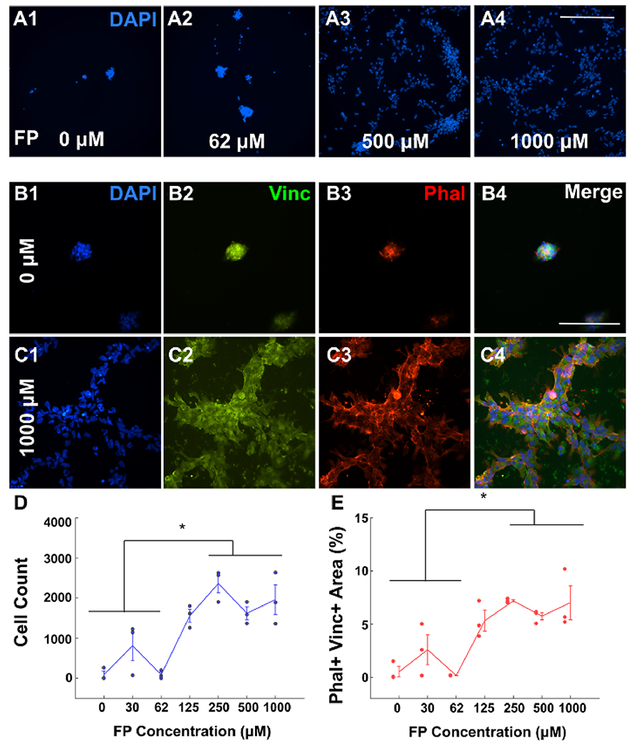
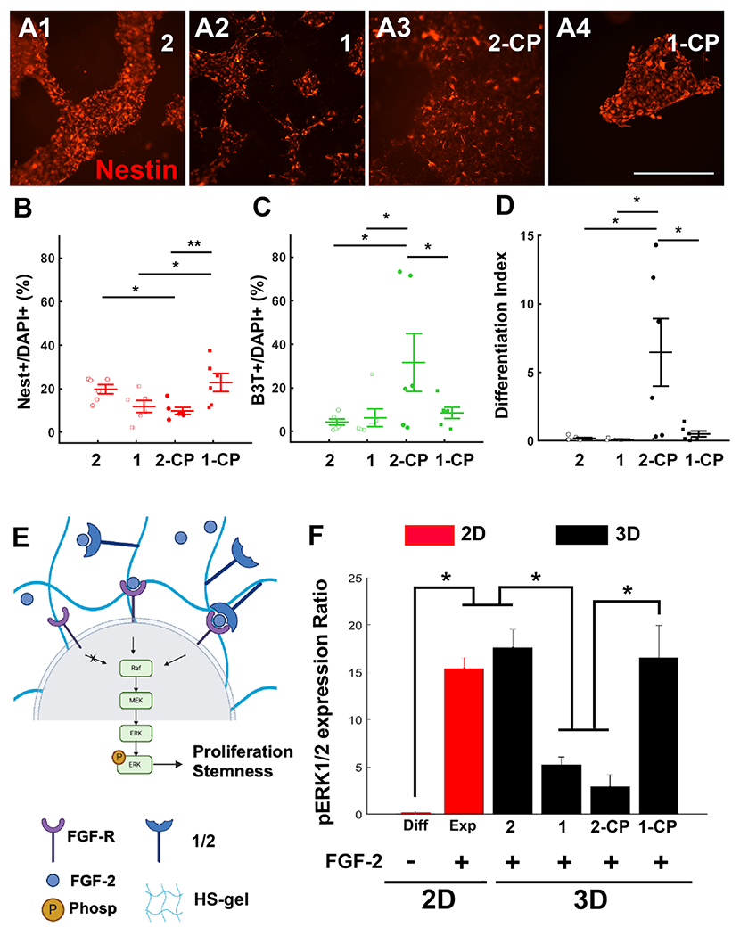
Three-dimensional (3D) synthetic heparan sulfate (HS) constructs possess promising attributes for neural tissue engineering applications. However, their sulfation-dependent ability to facilitate molecular recognition and cell signaling has not yet been investigated. We hypothesized that fully sulfated synthetic HS constructs (bearing compound 1) that are functionalized with neural adhesion peptides will enhance fibroblast growth factor-2 (FGF2) binding and complexation with FGF receptor-1 (FGFR1) to promote the proliferation and neuronal differentiation of human neural stem cells (hNSCs) when compared to constructs with unsulfated controls (bearing compound 2). We tested this hypothesis in vitro using 2D and 3D substrates consisting of different combinations of HS tetrasaccharides (compounds 3 and 4) and an engineered integrin-binding chimeric peptide (CP), which were assembled using strain-promoted alkyne-azide cycloaddition (SPAAC) chemistry. Results indicated that the adhesion of hNSCs increased significantly when cultured on 2D glass substrates functionalized with chimeric peptide. hNSCs encapsulated in 1-CP hydrogels and cultured in media containing the mitogen FGF2 exhibited significantly higher neuronal differentiation when compared to hNSCs in 2-CP hydrogels. These observations were corroborated by Western blot analysis, which indicated the enhanced binding and retention of both FGF2 and FGFR1 by 1 as well as downstream phosphorylation of extracellular signal-regulated kinases (ERK1/2) and enhanced proliferation of hNSCs. Lastly, calcium activity imaging revealed that both 1 and 2 hydrogels supported the neuronal growth and activity of pre-differentiated human prefrontal cortex neurons. Collectively, these results demonstrate that synthetic HS hydrogels can be tailored to regulate growth factor signaling and neuronal fate and activity.

Keywords: ERK; FGF2; click chemistry; neural stem cells; synthetic heparan sulfate hydrogel.


Conflict of interest statement

The authors declare no competing financial interest.

Figures

Figure 1.
Figure 1.
Chemical synthesis of HS-hydrogel components. (A) Microwave-assisted solid phase synthesis (MW-SPPS) of adhesion peptides. Reagents and conditions: (i) 20% 4-methylpiperidine, DMF, MW, 3 min; (ii) Fmoc-AA-OH, HOBt, HBTU, DIPEA, DMF, MW, 5 min; and (iii) TFA/H2O/TIPS (95/2.5/2.5), RT, 2 h. (B) For modular synthesis of HS tetrasaccharides triazide conjugates (1 and 2) from their corresponding HS tetrasaccharides (3 and 4), disaccharide building blocks (5 and 6) were used to generate tetrasaccharide (7). Tetramer 7 was subjected to partial deprotection and then either to a sequence of O- and N-sulfation or N-acetylation followed by global deprotection to obtain 3 and 4, which upon reaction with an NHS-activated triazide linker affords their corresponding triazide conjugates 1 and 2, respectively. The Supporting Information provides the complete details of synthesis and analysis. (C) Synthesis of DIBO-functionalized 4-arm PEG polymer (8). Reagents and conditions: (i) DCM, Et3N, 0 °C to RT, 16 h.
Figure 2.
Figure 2.
DIBO functionalization allows controlled peptide coating of 2D glass for the adhesion assay. (A) Integrin binding sites of laminin-1 can be combined in a shorter efficient chimeric peptide that promotes cellular adhesion to a surface. We used a chimeric peptide (CP) previously designed for enhanced cell adhesion combining RGDS and IKVAV. The laminin graphic was created by biorender.com. Inset: the I-TASSER protein model is shown. (B) Functionalization steps include covalent attachment of amine-(PEG)3-DIBO to the NHS-activated glass slide followed by a click-reaction of various concentrations of azide-modified peptides. Post-functionalization, human-induced pluripotent (hIP)-derived neural stem cells (NSCs) were seeded for attachment. The summary graphic was created by biorender.com. (C) Validation of DIBO group attachment to a glass surface. The presence of DIBO is confirmed by sequential addition of biotin-azide and streptavidin-AlexaFluor-635. Fluorescence was observed only when all required components are present. (D) Validation of CP functionalization on a glass slide using a fluoresceine-modified azide-CP (FL-azide-CP, top panel). Concentration-dependent binding of adhesion peptide; triplicates for each condition. * indicates p < 0.05. Representative fluorescence intensity for each concentration-dependent binding of FL-azide-CP (bottom panel). The error bars indicate ± s.e.m.
Figure 3.
Figure 3.
Concentration-dependent adhesion of NSCs to chimeric peptide. (A) Representative figure showing the NSC presence 24 h post-seeding with DAPI staining for 0 μM (A1), 62 μM (A2), 500 μM (A3), and 1000 μM (A4) CP-coated glass. Scale bar: 500 μm. (B) Representative image showing the cell presence 24 h post-seeding for 0 μM CP-coated glass (negative control); DAPI (B1, blue), Vinculin (B2, green), Phalloidin-TX (B3, red), and merge channels (B4) are shown. Scale bar: 100 μm. (C) Representative image showing the cell presence 24 h post-seeding for 1000 μM CP-coated glass; DAPI (C1, blue), Vinculin (C2, green), Phalloidin-TX (C3, red), and merge channels (C4) are shown. Scale bar: 100 μm. (D) Count for DAPI+ cells in each condition. (E) Area coverage of Phalloidin+ and Vinculin+ as a percentage of the total image area for DAPI+ cells in each condition. * indicates p < 0.05. All experiments are performed in triplicates for each condition. The error bars indicate ± s.e.m.
Figure 4.
Figure 4.
Fully sulfated HS tetrasaccharides form a ternary complex with FGF-2 and FGFR (A) Fully sulfated HS (1 or 3) and a control tetrasaccharide lacking sulfation (2 or 4). R represents the anomeric linker, R = O(CH2)5NHCO[Ph(O-PEG-N3)3] for 1 and 2 covalent attachments to the DIBO-functionalized glass slide and R = O(CH2)5NH2 for 3 and 4 printing on the NHS-activated glass slide. (B) Fluorescence intensity from sub-arrays modified with 1 (100 μM) and 2 (100 μM), incubated with FGF-2 (3.0 μg/mL). Bound FGF-2 was detected by first incubating with anti-FGF2 antibody (1:300) followed by anti-rabbit-AlexaFluor-647 (1:300). ** indicates p < 0.01. Intensity values in 1 were normalized relative to 2. (C) Fluorescence intensity from sub-arrays printed with 2 (100 μM) and 1 (100 μM), incubated with His-tagged FGFR1 (10.0 μg/mL). Bound FGFR1 was detected using anti-His-tag-AlexaFluor-635 (5.0 μg/mL). ** indicates p < 0.01. (D) Fluorescence intensity from subarrays modified with 4 (100 μM) and 3 (100 μM), incubated with His-tagged FGFR1 (10.0 μg/mL). Bound FGFR1 was detected using anti-His-tag-AlexaFluor-635 (5.0 μg/mL); after washing, the remaining fluorescence would indicate the presence of FGFR1. * indicates p < 0.05. Intensity normalized relative to no HS control. All experiments were performed in triplicates (SPAAC arrays) or six replicates (printed arrays) for each condition. The error bars indicate ± s.e.m.
Figure 5.
Figure 5.
Heparan sulfate-chimeric peptide-mediated regulation of NSC maintenance. (A) Representative images showing Nestin staining of NSCs at week 1 post-seeding in HS hydrogels with 2 (A1), 1 (A2), 2-CP (A3), and 1-CP (A4) conditions. Scale bar: 500 μm. (B) Nestin+ cells as a percentage of DAPI+ in each condition. (C) B3T+ cells as a percentage of DAPI+ in each condition. (D) Differentiation index in each condition is estimated as the ratio of the cumulative number of differentiated cells (B3T+ and GFAP+) and the total number of cells (DAPI+). (E) FGF-2 binding to its receptor FGFR triggers the downstream expression of the Raf-MEK-ERK1/2 pathway. Phosphorylation of ERK1/2 results in cellular proliferation and maintenance of stemness. The summary graphic was created by biorender.com. (F) Quantified p-ERK1/2 to ERK1/2 ratio. 2D condition (red) used laminin (20 μg/mL) as the cell adhesion substrate. 3D HS-GAG (black) condition used CP (1 mM) the cell adhesion substrate. * indicates p < 0.05. All experiments were performed in six replicates for each condition. The error bars indicate ± s.e.m.
Figure 6.
Figure 6.
NSCs differentiated into neurons only in chimeric peptide-functionalized HS constructs and in the absence of FGF-2. (A) Representative images showing lineage commitment of NSCs 1 week post-seeding in the 3D HS hydrogel with 2; DAPI (A1), BIII-Tubulin (A2), Nestin (A3), and GFAP (A4) staining are shown. (B) Representative images showing lineage commitment of NSCs 1 week post-seeding in the 3D HS hydrogel with 1; DAPI (B1), BIII-Tubulin (B2), Nestin (B3), and GFAP (B4) staining are shown. (C) Representative images showing lineage commitment of NSCs 1 week post-seeding in the 3D HS hydrogel with 2-CP; DAPI (C1), BIII-Tubulin (C2), Nestin (C3), and GFAP (C4) staining are shown. (D) Representative images showing lineage commitment of NSCs 1 week post-seeding in the 3D HS hydrogel with 1-CP; DAPI (D1), BIII-Tubulin (D2), Nestin (D3), and GFAP (D4) staining are shown. Scale bar for (A–D): 500 μm. (E) B3T+ cells as a percentage of DAPI+ in each condition. (F) Nestin+ cells as a percentage of DAPI+ in each condition. *** indicates p < 0.001. (G) Differentiation index in each condition. * indicates p < 0.05. All experiments were performed in six replicates for each condition. The error bars indicate ± s.e.m.
Figure 7.
Figure 7.
Chimeric peptide-functionalized HS hydrogels support the long-term activity of a neuronal network. (A) Experimental schedule showing PFC neuron differentiation steps from Mil6 human embryonic stem cells. At day 30 of differentiation, cells are reseeded in either MEA plates, 2D glass bottom plates (laminin; 20 μg/mL), or HS hydrogel with chimeric peptide (1 mM; 1 and 2). The summary graphic was created by biorender.com. (B) Representative 2D culture of PFC neurons showing the dark field image (left), maximum intensity (TRITC, middle), and extracted calcium traces from Fluo4-AM recorded time lapses (right). Total recording duration: 5 min. * indicates the detection of a calcium spike from the ΔF/F processed traces. Scale bar: 100 μm. (C) Representative 3D culture of PFC neurons (1-CP) showing the dark field image (left), maximum intensity (TRITC, middle), and extracted calcium traces from Fluo4-AM recorded time lapses (right). Total recording duration: 5 min. * indicates the detection of a calcium spike from the ΔF/F processed traces. Scale bar: 100 μm. (D) Estimated % of active sites from recording. An active site was designated for an electrode or an ROI showing at least one spike during the 5 min recording. ** indicates a p < 0.01. (E) log of the weighted firing rate (wFR). wFR was estimated as the average firing rate in spikes/min from the active electrodes or ROI obtained from MEA and Fluo4-AM recordings, respectively. Inset numbers indicate the wFR for each condition. ** indicates a p < 0.01. All experiments were performed in 8, 4, 6, and 9 replicates for the MEA, 2D, 3D 2-CP, and 3D 1-CP, respectively. Error bars indicate ± s.e.m.

Similar articles

Cited by

References

    1. Bishop JR; Schuksz M; Esko JD Heparan Sulphate Proteoglycans Fine-Tune Mammalian Physiology. Nature 2007, 446, 1030–1037. - PubMed
    1. Leu S-J; Chen N; Chen C-C; Todorović V; Bai T; Juric V; Liu Y; Yan G; Lam SC-T; Lau LF Targeted Mutagenesis of the Angiogenic Protein CCN1 (CYR61) Selective Inactivation of Integrin α6β1-Heparan Sulfate Proteoglycan Coreceptor-Mediated Cellular Functions. J. Biol. Chem 2004, 279, 44177–44187. - PubMed
    2. Suarez SC; Pieren M; Cariolato L; Arn S; Hoffmann U; Bogucki A; Manlius C; Wood J; Ballmer-Hofer K A VEGF-A Splice Variant Defective for Heparan Sulfate and Neuropilin-1 Binding shows Attenuated Signaling through VEGFR-2. Cell. Mol. Life Sci 2006, 63, 2067–2077. - PMC - PubMed
    1. Yamaguchi Y Heparan Sulfate Proteoglycans in the Nervous System: Their Diverse Roles in Neurogenesis, Axon Guidance, and Synaptogenesis. In Semin. Cell Dev. Biol, 2001; Elsevier: Vol. 12, pp. 99–106. - PubMed
    1. Yu C; Griffiths LR; Haupt LM Exploiting Heparan Sulfate Proteoglycans in Human Neurogenesis—Controlling Lineage Specification and Fate. Front. Integr. Neurosci 2017, 11, 28. - PMC - PubMed
    1. Ibrahimi OA; Zhang F; Hrstka SC; Mohammadi M; Linhardt RJ Kinetic model for FGF, FGFR, and proteoglycan signal transduction complex assembly. Biochemistry 2004, 43, 4724–4730. - PubMed
    2. Walker A; Turnbull JE; Gallagher JT Specific heparan sulfate saccharides mediate the activity of basic fibroblast growth factor. J. Biol. Chem 1994, 269, 931–935. - PubMed
    3. Asada M; Shinomiya M; Suzuki M; Honda E; Sugimoto R; Ikekita M; Imamura T Glycosaminoglycan affinity of the complete fibroblast growth factor family. Biochim. Biophys. Acta 2009, 1790, 40–48. - PubMed

MeSH terms