Review
. 2022 Jun 15;149(12):dev199819.
doi: 10.1242/dev.199819. Epub 2022 Jun 16.

The regenerative capacity of neonatal tissues

Affiliations
Review

The regenerative capacity of neonatal tissues

Angela M Montero et al. Development. .

Abstract

It is well established that humans and other mammals are minimally regenerative compared with organisms such as zebrafish, salamander or amphibians. In recent years, however, the identification of regenerative potential in neonatal mouse tissues that normally heal poorly in adults has transformed our understanding of regenerative capacity in mammals. In this Review, we survey the mammalian tissues for which regenerative or improved neonatal healing has been established, including the heart, cochlear hair cells, the brain and spinal cord, and dense connective tissues. We also highlight common and/or tissue-specific mechanisms of neonatal regeneration, which involve cells, signaling pathways, extracellular matrix, immune cells and other factors. The identification of such common features across neonatal tissues may direct therapeutic strategies that will be broadly applicable to multiple adult tissues.

Keywords: Mouse regeneration; Neonatal healing; Neonatal regeneration.


Conflict of interest statement

Competing interests The authors declare no competing or financial interests.

Figures
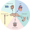
Fig. 1.
Fig. 1.
Regenerative or improved healing is observed in select tissues in neonatal mice. The recent literature shows that neonatal mice have the capacity to regenerate or display improve healing of multiple tissues that either fail to heal or heal by scar formation in adult mice. These tissues include the neonatal heart, cochlear hair cells, the Achilles tendon, the annulus fibrosus of the intervertebral disc, and the brain and spinal cord. Although not all of these neonatal tissues regenerate fully, all show distinctive mechanisms of healing compared with their adult counterparts.
Fig. 2.
Fig. 2.
Heart tissue is composed of specialized layers and cell types. Cardiomyocytes within the heart myocardium are the primary source of regenerative cells during neonatal cardiac healing. Other cell types within the heart include endothelial cells and cardiac fibroblasts; however, their potential functions during neonatal regeneration have not been studied.
Fig. 3.
Fig. 3.
Supporting cell types within the organ of Corti. Although hair cells of the cochlea do not regenerate, neonatal regeneration is possible via transdifferentiation of supporting cell types housed within the organ of Corti. These cells include inner phalangeal cells, inner piller cells, outer piller cells, Deiters' cells, Clausius cells and Hensen cells.
Fig. 4.
Fig. 4.
Factors associated with loss of regenerative potential during adult maturation. We propose that features associated with adult maturation (including loss of cells and cell potential, and a changing extracellular matrix) may be universal features underlying loss of neonatal regenerative potential.

Similar articles

Cited by

References

    1. Abbas, N., Perbellini, F. and Thum, T. (2020). Non-coding RNAs: emerging players in cardiomyocyte proliferation and cardiac regeneration. Basic Res. Cardiol. 115, 52. 10.1007/s00395-020-0816-0 - DOI - PMC - PubMed
    1. Abdul-Aziz, D., Hathiramani, N., Phung, L., Sykopetrites, V. and Edge, A. S. B. (2021). HIC1 represses Atoh1 transcription and hair cell differentiation in the cochlea. Stem Cell Rep. 16, 797-809. 10.1016/j.stemcr.2021.02.022 - DOI - PMC - PubMed
    1. Aguirre, A., Montserrat, N., Zacchigna, S., Nivet, E., Hishida, T., Krause, M. N., Kurian, L., Ocampo, A., Vazquez-Ferrer, E., Rodriguez-Esteban, C.et al. (2014). In vivo activation of a conserved MicroRNA program induces mammalian heart regeneration. Cell Stem Cell 15, 589-604. 10.1016/j.stem.2014.10.003 - DOI - PMC - PubMed
    1. Andersen, D. C., Ganesalingam, S., Jensen, C. H. and Sheikh, S. P. (2014). Do neonatal mouse hearts regenerate following heart apex resection? Stem Cell Rep. 2, 406-413. 10.1016/j.stemcr.2014.02.008 - DOI - PMC - PubMed
    1. Arvind, V., Howell, K. and Huang, A. H. (2021). Reprogramming adult tendon healing using regenerative neonatal regulatory T cells. biorXiv. 10.1101/2021.05.12.443424 - DOI

Publication types