The effect of exercise intensity and cardiorespiratory fitness on the kinetic response of middle cerebral artery blood velocity during exercise in healthy adults
- PMID: 35708705
- PMCID: PMC9291408
- DOI: 10.1152/japplphysiol.00862.2021
The effect of exercise intensity and cardiorespiratory fitness on the kinetic response of middle cerebral artery blood velocity during exercise in healthy adults
Abstract
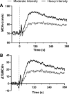
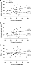
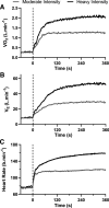
The aim of this study was to compare the kinetic response of middle cerebral artery blood velocity (MCAv) to moderate- and heavy-intensity cycling in adults, and explore the relationship between maximal oxygen uptake (V̇o2max) and MCAv kinetics. Seventeen healthy adults (23.8 ± 2.4 yr, 9 females) completed a ramp incremental test to exhaustion on a cycle ergometer to determine V̇o2max and the gas exchange threshold (GET). Across six separate visits, participants completed three 6-min transitions at a moderate intensity (90% GET) and three at a heavy intensity (40% of the difference between GET and V̇o2max). Bilateral MCAv was measured using transcranial Doppler (TCD) ultrasonography and analyzed using a monoexponential model with a time delay. The time constant (τ) of the MCAv response was not different between moderate- and heavy-intensity cycling (25 ± 10 vs. 26 ± 8 s, P = 0.82), as was the time delay (29 ± 11 vs. 29 ± 10 s, P = 0.95). The amplitude of the exponential increase in MCAv from baseline was greater during heavy-intensity cycling (23.9 ± 10.0 cm·s-1, 34.1 ± 14.4%) compared with moderate-intensity cycling (12.7 ± 4.4 cm·s-1, 18.7 ± 7.5%; P < 0.01). Following the exponential increase, a greater fall in MCAv was observed during heavy-intensity exercise compared with moderate-intensity exercise (9.5 ± 6.9 vs. 2.8 ± 3.8 cm·s-1, P < 0.01). MCAv after 6 min of exercise remained elevated during heavy-intensity exercise compared with moderate-intensity exercise (85.2 ± 9.6 vs. 79.3 ± 7.7 cm·s-1, P ≤ 0.01). V̇o2max was not correlated with MCAv τ or amplitude (r = 0.11-0.26, P > 0.05). These data suggest that the intensity of constant-work rate exercise influences the amplitude, but not time-based, response parameters of MCAv in healthy adults, and found no relationship between cardiorespiratory fitness and MCAv kinetics.NEW & NOTEWORTHY This is the first study to model the MCAv kinetic response to moderate- and heavy-intensity cycling in healthy adults. This study found that the amplitude of the exponential rise in MCAv at exercise onset was greater during heavy-intensity exercise (∼34%) compared with moderate-intensity exercise (∼19%), but the time-based characteristics of the responses were similar between intensities. Higher cardiorespiratory fitness was not associated with a greater or faster MCAv response to moderate- or heavy-intensity exercise.
Keywords: cerebral blood flow; exercise; kinetics.
Conflict of interest statement
No conflicts of interest, financial or otherwise, are declared by the authors.
Figures


References
Publication types
MeSH terms
LinkOut - more resources
Full Text Sources

