. 2022 Jun 16;7(14):e158571.
doi: 10.1172/jci.insight.158571.

JAML promotes acute kidney injury mainly through a macrophage-dependent mechanism

Affiliations

JAML promotes acute kidney injury mainly through a macrophage-dependent mechanism

Wei Huang et al. JCI Insight. .

Abstract

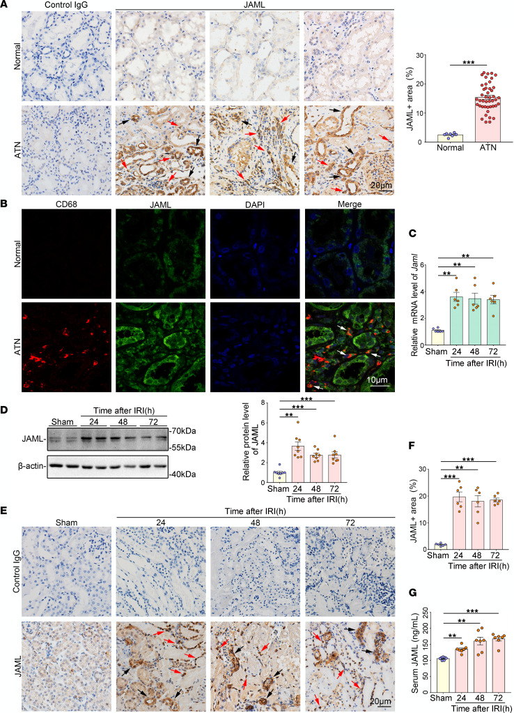
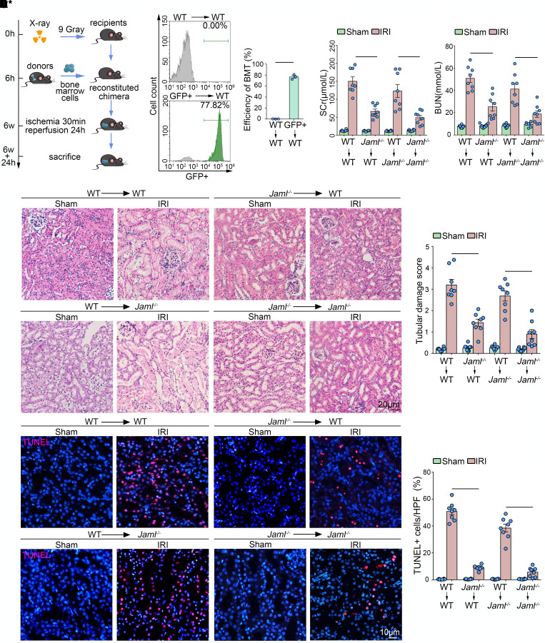
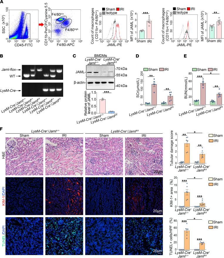
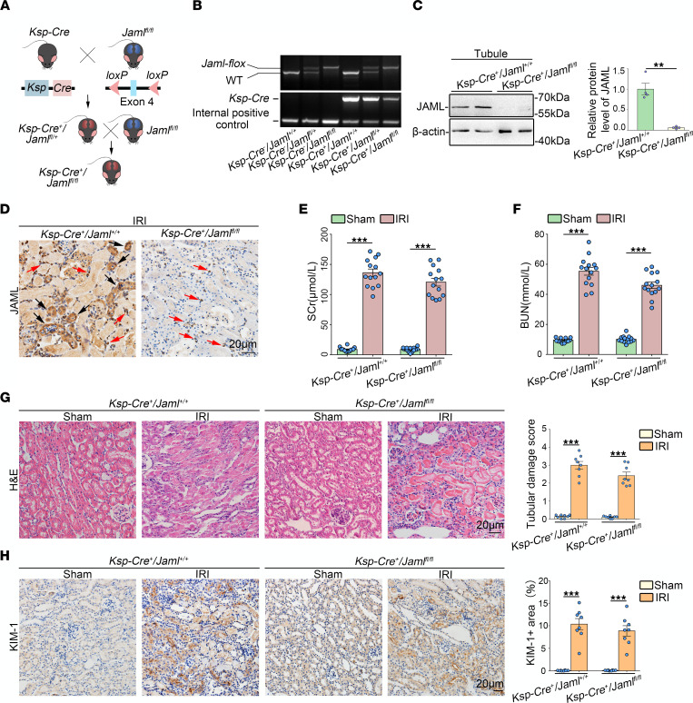
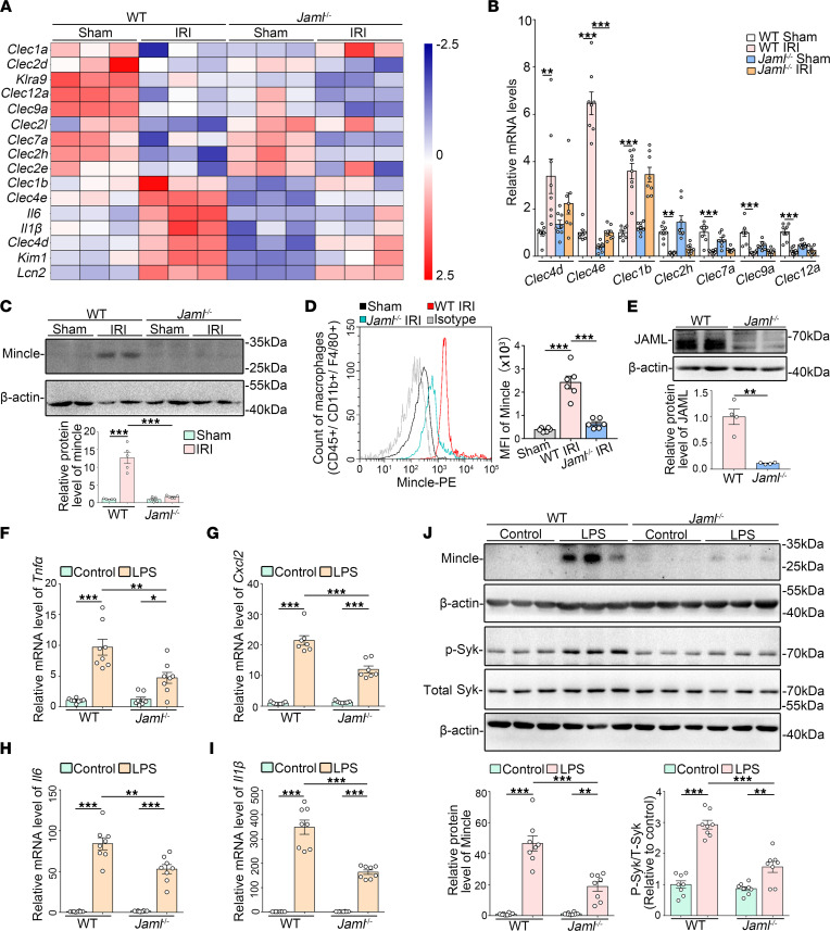
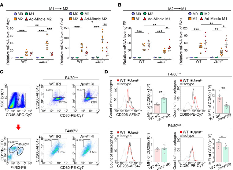
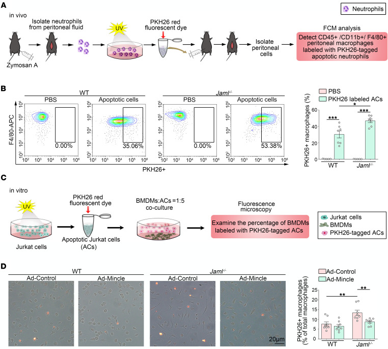
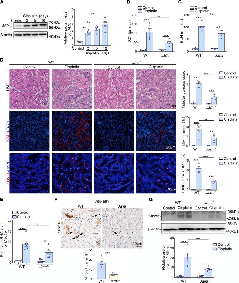
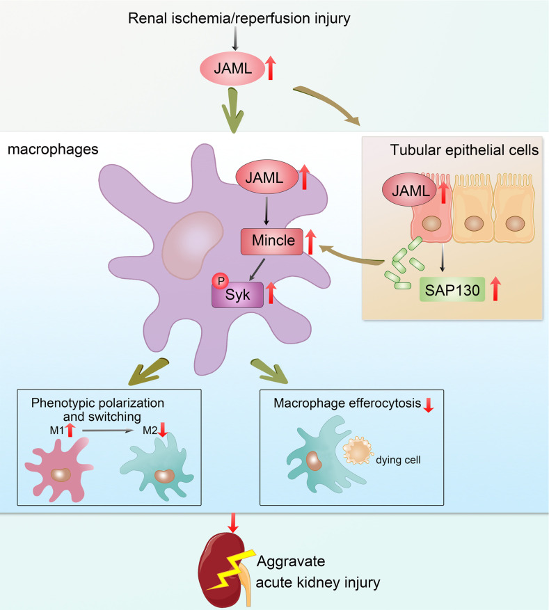
Although macrophages are undoubtedly attractive therapeutic targets for acute kidney injury (AKI) because of their critical roles in renal inflammation and repair, the underlying mechanisms of macrophage phenotype switching and efferocytosis in the regulation of inflammatory responses during AKI are still largely unclear. The present study elucidated the role of junctional adhesion molecule-like protein (JAML) in the pathogenesis of AKI. We found that JAML was significantly upregulated in kidneys from 2 different murine AKI models including renal ischemia/reperfusion injury (IRI) and cisplatin-induced AKI. By generation of bone marrow chimeric mice, macrophage-specific and tubular cell-specific Jaml conditional knockout mice, we demonstrated JAML promoted AKI mainly via a macrophage-dependent mechanism and found that JAML-mediated macrophage phenotype polarization and efferocytosis is one of the critical signal transduction pathways linking inflammatory responses to AKI. Mechanistically, the effects of JAML on the regulation of macrophages were, at least in part, associated with a macrophage-inducible C-type lectin-dependent mechanism. Collectively, our studies explore for the first time to our knowledge new biological functions of JAML in macrophages and conclude that JAML is an important mediator and biomarker of AKI. Pharmacological targeting of JAML-mediated signaling pathways at multiple levels may provide a novel therapeutic strategy for patients with AKI.

Keywords: Macrophages; Nephrology.


Conflict of interest statement

Conflict of interest: The authors have declared that no conflict of interest exists.

Figures

Figure 1
Figure 1. JAML is upregulated in kidneys from patients with AKI and mice with renal IRI.
(A) Representative IHC images and quantification of JAML in kidneys from human normal kidney poles (n = 7) and patients with biopsy-proven acute tubular necrosis (ATN) (n = 44). Red arrows indicate representative positive cells in renal interstitium; black arrows indicate renal parenchymal cells. Human kidneys stained with normal IgG in place of the corresponding primary antibodies were a negative control. Scale bar: 20 μm. (B) Representative fluorescence multiplexed IHC images of JAML (green) and CD68 (red) in kidneys from human normal kidney poles and patients with ATN (n = 6). Arrows indicate the expression of JAML in macrophages in renal interstitium. Scale bar: 10 μm. (C) Relative mRNA level of Jaml in kidneys from mice with renal IRI (n = 6). (D) Representative Western blot and quantifications of JAML expression in kidneys from mice with renal IRI (n = 8). (E) Representative IHC images and (F) quantification of JAML in kidneys from mice with renal IRI (n = 6). Red and black arrows indicate the same as in A. Scale bar: 20 μm. (G) Serum level of JAML in mice with renal IRI (n = 7). Data are shown as mean ± SEM. **P < 0.01, ***P < 0.001. Two-tailed Student’s unpaired t test (A), 1-way ANOVA test (C, D, F, and G). See complete unedited blots in the supplemental material.
Figure 2
Figure 2. JAML in BM-derived immune cells primarily contributes to renal IRI.
(A) A schematic diagram showing the procedure of BM transplantation for experimental mice. (B) Representative GFP expression profile of recipient BM cells 6 weeks after BM transplantation by flow cytometry analysis. The proportion of GFP+ cells in the reconstructed BM was about 80% (n = 3). (C) Serum creatinine (SCr) concentration in different groups of mice after renal IRI (n = 8). (D) Blood urea nitrogen (BUN) levels of different groups of mice after renal IRI (n = 8). (E) Representative images of H&E staining showing the morphology of kidney and quantitative assessment of tubular damage in the kidney from different groups of mice (n = 8). Scale bar: 20 μm. (F) In situ TUNEL assays and quantification were performed to assess renal cell death in the kidney from different groups of mice (n = 8). Scale bar: 10 μm. Data are mean ± SEM. *P < 0.05, **P < 0.01, ***P < 0.001. Two-tailed Student’s unpaired t test (B), 2-way ANOVA test (CF).
Figure 3
Figure 3. Macrophage JAML is required in the pathogenesis of IRI-induced renal injury.
(A) Flow cytometry analysis of macrophages freshly isolated from the kidney in mice with renal IRI. CD45-positive cells were divided into F4/80lo and F4/80hi macrophages. Representative histogram showing cell surface JAML expression on 2 subsets of macrophages and quantitative analysis of the MFI of JAML-phycoerythrin (n = 6). (B) Genotyping was confirmed by tail preparation and PCR at 2 weeks of age (n = 8). (C) Representative Western blot and quantifications of JAML expression in the BM-derived macrophages from LysM-Cre+ Jamlfl/fl mice (n = 3). (D) SCr concentration in different groups of mice after renal IRI (n = 5). (E) BUN levels of different groups of mice after renal IRI (n = 5). (F) Representative images of H&E staining and quantitative assessment of tubular damage in the kidney from different groups of mice (upper). Representative images and quantification of immunofluorescence staining of kidney injury molecule 1 (KIM-1) (red) (middle). In situ TUNEL assays and quantification were performed to assess renal cell death (lower) (n = 6). Scale bar: 20 μm. HPF, high power field. Data are mean ± SEM. *P < 0.05, **P < 0.01, ***P < 0.001. Two-tailed Student’s unpaired t test (A and C), 2-way ANOVA test (DF). See complete unedited blots in the supplemental material.
Figure 4
Figure 4. Tubule-specific JAML deletion in mice only slightly ameliorates renal IRI.
(A and B) Experimental scheme for generating conditional knockout mice in which Jaml is specifically ablated in renal tubular cells (Ksp-Cre+ Jamlfl/fl) by using Cre-loxP recombination system. Exon 4 is deleted upon Ksp-Cre–mediated recombination (A). Genotyping was confirmed by tail preparation and PCR at 2 weeks of age (n = 8) (B). (C) Representative Western blot and quantification of JAML expression in isolated tubules from Ksp-Cre+ Jaml+/+ and Ksp-Cre+ Jamlfl/fl mice (n = 4). (D) Representative IHC images of JAML in kidneys from Ksp-Cre+ Jaml+/+ and Ksp-Cre+ Jamlfl/fl mice with IRI (n = 5). Red arrows indicate representative JAML positive interstitial cells; black arrows indicate renal parenchymal cells. (E) SCr concentration in different groups of mice (n = 14). (F) BUN levels of different groups of mice (n = 14). (G) Representative images of H&E staining and quantitative assessment of tubular damage in the kidney from different groups of mice (n = 8). (H) Representative images and quantifications of IHC staining of KIM-1 in the kidney from different groups of mice (n = 8). Scale bar: 20 μm. Data are mean ± SEM. **P < 0.01, ***P < 0.001. Two-way ANOVA test (EH), 2-tailed Student’s unpaired t test analysis (C). See complete unedited blots in the supplemental material.
Figure 5
Figure 5. Upregulation of JAML facilitates macrophage-inducible C-type lectin signaling.
(A) Representative heatmap of gene expression levels in the kidney from different groups of mice with IRI by microarray analysis (n = 3). (B) Relative mRNA levels of C-type lectin members including Clec4d, Clec4e (macrophage-inducible C-type lectin, Mincle), Clec1b, Clec2h, Clec7a, Clec9a, and Clec12a in kidneys from different groups of mice (n = 8). (C) Representative Western blots and quantifications of Mincle in kidneys from different groups of mice (n = 5). (D) Representative flow cytometry histogram showing cell surface Mincle expression on macrophages freshly isolated from the kidney in different groups of mice and quantitative analysis of MFI of Mincle-phycoerythrin (n = 6). (E) Representative Western blot and quantifications of JAML expression in BMDMs from WT or Jaml–/– mice (n = 4). (FI) Relative mRNA levels of proinflammatory mediators including Tnfα (F), Cxcl2 (G), Il6 (H), and Il1β (I) in LPS-treated BMDMs (n = 8). (J) Representative Western blots and quantifications of Mincle and phosphorylated and total spleen tyrosine kinase (Syk) in different groups of BMDMs (n = 8). Data are mean ± SEM. *P < 0.05, **P < 0.01, ***P < 0.001. Two-tailed Student’s unpaired t test (E), 2-way ANOVA test (C, D, FJ). See complete unedited blots in the supplemental material.
Figure 6
Figure 6. JAML regulates macrophage phenotypic polarization via a Mincle-dependent mechanism.
(A and B) BMDMs from WT or Jaml–/– mice were serum starved for 4 hours (M0) and then polarized to M1 (LPS) or M2 (IL-4) for 8 hours. Media were removed, M1 macrophages were treated with M2 stimuli (IL-4), and M2 macrophages were treated with M1 stimuli (LPS) for an additional 8 hours. Real-time PCR was performed to measure Arg1 and Ccl8 expression in M1 macrophages polarized to M2 (A). Il6 and iNos gene expression was measured in M2 macrophages polarized to M1 (B) (n = 7). (C) Flow cytometry analysis of renal macrophages in the injured kidney after IRI. CD45-positive cells were divided into the F4/80lo and F4/80hi groups. Representative flow cytometry analysis of M1 (CD80hi) and M2 (CD206hi) cell populations in F4/80lo and F4/80hi macrophages isolated from the kidney in different groups of mice. SSC, side scatter. (D) Representative flow cytometry histogram showing cell surface marker CD206 (M2) and CD80 (M1) expression on 2 subtypes of macrophages and quantitative analysis of MFI of CD206-AF647 or CD80-PE-Cy7 (n = 6). Data are mean ± SEM. *P < 0.05, **P < 0.01, ***P < 0.001. Two-way ANOVA test (A and B), 2-tailed Student’s unpaired t test (D).
Figure 7
Figure 7. JAML regulates macrophage efferocytosis via a Mincle-dependent mechanism.
(A) Schematic diagrams showing the procedure of macrophage efferocytosis assay in vivo. FCM, flow cytometry. (B) Mice were injected intraperitoneally with PKH26-labeled apoptotic cells, and 45 minutes later lavage fluid was analyzed by flow cytometry for the percentage of F4/80+ macrophages that had incorporated the labeled neutrophils (n = 7). (C) Schematic diagrams showing the procedure of macrophage efferocytosis assay in vitro. (D) Overlay images of phase and fluorescence microscopy images of cultured BMDMs treated for 2 hours with UV-exposed PKH26-labeled Jurkat cells. Quantitative analysis of percentage of PKH26+ macrophages (n = 8). Data are mean ± SEM. *P < 0.05, **P < 0.01, ***P < 0.001. Two-way ANOVA test (B and D).
Figure 8
Figure 8. JAML deficiency protects against AKI induced by cisplatin in mice.
(A) Representative Western blot and quantifications of JAML expression in kidneys from different groups of mice (n = 5). (B and C) Levels of SCr (B) and BUN (C) in different groups of mice (n = 5). (D) Representative images of H&E staining showing the morphology of kidneys and quantitative assessment of tubular damage (n = 6). Scale bar: 20 μm. Representative images and quantifications of immunofluorescence staining of KIM-1 (red) in kidneys from different groups of mice (n = 6). Scale bar: 20 μm. In situ TUNEL assays and quantification were performed to assess renal cell death (n = 6). Scale bar: 10 μm. (E) Relative mRNA levels of Clec4e in kidneys from different groups of mice (n = 6). (F) Representative IHC images and quantification of Mincle in kidneys from different groups of mice. Arrows indicate macrophages that express Mincle (n = 6). Scale bar: 20 μm. (G) Representative Western blot and quantifications of Mincle expression in kidneys from different groups of mice (n = 6). Data are mean ± SEM. *P < 0.05, **P < 0.01, ***P < 0.001. One-way ANOVA test (A), 2-way ANOVA test (BG). See complete unedited blots in the supplemental material.
Figure 9
Figure 9. Schematic depicting that JAML promotes AKI mainly through regulating macrophage polarization and efferocytosis via C-type lectin Mincle.

References

    1. Ronco C, et al. Acute kidney injury. Lancet. 2019;394(10212):1949–1964. doi: 10.1016/S0140-6736(19)32563-2. - DOI - PubMed
    1. Yang L, et al. Acute kidney injury in China: a cross-sectional survey. Lancet. 2015;386(10002):1465–1471. doi: 10.1016/S0140-6736(15)00344-X. - DOI - PubMed
    1. Levey AS, James MT. Acute kidney injury. Ann Intern Med. 2017;167(9):ITC66–ITC80. doi: 10.7326/AITC201711070. - DOI - PubMed
    1. Rabb H, et al. Inflammation in AKI: current understanding, key questions, and knowledge gaps. J Am Soc Nephrol. 2016;27(2):371–379. doi: 10.1681/ASN.2015030261. - DOI - PMC - PubMed
    1. Jang HR, Rabb H. Immune cells in experimental acute kidney injury. Nat Rev Nephrol. 2015;11(2):88–101. doi: 10.1038/nrneph.2014.180. - DOI - PubMed

Publication types

Substances