. 2023 Jan 1;72(1):59-70.
doi: 10.2337/db21-1172.

Autoantibodies to Perilipin-1 Define a Subset of Acquired Generalized Lipodystrophy

Affiliations

Autoantibodies to Perilipin-1 Define a Subset of Acquired Generalized Lipodystrophy

Caleigh Mandel-Brehm et al. Diabetes. .

Abstract

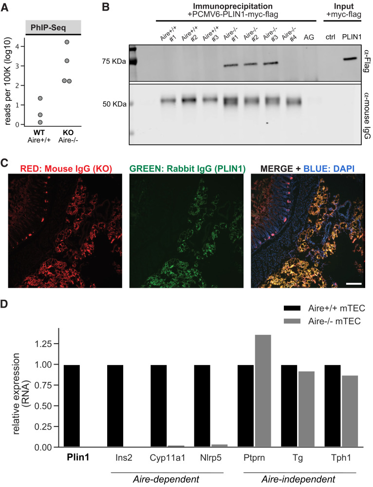
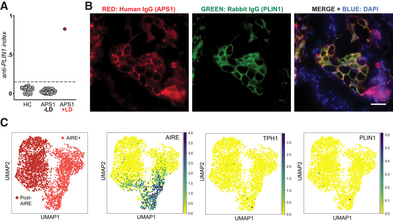
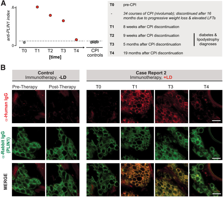
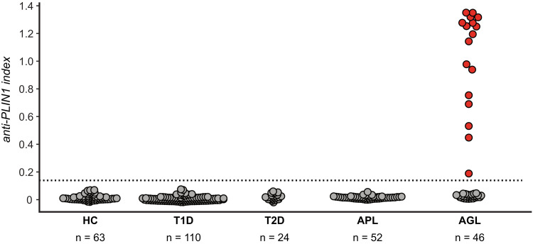
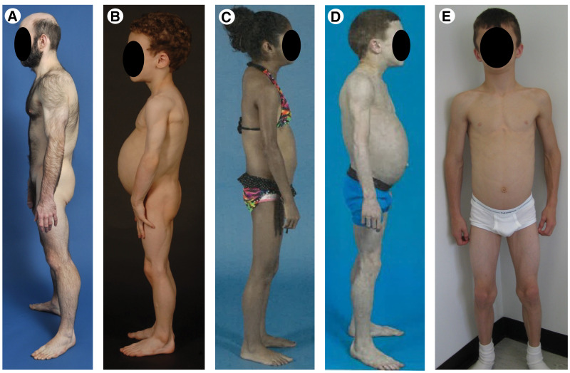
Acquired lipodystrophy is often characterized as an idiopathic subtype of lipodystrophy. Despite suspicion of an immune-mediated pathology, biomarkers such as autoantibodies are generally lacking. Here, we used an unbiased proteome-wide screening approach to identify autoantibodies to the adipocyte-specific lipid droplet protein perilipin 1 (PLIN1) in a murine model of autoimmune polyendocrine syndrome type 1 (APS1). We then tested for PLIN1 autoantibodies in human subjects with acquired lipodystrophy with two independent severe breaks in immune tolerance (including APS1) along with control subjects using a specific radioligand binding assay and indirect immunofluorescence on fat tissue. We identified autoantibodies to PLIN1 in these two cases, including the first reported case of APS1 with acquired lipodystrophy and a second patient who acquired lipodystrophy as an immune-related adverse event following cancer immunotherapy. Lastly, we also found PLIN1 autoantibodies to be specifically enriched in a subset of patients with acquired generalized lipodystrophy (17 of 46 [37%]), particularly those with panniculitis and other features of autoimmunity. These data lend additional support to new literature that suggests that PLIN1 autoantibodies represent a marker of acquired autoimmune lipodystrophies and further link them to a break in immune tolerance.


Figures

Figure 1
Figure 1
Discovery and validation of autoantibodies to perilipin 1 in Aire−/− mouse sera. A: PhIP-Seq analysis in Aire−/− and Aire+/+ mice. Aggregated Plin1 PhIP-Seq data from Aire−/− (n = 4) and Aire+/+ (n = 3) mice. B: Whole cell lysates generated from 293T cells expressing full-length mouse PLIN1 were incubated with sera from Aire−/− (n = 4) or Aire+/+ (n = 3) mice. Antibodies were immunoprecipitated with use of AG beads, and IP elutions were subject to SDS-PAGE immunoblotting. AG lane indicates an IP with use of protein A/G beads only—no sera. Input lane indicates loading of whole cell lysate with and without (−) transfection of PLIN1-myc-flag plasmid. IP elutions and input were immunostained with either anti-Flag IgG to identify positive anti-Plin1 signal, or anti-mouse IgG to show qualitative capture of IgG from sera. C: Representative image of immunohistochemistry on mouse omental adipose tissue showing positive colocalization of antibodies from Aire−/− sera and commercial anti-Plin1 IgG. Primary antibodies from Aire−/− mouse sera and commercial antibody to PLIN1 were visualized with secondaries to mouse IgG (Alexa Fluor 567 [red]) and rabbit IgG (Alexa Fluor 488 [green]), respectively. DAPI (blue) stains nuclei. Scale bar indicates 150 μm. D: Examination of RNA expression of Plin1 and genes with known Aire-dependent thymic expression using a publicly available murine thymic RNA sequencing data from Aire−/− and Aire+/+ mTECs (Sansom et al. [21]). ctrl, control; KO, knockout.
Figure 2
Figure 2
Autoantibodies to PLIN1 in sera from a patient with APS1 and acquired lipodystrophy. A: RLBA for detection of anti-PLIN1 antibodies. Radiolabeled PLIN1 protein was incubated with sera from healthy control subjects (HC) (n = 54) or from APS1 patients with (n = 1) or without (n = 68) lipodystrophy (LD). Dotted line indicates mean ± 3 SD healthy control subjects. B: Validation of autoantibodies to PLIN1 in orthogonal cell–based assay. Fixed stomach tissue of mice was mounted and immunostained with sera from case report patient 1 and commercial antibody to PLIN1. A mix of secondary antibodies anti-human IgG Alexa Fluor 547 and anti-rabbit Alexa Fluor 488 at 1:2,000 was used to visualize human IgG and PLIN1 antibodies, respectively. A merge is provided on the right of individual images. Images taken at ×20 magnification. Note yellow in merge indicating colocalization of PLIN1 (green) and human IgG (red). DAPI is blue and indicates nuclei. Scale bar indicates 100 μm. C: Examination of RNA expression of AIRE, PLIN1,and TPH1 genes in human mTECs using a publicly available human thymic RNA sequencing data set (Park et al. [16] and Bautista et al. [17]).
Figure 3
Figure 3
Autoantibodies to PLIN1 in sera from a patient with autoimmune AGL following cancer immunotherapy. A: RLBA screening for PLIN1 antibodies in sera from all time points from index patient 2 as well as checkpoint-treated control patients without lipodystrophy (n = 7), as done for case report 1. Dotted line indicates mean ± 3 SD healthy control subjects (n = 11). Numbers to the right of circles indicate the time point series, for reference in panel B. The left panel describes the clinical timeline corresponding to time points T0–T4. Checkpoint inhibitor therapy (CPI) was discontinued at 34 cycles (16 months) due to progressive weight loss and elevated liver function tests (LFTs), with further workup over subsequent months. B: Validation of autoantibodies to PLIN1 with use of immunohistochemistry on mouse omental adipose tissue, as in Fig. 2B. Sera were used from either a control subject (immunotherapy but no AGL) or case report 2 patient (immunotherapy with AGL), with various time points. Each column represents an individual sample; from left to right: sera from checkpoint control pretreatment, sera from checkpoint control posttreatment with no autoimmunity, case report 1 pretherapy, case report 1 posttherapy 1, case report 1 posttherapy 3, case report 1 posttherapy 4. Images are 600 × 600 pixel insets from original ×40 image. Scale bar represents 100 μm. LD, lipodystrophy.
Figure 4
Figure 4
Autoantibodies to PLIN1 are specific to the generalized subtype of acquired lipodystrophy. RLBA screening for PLIN1 antibodies in sera from healthy control subjects (HC) (n = 63) or individuals with either lipodystrophy (AGL n = 46, APL n = 52) or autoimmune metabolic disorders not involving lipodystrophy (type 1 diabetes [T1D] n = 110, type 2 diabetes [T2D] n = 24). Dotted line represents 3 SD above the mean from healthy control subjects. Red dots depict 17 of 46 (37%) AGL patients scoring positive for PLIN1 autoantibodies.
Figure 5
Figure 5
Clinical features of patients with AGL. A: A 20-year-old male with panniculitis diagnosed in early childhood. B: A 5-year-old male with hepatomegaly, hepatic steatosis, splenomegaly, diabetes, and celiac disease. C: A 12-year-old female with diabetes, dermatomyositis, hepatomegaly, and celiac disease. D: An 8-year-old male with panniculitis, fatty liver, hypothyroidism, and vitiligo. E: A 9-year-old male with IgA deficiency, hepatomegaly, and splenomegaly.

Comment in

References

    1. Garg A. Clinical review#: lipodystrophies: genetic and acquired body fat disorders. J Clin Endocrinol Metab 2011;96:3313–3325 - PMC - PubMed
    1. Araújo-Vilar D, Santini F. Diagnosis and treatment of lipodystrophy: a step-by-step approach. J Endocrinol Invest 2019;42:61–73 - PMC - PubMed
    1. Özen S, Akıncı B, Oral EA. Current diagnosis, treatment and clinical challenges in the management of lipodystrophy syndromes in children and young people. J Clin Res Pediatr Endocrinol 2020;12:17–28 - PMC - PubMed
    1. Misra A, Peethambaram A, Garg A. Clinical features and metabolic and autoimmune derangements in acquired partial lipodystrophy: report of 35 cases and review of the literature. Medicine (Baltimore) 2004;83:18–34 - PubMed
    1. Pope E, Janson A, Khambalia A, Feldman B. Childhood acquired lipodystrophy: a retrospective study. J Am Acad Dermatol 2006;55:947–950 - PubMed

Publication types