. 2022 Jun 16;17(6):e0269499.
doi: 10.1371/journal.pone.0269499. eCollection 2022.

Evaluation of perfusion-driven cell seeding of small diameter engineered tissue vascular grafts with a custom-designed seed-and-culture bioreactor

Affiliations

Evaluation of perfusion-driven cell seeding of small diameter engineered tissue vascular grafts with a custom-designed seed-and-culture bioreactor

Sarah K Saunders et al. PLoS One. .

Abstract

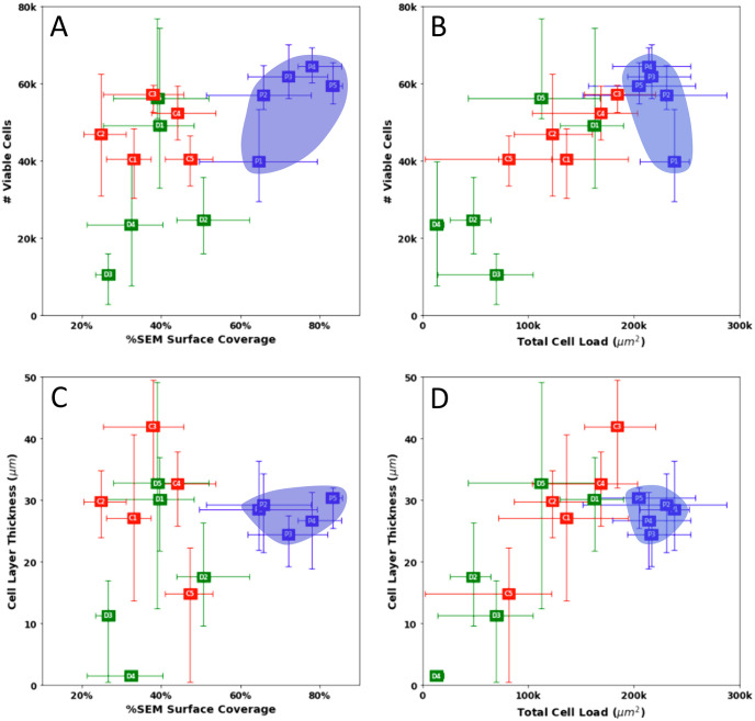
Tissue engineering commonly entails combining autologous cell sources with biocompatible scaffolds for the replacement of damaged tissues in the body. Scaffolds provide functional support while also providing an ideal environment for the growth of new tissues until host integration is complete. To expedite tissue development, cells need to be distributed evenly within the scaffold. For scaffolds with a small diameter tubular geometry, like those used for vascular tissue engineering, seeding cells evenly along the luminal surface can be especially challenging. Perfusion-based cell seeding methods have been shown to promote increased uniformity in initial cell distribution onto porous scaffolds for a variety of tissue engineering applications. We investigate the seeding efficiency of a custom-designed perfusion-based seed-and-culture bioreactor through comparisons to a static injection counterpart method and a more traditional drip seeding method. Murine vascular smooth muscle cells were seeded onto porous tubular electrospun polycaprolactone scaffolds, 2 mm in diameter and 30 mm in length, using the three methods, and allowed to rest for 24 hours. Once harvested, scaffolds were evaluated longitudinally and circumferentially to assess the presence of viable cells using alamarBlue and live/dead cell assays and their distribution with immunohistochemistry and scanning electron microscopy. On average, bioreactor-mediated perfusion seeding achieved 35% more luminal surface coverage when compared to static methods. Viability assessment demonstrated that the total number of viable cells achieved across methods was comparable with slight advantage to the bioreactor-mediated perfusion-seeding method. The method described is a simple, low-cost method to consistently obtain even distribution of seeded cells onto the luminal surfaces of small diameter tubular scaffolds.


Conflict of interest statement

The authors have declared that no competing interests exist.

Figures

Fig 1
Fig 1. Bioreactor design.
(A) The fully assembled bioreactor chamber. Intake and outtake tube holders are found on each side of the individual ETVG chambers. (B) An electrospun PCL tubular scaffold mounted with collet chucks in a four-pronged stainless-steel bracket. The 2mm cannulas affixed to either end of the scaffold form a tight seal with the tightened collet chucks. This connection makes tensile stretch and luminal pressurization possible while also leaving the lumen of the ETVG in direct contact with media flow. (C) Flow diagram within the bioreactor chamber. The red arrow shows media traveling from a reservoir to the ETVG lumen, the green arrow shows media movement out of the ETVG, out of the bioreactor, through the pinch valve, and back into the bioreactor. The blue arrow represents media being drawn from the bioreactor chamber to return to the reservoir. (D) Full schematic of bioreactor flow loop and intended features. Color coded as in panel C.
Fig 2
Fig 2. Seeding methodology.
Seeding method diagrams for (A) the bioreactor mediated perfusion seeding method, (B) static injection counterpart seeding method and (C) drip seeding. (D) Operating principle of bioreactor mediated perfusion seeding method. Numbers indicate sample organization for analysis.
Fig 3
Fig 3. AlamarBlue viability assay results.
Section average viable cell numbers for (A) perfusion bioreactor seeding, (B) static counterpart seeding, and (C) drip seeding. (n = 4; mean with 95% confidence intervals).
Fig 4
Fig 4. Spatial live/dead cell imaging.
The middle section of a perfusion seeded scaffold is represented in 3rds. The left most portion is the top right edge of the scaffold, the middle portion is the bottom of the scaffold, and the right most portion is the top left side of the scaffold. Each section length was approximately 5 mm, and its circumference was 6.28 mm.
Fig 5
Fig 5. H&E cross-sections.
Representative haematoxylin and eosin (H&E) stained cross sections of each seeding method 24 hrs after seeding in longitudinal order.
Fig 6
Fig 6. DAPI cross-sections.
DAPI stained nuclei of cells in cross sections of each seeding method 24 hrs after seeding in longitudinal order.
Fig 7
Fig 7. Quantitative assessment of uniformity.
Quantitative analysis of H&E images for bioreactor mediated perfusion and static injection counterpart seeding methods preformed with rVSMCs onto electrospun PCL scaffolds. Uniformity judged in terms of total cell load, average cell layer thickness, and % circumferential coverage per section. (n = 20, n = 15; mean ± standard deviation).
Fig 8
Fig 8. Interpretation of SEM images.
(A) Representative SEM images of luminal surface of seeded electrospun PCL scaffolds for each seeding method, (B) Sample images representing quantitative analysis of luminal surface coverage per method. (C) Zoomed in view of perfusion seeded scaffold.
Fig 9
Fig 9. Quantitative analysis of surface coverage.
Color map of surface coverage and section averages respectively for (A,B) perfusion bioreactor seeding, (C,D) static counterpart seeding, and (E,F) drip seeding (n = 36; mean with 95% confidence intervals).
Fig 10
Fig 10. Scatter plots of combined data.
The top left graph compares the average number of viable cells present on each scaffold section to the surface coverage determined with SEM analysis. The top right graph compares the number of viable cells and the cell load area as determined by H&E analysis. The bottom left graph compares the average cell layer thickness to SEM surface coverage and the bottom right graph compares cell layer thickness to cell load. Bioreactor mediated perfusion data is highlighted in blue.

References

    1. Mensah GA, Brown DW. An overview of cardiovascular disease burden in the United States. Health Aff. 2007;26(1):38–48. doi: 10.1377/hlthaff.26.1.38 - DOI - PubMed
    1. Conklin BS, Richter ER, Kreutziger KL, Zhong DS, Chen C. Development and evaluation of a novel decellularized vascular xenograft. Med Eng Phys. 2002;24(3):173–83. doi: 10.1016/s1350-4533(02)00010-3 - DOI - PubMed
    1. Dean EW, Udelsman B, Breuer CK. Current Advances in the Translation of Vascular Tissue Engineering to the Treatment of Pediatric Congenital Heart Disease. Yale J Biol Med. 2012;85(2):229–38. - PMC - PubMed
    1. Soletti L, Nieponice A, Guan J, Stankus JJ, Wagner WR, Vorp DA. A seeding device for tissue engineered tubular structures. Biomaterials. 2006;27(28):4863–70. doi: 10.1016/j.biomaterials.2006.04.042 - DOI - PubMed
    1. Tara S, Rocco KA, Hibino N, Sugiura T, Kurobe H, Breuer CK, et al.. Vessel bioengineering—Development of small-diameter arterial grafts -. Circ J. 2014;78(1):12–9. doi: 10.1253/circj.cj-13-1440 - DOI - PubMed

Publication types