. 2022 Jun 16;18(6):e1010588.
doi: 10.1371/journal.ppat.1010588. eCollection 2022 Jun.

RANBP2 and USP9x regulate nuclear import of adenovirus minor coat protein IIIa

Affiliations

RANBP2 and USP9x regulate nuclear import of adenovirus minor coat protein IIIa

Ashrafali M Ismail et al. PLoS Pathog. .

Abstract

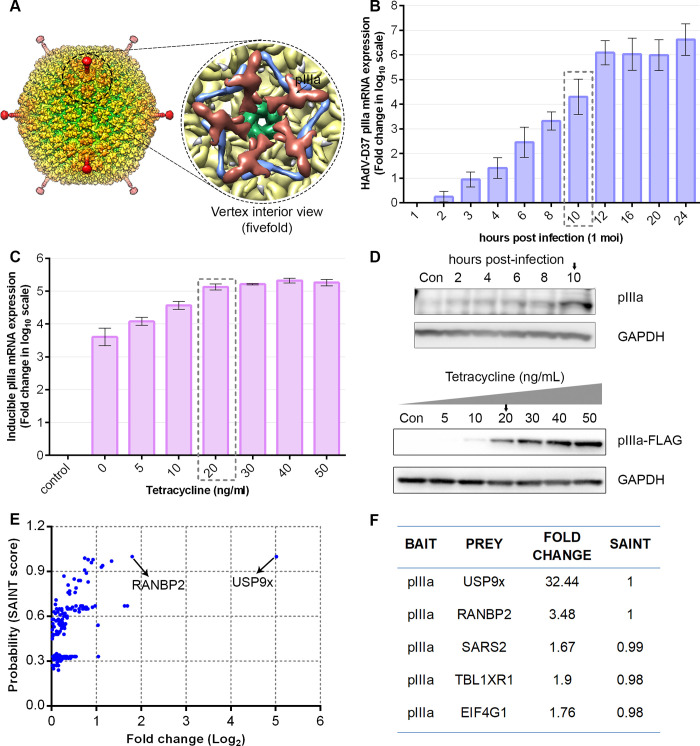
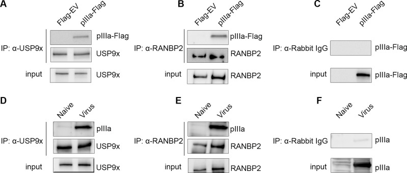
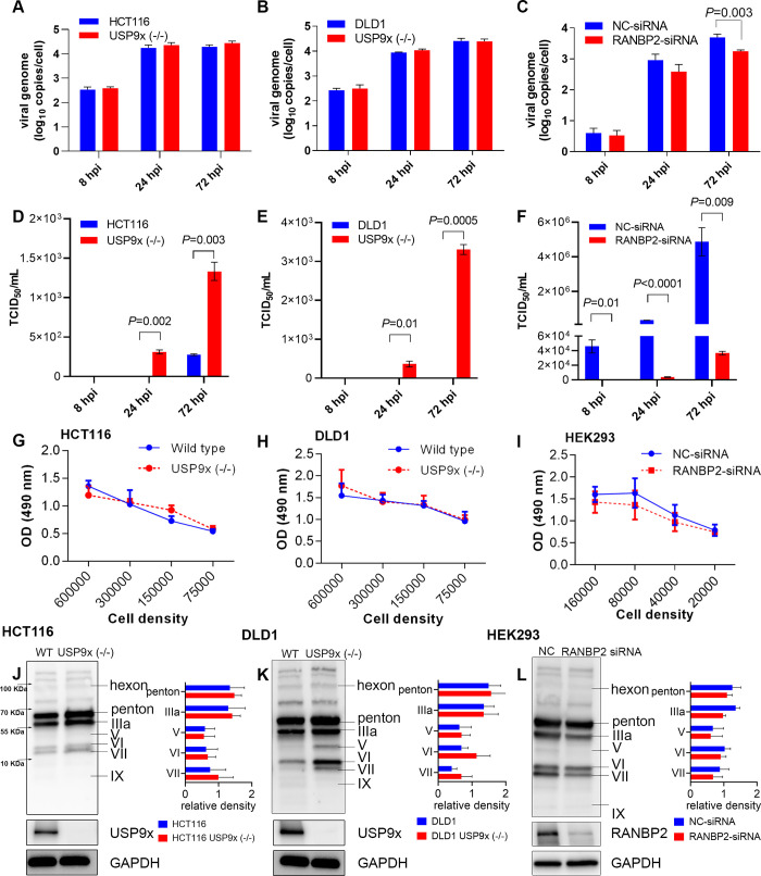
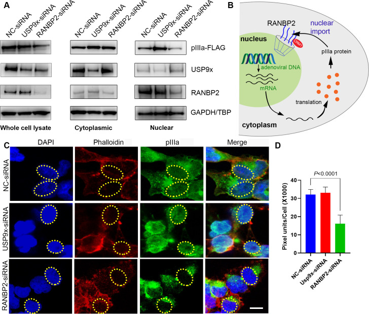
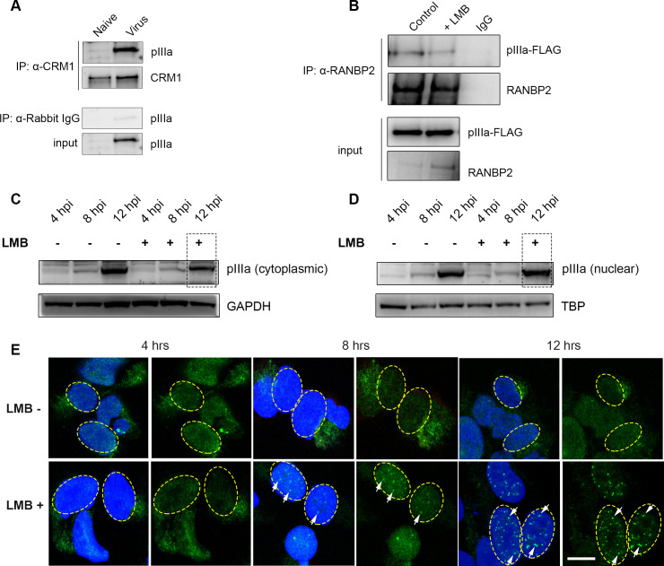
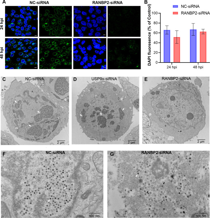
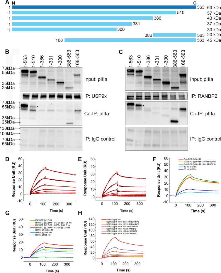
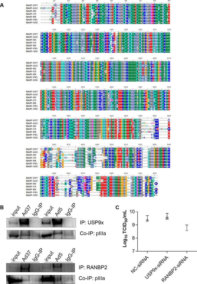
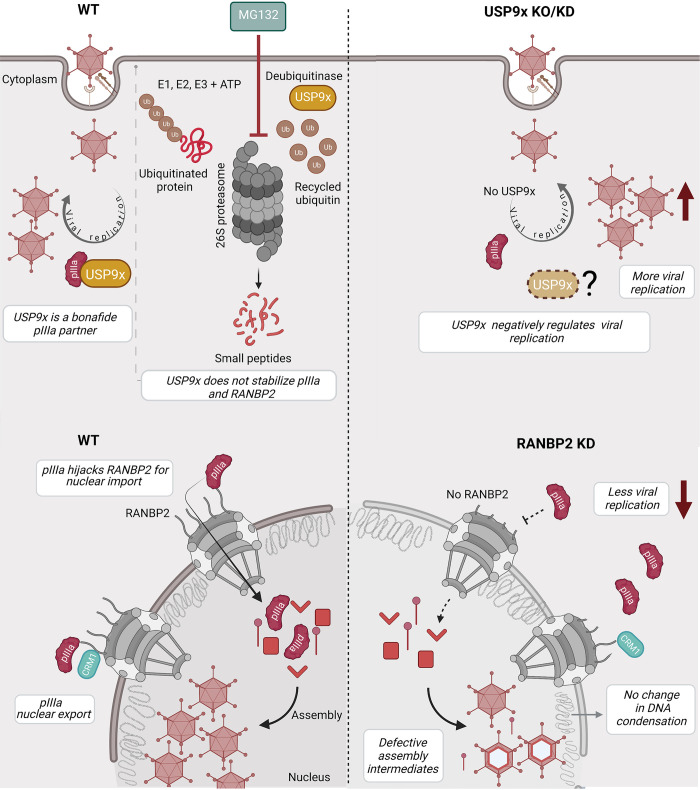
As intracellular parasites, viruses exploit cellular proteins at every stage of infection. Adenovirus outbreaks are associated with severe acute respiratory illnesses and conjunctivitis, with no specific antiviral therapy available. An adenoviral vaccine based on human adenovirus species D (HAdV-D) is currently in use for COVID-19. Herein, we investigate host interactions of HAdV-D type 37 (HAdV-D37) protein IIIa (pIIIa), identified by affinity purification and mass spectrometry (AP-MS) screens. We demonstrate that viral pIIIa interacts with ubiquitin-specific protease 9x (USP9x) and Ran-binding protein 2 (RANBP2). USP9x binding did not invoke its signature deubiquitination function but rather deregulated pIIIa-RANBP2 interactions. In USP9x-knockout cells, viral genome replication and viral protein expression increased compared to wild type cells, supporting a host-favored mechanism for USP9x. Conversely, RANBP2-knock down reduced pIIIa transport to the nucleus, viral genome replication, and viral protein expression. Also, RANBP2-siRNA pretreated cells appeared to contain fewer mature viral particles. Transmission electron microscopy of USP9x-siRNA pretreated, virus-infected cells revealed larger than typical paracrystalline viral arrays. RANBP2-siRNA pretreatment led to the accumulation of defective assembly products at an early maturation stage. CRM1 nuclear export blockade by leptomycin B led to the retention of pIIIa within cell nuclei and hindered pIIIa-RANBP2 interactions. In-vitro binding analyses indicated that USP9x and RANBP2 bind to C-terminus of pIIIa amino acids 386-563 and 386-510, respectively. Surface plasmon resonance testing showed direct pIIIa interaction with recombinant USP9x and RANBP2 proteins, without competition. Using an alternative and genetically disparate adenovirus type (HAdV-C5), we show that the demonstrated pIIIa interaction is also important for a severe respiratory pathogen. Together, our results suggest that pIIIa hijacks RANBP2 for nuclear import and subsequent virion assembly. USP9x counteracts this interaction and negatively regulates virion synthesis. This analysis extends the scope of known adenovirus-host interactions and has potential implications in designing new antiviral therapeutics.


Conflict of interest statement

The authors have declared that no competing interests exist.

Figures

Fig 1
Fig 1. Human adenovirus (HAdV) pIIIa expression and affinity purification-MS/MS analysis.
(A) Surface representation of HAdV-D26 (PDB: 5TX1) colored by radius [33] with expanded zoom into the vertex interior view, rendered using UCSF Chimera v1.14. Protein IIIa (pIIIa) (brick red), penton base (green), peripentonal hexons (pale gold), and protein VIII (purple). (B) HAdV-D37 pIIIa mRNA expression levels in HEK293 cells at multiplicity of infection (MOI) of 1 through 24 hrs post infection (hpi). (B and C) Viral pIIIa mRNA expression levels seen in virus-infected cells and stable pIIIa expression in Flp-In 293 T-REx cell lines with tetracycline induction. B and C data shown represents the mean ± standard deviation. (D) Western blot showing pIIIa expression in HEK293 cells infected at an MOI of 1 (upper panel), and pIIIa-Flag protein expression in Flp-In 293 T-Rex cells upon tetracycline induction (lower panel). (E) pIIIa-FLAG and FLAG-control immunoprecipitated protein complexes were TCA precipitated, and LC-MS/MS performed. The obtained mass spectrometry protein interactions analyzed using the CRAPome database identified two high confidence interactors: USP9x and RANBP2. (F) Compared to the experimental and database controls, further analysis of FLAG-pIIIa interactions showed a 32.4 and 3.5 fold change (FC) difference for USP9x and RANBP2, respectively, with a SAINT probability score 1 (100%). Data for B to F were obtained from at least 3 replicate experiments.
Fig 2
Fig 2. Reciprocal co-immunoprecipitation validation of pIIIa-host protein interactions.
(A, B) Viral pIIIa expressing Flp-In 293 T-Rex and empty vector control cell extracts subjected to co-immunoprecipitation assay. (D, E) HAdV-D37 infected HEK293 cells (MOI of 1, 15 hpi), and naïve control cell lysates were immunoprecipitated with anti-USP9x and anti-RANBP2 antibodies. Blots were developed with α-USP9x, α-RANBP2, α-pIIIa-Flag, and α-pIIIa (B) as indicated. Immunoprecipitations with IgG controls (C, F) are shown for both virus infected and inducible systems. Data is representative of 3 biological replicates.
Fig 3
Fig 3. USP9x has no role in pIIIa or RANBP2 deubiquitination but deregulates their expression.
(A) Flp-In 293 T-Rex cells were treated with negative control (NC) or USP9x-siRNA for 48 hours. Following MG132 proteasome inhibitor treatment (10 μmol/L for 4 hrs), pIIIa expression was induced by tetracycline (20 ng/mL for 8hrs). GAPDH control is shown for each set. (B) HEK293 cells were treated with negative control (NC) and USP9x-siRNA for 48 hours. Following MG132 proteasome inhibitor treatment (10 μmol/L for 4 hrs), cells were infected with HAdV-D37 at an MOI of 1 for 4 hrs. USP9x-siRNA treated cells showed increased pIIIa and RANBP2 expressions compared to NC-siRNA treated cells, irrespective of MG132 treatment. (C) HCT116 wild-type and (D) HCT116 USP9x (-/-) cells were treated with MG132 and infected (MOI of 5) for 4 and 8 hrs, respectively. Neither condition significantly stabilized pIIIa or RANBP2 steady state concentrations. The final data are presented as the mean ± SD of at least triplicate experiments. Statistical significance was performed with unpaired t-test (two-tailed). Only comparisons where P<0.05 are shown.
Fig 4
Fig 4. Human adenovirus replication and protein expression in USP9x knockout and RANBP2-siRNA knockdown cells.
Quantification of virus genome copies/cell (normalized to ACTG). at an MOI of 5 at 8, 24, and 72 hours post-infection (hpi) in (A) HCT116 and (B) DLD1 wild type cells and their USP9x knockouts (-/-), each with deletions of exons 7 and 8 [87] (C) HEK293 cells were treated with NC-siRNA and RANBP-siRNA and infected at an MOI of 1 for 8, 24, and 72 hrs. Viral DNA replication was determined by q-PCR using HAdV-D37 E1A and human ACTG gene quantitation standards. Viral titers were measured in (D) HCT116 and (E) DLD1 wild type and USP9x (-/-) cells, and (F) in HEK293 cells pretreated with NC-siRNA or RANBP2 siRNA for 48 hrs, and HAdV-D37 infected at an MOI of 1 for 8, 24, and 72 hrs, using a TCID50/mL assay for measuring the viral titer. MTS cell proliferation assay: (G) HCT116 cells, (H) DLD1 cells, and their USP9x knockouts, and (I) HEK293 cells treated with negative control (NC) or RANBP2-siRNA for 48 hours, were seeded at different densities in 96 well plates in triplicate wells and incubated for 24 hours. Spectrometric readings were taken following MTS reagent addition and incubation for 3 hours. Western blots using a pan-adenovirus antibody show relative viral protein expression levels in wild type and USP9x (-/-) or RANBP2-siRNA knockdown cells: (J) HCT116 cells infected at an MOI of 5, (K) DLD1 cells infected at an MOI of 5, and (L) HEK293 cells infected at an MOI of 1, all at 72 hpi. Molecular weight markers are indicated in (J). The lower blots show USP9x -/- and RANBP2-siRNA knockdown efficiency after 72 hpi with GAPDH controls. WT: wild type. The final data are presented as the mean ± SD of at least triplicate experiments. Statistical significance was performed with unpaired t-test (two-tailed). Only comparisons where P<0.05 are shown.
Fig 5
Fig 5. Human adenovirus pIIIa exploits RANBP2 nuclear import function.
(A) Cytoplasmic and nuclear fractions of stable pIIIa expressing Flp-In 293 T-Rex cells treated with USP9x or RANBP2 siRNA for 48 hours, and then induced with tetracycline (20 ng/μL), and blotted for, pIIIa-Flag, RANBP2, USP9x and GAPDH protein expression (B) Schematic of the role of pIIIa-RANBP2 interaction in the adenovirus replication cycle. (C) Following Flp-In 293 T-Rex cells pretreated with USP9x or RANBP2-siRNA were tetracycline induced (20ng/mL) for 8 hrs for pIIIa expression. FLAG-pIIIa (green), phalloidin (red), and nuclei (blue) The dotted ellipse corresponds to the nucleus area. Scale bar: 10 μm. (D) ImageJ analysis done on 30 cells per group and green fluorescence intensity was measured in the nucleus of each cell. The final data are presented as the mean ± SD of at least triplicate experiments. Statistical significance was performed with two-way ANOVA followed by Tukey multiple comparison test. Only comparisons where P<0.05 are shown.
Fig 6
Fig 6. CRM1 nuclear export signal blocking retains human adenovirus pIIIa in the nucleus.
(A) Immunoprecipitation of CRM1 in infected HEK293 cells shows pIIIa interaction with CRM1. (B) Viral pIIIa protein expression after infection of cells pretreated with 20 nmol/L leptomycin B (LMB) for 4 hrs, a specific inhibitor of CRM1 nuclear export signal, or methanol control. Cell lysates were immunoprecipitated with α-RANBP2 and immunoblot developed with α-pIIIa-FLAG antibodies. (C) Cytoplasmic and nuclear extract of extracts of HEK293 cells infected with HAdV-D37 at an MOI of 1 for 4, 8, and 12 hours post-infection (hpi), and treated with LMB or methanol control for 4 hours. GAPDH and TATA-binding proteins served as load controls for cytoplasmic and nuclear fractions, respectively. (E) Confocal images of Flp-In 293 TRex cells induced with tetracycline (20ng/mL) for 8 hrs, and treated with LMB (20 nmol/L) for 4 hrs or methanol control. DAPI nuclear (blue), pIIIa-Flag (green). Scale bar: 10 μm. These experiments were repeated at least 3 times.
Fig 7
Fig 7. Human adenovirus interacts with RANBP2 for nuclear import and viral assembly.
(A) DAPI nuclear (blue) and RANBP2 (green) staining of NC-siRNA and RANBP2-siRNA treated HEK293 cells, infected with HAdV-D37 at an MOI of 0.1 for 24 and 48 hrs, respectively. Scale bar = 10 μm. (B) ImageJ quantification of DAPI fluorescence per cell, normalized to uninfected control cells (n = 100) and plotted as the percent of uninfected control fluorescence. (C) Transmission electron microscopy of NC-siRNA and (D) USP9x siRNA, and (E) RANBP2-siRNA treated HEK293 cells infected with HAdV-D37 at an MOI of 0.1 for 72 hrs. The specific MOI and time-points were chosen to establish viral replication while minimizing early cell death; HAdV-D37 infection at an MOI of ≥1 in HEK293 cells leads to significant cytopathic effect within 24 hpi. White arrows over the nuclei show large paracrystalline viral arrays. Higher magnifications are shown for NC-siRNA (F) and RANBP2-siRNA (G) treated cells. The final data in (B) are presented as the mean ± SD of triplicate experiments. Statistical significance was performed with unpaired t-test (two-tailed). No statistically significant differences were seen.
Fig 8
Fig 8. USP9x and RANBP2 bind to different sites of C-terminal pIIIa domain.
(A) Full-length pIIIa (1–563 aa; HAdV-D37, per GenBank: DQ900900) and six partial pIIIa deletion mutants were constructed in a pCDNA 3.1 vector backbone. After transfection of HEK293 cells with the pIIIa constructs, immunoprecipitations were performed using antibodies against USP9x (B) and RANBP2 (C). Both USP9x and RANBP2 interacted with full-length pIIIa (1–563 aa), and pIIIa fragments 386–563 aa, and 168–563 aa. The pIIIa coprecipitation was weak for fragment 1–510 (asterisk: *). (D) SPR sensorgrams of USP9x with pIIIa. Black curves are the sensorgrams and red curves are the fitted cycle. Two-fold series of dilutions of USP9X ranging from 7.8 nM to 250 nM were injected with a 2 min injection time and 230s dissociation time. The affinity was KD = 32.01±4.92 nM, Kon = 3.717x10⁴ ±120 (1/Ms) Koff = 1.19x 10⁻³±5.9x10⁻⁶ (1/s). (E) SPR sensorgrams of RANBP2 with pIIIa. Two-fold dilutions of RANBP2 ranging from 1.79nM to 57.5 nM were injected with a 2 min injection time and 230s dissociation time. The affinity was KD = 13.69±9.75nM, Kon = 4.803x10⁴±400 (1/Ms) Koff = 6.576x10⁻⁴±3.9x10⁻⁶ (1/s). (F-H) Competition assay using a fixed concentration of RANBP2 with increasing concentrations of USP9x. (F) RANBP2 was fixed at 80nM. Sensorgrams were collected with RANBP2 alone, RANBP2 with 40 nM USP9x, RANBP2 with 80 nM USP9x, 40 nM USP9x alone, 80 nM USP9x alone (G) RANBP2 was fixed at 2.5nM. Sensorgrams were collected with RANBP2 alone, RANBP2 with 31.25 nM USP9x, RANBP2 with 62.5 nM USP9x, RANBP2 with 125 nM USP9x, 31.25 nM USP9x alone, 62.5 nM USP9x alone and 125 nM USP9x alone. (H) USP9x was fixed at 40 nM and data was collected with two-fold dilutions of RANBP2 ranging from 9.75 to 300 nM. Data was fitted using heterogeneous analytes model by BIAevaluation software.
Fig 9
Fig 9. Identified pIIIa-host interactions are crucial across different HAdV species.
(A) Multiple sequence alignment of representative pIIIa amino acids across HAdV A-G species using BioEdit. Sequence conservation across types are color coded. (B) Co-immunoprecipitation of HAdV-D37 and HAdV-C5 infected cells (MOI of 1 and 5, respectively, 24 hpi) show pIIIa-USP9x and pIIIa-RANBP2 interactions for both viruses. (C) HAdV-C5 viral replication in USP9x and RANBP2-siRNA treated HEK293 cells at an MOI of 5 and 24 hpi. Data shown are mean ± standard deviation for 3 replicates (P>0.05 by unpaired t-test, two-tailed).
Fig 10
Fig 10. Schematic overview of pIIIa, USP9x, and RANBP2 interactions.
During infection, viral pIIIa bound to host USP9x. MG132 proteasome inhibitor treatment did not stabilize pIIIa or RANBP2 steady state concentrations. On the other hand, viral replication increases in USP9x knockout (KO) or knockdown (KD) cells suggesting that USP9x favors the host during infection by negatively regulating viral replication. Viral pIIIa binds to nucleoporin RANBP2, using its nuclear import function for translocation to the nucleus. RANBP2 knockdown does not impact the early stages of infection–there is no change in nuclear DNA entry or nuclear chromatin condensation. However, RANBP2 knockdown reduces viral replication, leading to accumulation of defective assembly products in the infected cells. CRM1 modulates RANBP2-pIIIa interactions and cytoplasmic transport. USP9x and RANBP2 bind to different sites of C-terminus pIIIa. These interactions are important across different HAdV species.

References

    1. Bludau I, Aebersold R. Proteomic and interactomic insights into the molecular basis of cell functional diversity. Nat Rev Mol Cell Biol. 2020. doi: 10.1038/s41580-020-0231-2 . - DOI - PubMed
    1. Hashimoto Y, Sheng X, Murray-Nerger LA, Cristea IM. Temporal dynamics of protein complex formation and dissociation during human cytomegalovirus infection. Nat Commun. 2020;11(1):806. doi: 10.1038/s41467-020-14586-5 ; PubMed Central PMCID: PMC7010728. - DOI - PMC - PubMed
    1. Hernandez Duran A, Greco TM, Vollmer B, Cristea IM, Grunewald K, Topf M. Protein interactions and consensus clustering analysis uncover insights into herpesvirus virion structure and function relationships. PLoS Biol. 2019;17(6):e3000316. doi: 10.1371/journal.pbio.3000316 ; PubMed Central PMCID: PMC6594648. - DOI - PMC - PubMed
    1. Martinez-Martin N, Ramani SR, Hackney JA, Tom I, Wranik BJ, Chan M, et al. The extracellular interactome of the human adenovirus family reveals diverse strategies for immunomodulation. Nat Commun. 2016;7:11473. Epub 2016/05/06. doi: 10.1038/ncomms11473 ; PubMed Central PMCID: PMC4858740. - DOI - PMC - PubMed
    1. Lum KK, Cristea IM. Proteomic approaches to uncovering virus-host protein interactions during the progression of viral infection. Expert Rev Proteomics. 2016;13(3):325–40. doi: 10.1586/14789450.2016.1147353 ; PubMed Central PMCID: PMC4919574. - DOI - PMC - PubMed

Publication types