. 2022 Dec 9;118(15):3085-3096.
doi: 10.1093/cvr/cvac097.

Preferential uptake of SARS-CoV-2 by pericytes potentiates vascular damage and permeability in an organoid model of the microvasculature

Affiliations

Preferential uptake of SARS-CoV-2 by pericytes potentiates vascular damage and permeability in an organoid model of the microvasculature

Abdullah O Khan et al. Cardiovasc Res. .

Abstract

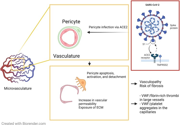
Aims: Thrombotic complications and vasculopathy have been extensively associated with severe COVID-19 infection; however, the mechanisms inducing endotheliitis and the disruption of endothelial integrity in the microcirculation are poorly understood. We hypothesized that within the vessel wall, pericytes preferentially take up viral particles and mediate the subsequent loss of vascular integrity.

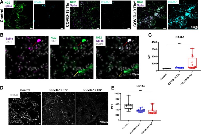
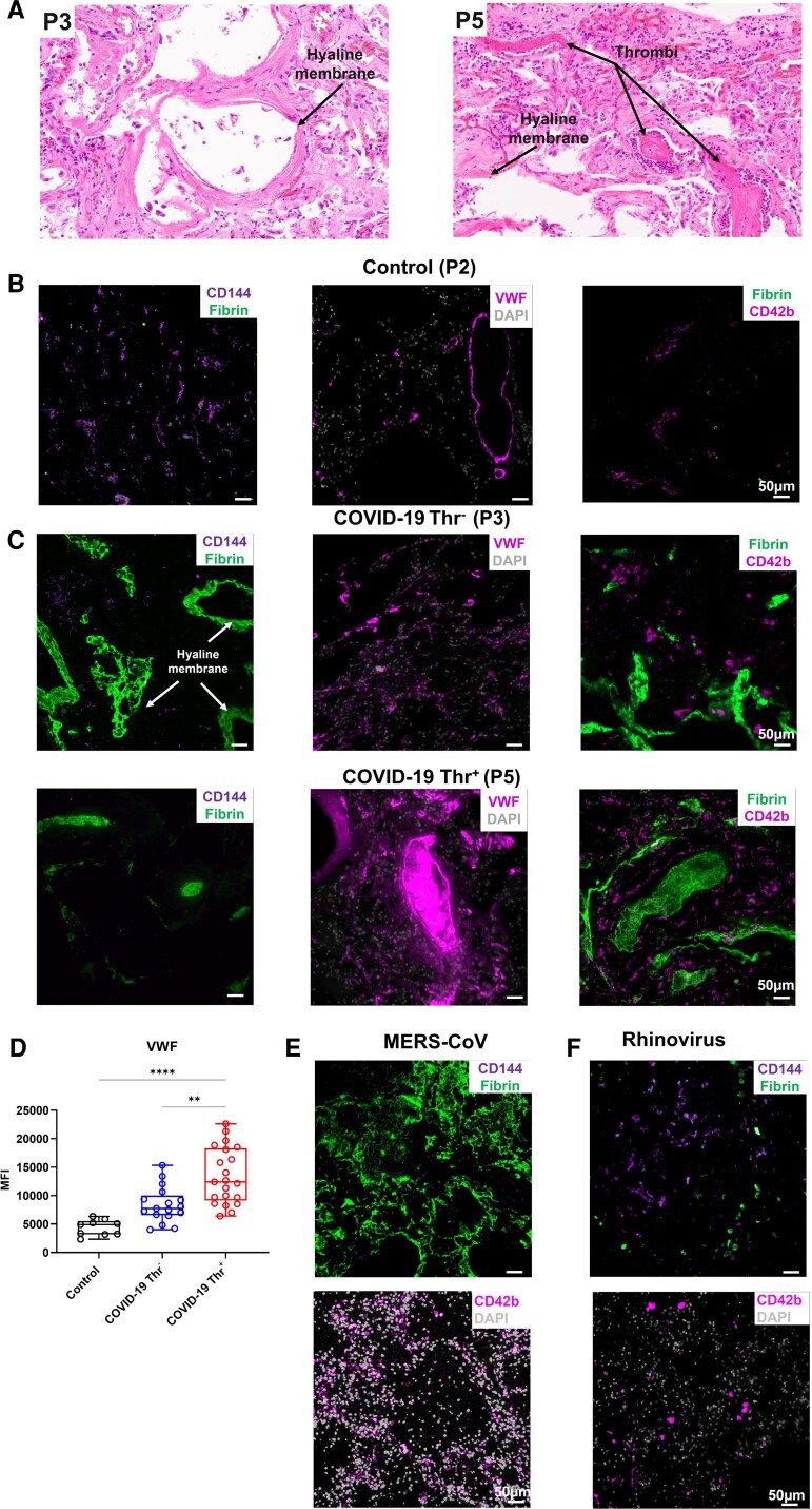
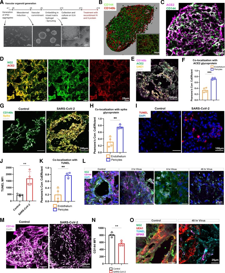
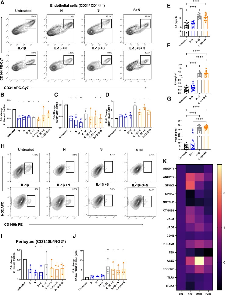
Methods and results: Immunofluorescence of post-mortem patient sections was used to assess pathophysiological aspects of COVID-19 infection. The effects of COVID-19 on the microvasculature were assessed using a vascular organoid model exposed to live viral particles or recombinant viral antigens. We find increased expression of the viral entry receptor angiotensin-converting enzyme 2 on pericytes when compared to vascular endothelium and a reduction in the expression of the junctional protein CD144, as well as increased cell death, upon treatment with both live virus and/or viral antigens. We observe a dysregulation of genes implicated in vascular permeability, including Notch receptor 3, angiopoietin-2, and TEK. Activation of vascular organoids with interleukin-1β did not have an additive effect on vascular permeability. Spike antigen was detected in some patients' lung pericytes, which was associated with a decrease in CD144 expression and increased platelet recruitment and von Willebrand factor (VWF) deposition in the capillaries of these patients, with thrombi in large vessels rich in VWF and fibrin.

Conclusion: Together, our data indicate that direct viral exposure to the microvasculature modelled by organoid infection and viral antigen treatment results in pericyte infection, detachment, damage, and cell death, disrupting pericyte-endothelial cell crosstalk and increasing microvascular endothelial permeability, which can promote thrombotic and bleeding complications in the microcirculation.

Keywords: COVID-19; Endothelial permeability; Organoids; SARS-CoV-2; Thrombosis; Vasculopathy.


Conflict of interest statement

Conflict of interest: none declared.

Figures

Graphical Abstract
Graphical Abstract
Pericyte infection by SARS-CoV-2 increases vascular damage and permeability. Preferential uptake of SARS-CoV-2 by pericytes induces their apoptosis leading to the disruption of endothelial cell junctions and the vascular network promoting vascular permeability. SARS-CoV-2-mediated endotheliopathy is associated with thrombotic complications in patients with COVID-19.
Figure 1
Figure 1
Detection of the spike glycoprotein in lung pericytes and decreased CD144 expression in COVID-19 patients. (A) Immunofluorescence imaging of NG2+, spike glycoprotein, and ICAM-1 in formalin-fixed and paraffin-embedded lung from a patient who died from COVID-19 and control COVID-19 (Control) lung sections. (B) High magnification of immunofluorescence imaging of NG2+ and spike glycoprotein in lung section. (C) Quantification of ICAM-1 expression using ImageJ in lung autopsies from two patients who died from non-respiratory-associated diseases, four COVID-19 patients without thrombosis and four patients with detectable thrombosis (3–6 areas per slide). (D) Representative immunofluorescence imaging of CD144 (VE-cadherin) in lung sections. (E) Quantification of CD144 levels using ImageJ. One-way ANOVA with multiple comparisons was performed for each statistical test with significance at (*** P = <0.001).
Figure 2
Figure 2
Decreased CD144 expression is associated with increased VWF deposition in the lung of patients with COVID-19. (A) H&E staining of lung sections from patients who died from COVID-19 with or without evidence of thrombosis. (B and C) Immunofluorescence imaging of CD144, VWF, platelets (CD42b), and fibrin in formalin-fixed and paraffin-embedded lung sections. (D) Quantification of VWF in lung sections. (E and F) Staining of CD144, CD42b, fibrin, and DAPI in lung sections from patients infected with (E) MERS-CoV and (F) rhinovirus. Images were captured using a Zeiss Observer 7 Epifluorescence microscope and slide scanner Axio Scan Z1. (Kruskal–Wallis test performed on a total of three to five lung sections from three control patients, three COVID-19 patients with thrombosis, and three without evidences of thrombosis (* P = <0.05, ** P = <0.01, *** P = <0.001, **** P = < 0.0001).
Figure 3
Figure 3
Pericyte preferential uptake of SARS-CoV-2 increases vascular permeability and endothelial and pericyte death in vascular organoids. (A) Vascular organoids were generated using a step-wise differentiation of human-induced pluripotent stem cells. (B) Cell composition and distribution in mature vascular organoids were assessed using immunofluorescence whole-mount microscopy. (C) Immunofluorescence imaging of ACE2 and CD144 positive cells in organoids (12 µm organoid sections. (D) Sections of vascular organoids were co-stained for pericytes (NG2) and ACE2. (E) Staining for UAE-1, ACE2, CD140b, and DAPI in vascular organoids. (F) Co-localization analysis between ACE2 and CD140b compared ACE2 co-localization with UAE1 as measured by the Pearson's correlation coefficient. (G) Co-staining for the spike glycoprotein in endothelial organoids with CD140b+ cells treated with SARS-CoV-2. (H) Quantification of co-localization of the spike glycoprotein between endothelium and pericytes. (I) Sections of vascular organoids were stained with apoptosis marker TUNEL and nuclei staining DAPI. (J) Quantification of TUNEL staining in control and SARS-CoV-2 treated vascular organoids. (K) Co-localization of the TUNEL staining with UAE-1 (endothelium) and pericyte (CD140b+). (L) Staining of control and SARS-CoV-2-infected organoids for CD144, pericytes (NG2), and nuclei (DAPI). (M) Immunofluorescence imaging of CD144 and nuclei (DAPI) in whole vascular organoids treated with SARS-CoV-2 (M.O.I = 0.5) for 48 h. (N) Quantification of CD144 staining in control and SARS-CoV-2 treated vascular organoids. (O) Staining for NG2, UAE-1, TUNEL, and spike in control and SARS-CoV-2 infected organoids (48 h). N = 4 for organoid experiments, where one replicate is a total of 8–10 individual organoids pooled for assays from each of four independent biological replicates. Each biological repeat was a separate differentiation protocol followed by viral infection. For co-localizations un-paired t-tests were performed (** P = <0.01) Schematic created on Biorender.com.
Figure 4
Figure 4
Recombinant viral antigens S and N increase vascular permeability and death without endothelial activation. (A–D) Vascular organoids were treated with spike glycoprotein (S) (100 nM), nucleocapsid (N), or a combination of both proteins for 72 h in the presence or absence of IL1-β (20 ng/mL) compared to control was measured by flow cytometry. (A) Representative flow cytometry blots of EC treated with S, N, and S and N antigens in the presence and absence of IL1-β. (B) Fold change in the live population of CD31+ cells from the total population using Live/Dead fixable dead cell stain. (C) Fold change in CD144 expression on CD31 + cells compared to untreated organoids and (D) fold change in ICAM-1 expression on CD31+ cells in treated organoids vs. untreated were measured by flow cytometry. (E) Detection of the levels of soluble IL6, (F) IL8, and (G) VWF in the supernatant of organoids treated for 72 h with viral antigens in the presence or absence of IL-1β by ELISA (n = 3 independent experiments, 3–4 replicate per experiment). (H) Representative flow cytometry plots of pericytes treated with S, N, and S and N antigens in the presence and absence of IL1-β (20 ng/mL). (I) Live/dead NG2+ cells from the total population, (J) fold change in ICAM-1 expression on CD140 + NG2+ cells in treated organoids vs. untreated were measured by flow cytometry. (K) qPCR of vascular organoids treated with combination S and N proteins for 0, 4, 24, and 72 h for genes regulating endothelial permeability. N = 3 for experiments, where one replicate is a total of 12–15 individual organoids pooled for assays from each of three independent biological replicates (independent differentiations). One-way ANOVA with multiple comparisons performed for each statistical test with significance at (* P = <0.05, ** P = <0.01, *** P = <0.001, **** P = <0.0001).

References

    1. Varga Z, Flammer AJ, Steiger P, Haberecker M, Andermatt R, Zinkernagel AS, Mehra MR, Schuepbach RA, Ruschitzka F, Moch H. Endothelial cell infection and endotheliitis in COVID-19. Lancet 2020;395:1417–1418. - PMC - PubMed
    1. Klok FA, Kruip M, van der Meer NJM, Arbous MS, Gommers D, Kant KM, Kaptein FHJ, van Paassen J, Stals MAM, Huisman MV, Endeman H. Incidence of thrombotic complications in critically ill ICU patients with COVID-19. Thromb Res 2020;191:145–147. - PMC - PubMed
    1. Rapkiewicz AV, Mai X, Carsons SE, Pittaluga S, Kleiner DE, Berger JS, Thomas S, Adler NM, Charytan DM, Gasmi B, Hochman JS, Reynolds HR. Megakaryocytes and platelet-fibrin thrombi characterize multi-organ thrombosis at autopsy in COVID-19: a case series. EClinicalMedicine 2020;24:100434. - PMC - PubMed
    1. Wichmann D, Sperhake JP, Lutgehetmann M, Steurer S, Edler C, Heinemann A, Heinrich F, Mushumba H, Kniep I, Schroder AS, Burdelski C, de Heer G, Nierhaus A, Frings D, Pfefferle S, Becker H, Bredereke-Wiedling H, de Weerth A, Paschen HR, Sheikhzadeh-Eggers S, Stang A, Schmiedel S, Bokemeyer C, Addo MM, Aepfelbacher M, Puschel K, Kluge S. Autopsy findings and venous thromboembolism in patients with COVID-19: a prospective cohort study. Ann Intern Med 2020;173:268–277. - PMC - PubMed
    1. Ackermann M, Mentzer SJ, Jonigk D. Pulmonary vascular pathology in Covid-19. Reply. N Engl J Med 2020;383:888–889. - PubMed

Publication types

Substances