Review
. 2022 Sep 29;9(1):285-306.
doi: 10.1146/annurev-virology-093020-013659. Epub 2022 Jun 16.

Liquid Phase Partitioning in Virus Replication: Observations and Opportunities

Affiliations
Review

Liquid Phase Partitioning in Virus Replication: Observations and Opportunities

Chao Wu et al. Annu Rev Virol. .

Abstract

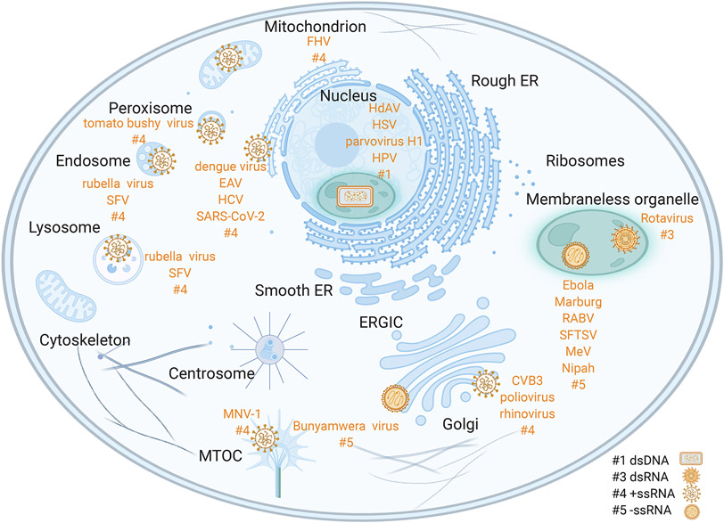
Viruses frequently carry out replication in specialized compartments within cells. The effect of these structures on virus replication is poorly understood. Recent research supports phase separation as a foundational principle for organization of cellular components with the potential to influence viral replication. In this review, phase separation is described in the context of formation of viral replication centers, with an emphasis on the nonsegmented negative-strand RNA viruses. Consideration is given to the interplay between phase separation and the critical processes of viral transcription and genome replication, and the role of these regions in pathogen-host interactions is discussed. Finally, critical questions that must be addressed to fully understand how phase separation influences viral replication and the viral life cycle are presented, along with information about new approaches that could be used to make important breakthroughs in this emerging field.

Keywords: NNSVs; host-pathogen interactions; inclusion bodies; phase separation; viral life cycle; virus transcription and replication.


Figures

Figure 1.
Figure 1.. Viral replication takes place in diverse membranous as well as membraneless organelles.
These viral replication compartments house essential steps of the viral life cycle and shield viral components from host defense systems. Sites of membrane-bound viral replication include mitochondria (Flock House virus, FHV (1)), peroxisomes (tomato bushy virus (2)), endosomes and lysosomes (rubella virus (3); Semliki Forest virus, SFV (3)), endoplasmic reticulum (ER) (dengue virus (4); equine arteritis virus, EAV (3), HCV (5), SARS-CoV-2 (5)), microtubule organizing center, MTOC (murine norovirus 1, MNV-1 (6)), Golgi (Bunyamwera virus (7); coxsackievirus B3, CVB3 (8), poliovirus (9), rhinovirus (10)). Sites of membraneless viral replication include cytosolic viral condensates (Ebola virus (10); Marburg virus (17); rabies virus, RABV (18); severe fever with thrombocytopenia syndrome virus, SFTSV (19); measles virus, MeV (20); Nipah virus (21)) and nuclear viral condensates (human adenovirus, HdAV (12); herpes simplex virus, HSV (13); parvovirus H1 (14); human papillomavirus, HPV (15)). The numbers at end of each group of viruses reflect the Baltimore classification (#1: dsDNA; #3: dsRNA; #4: +ssRNA; #5: -ssRNA). Created with Biorender.com.
Figure 2.
Figure 2.. Phase separation can lead to formation of membrane organelles.
A. Cell condensates formed through phase separation lack a membrane, and compartmentalization of biomolecules is driven by multivalent interactions that minimize the overall free energy such that chemically distinct cellular compartments form spontaneously (top right). Liquid-like condensates show many of the properties expected for liquids, including rapid intracondensate dynamics and the ability of two condensates to fuse rapidly into a single species (bottom right). B. Compartmentalization by phase separation enables diverse functions that can control subcellular biochemistry and information flow. C. Phase separation depends on multivalency that are encoded in biomolecules. These include modular binding domains connected in tandem, stoichiometric oligomerization domains that enable the formation of multivalent oligomers, intrinsically disordered regions with distributed sticker regions or residues, and long nucleic acid molecules that can function as platforms for higher-order assembly. Created with Biorender.com.
Figure 3.
Figure 3.. Viral replication compartments in NNSVs.
A. The genomic structure of RABV as an example of an NNSV. The genome of RABV is a single-stranded, linear RNA of negative polarity with inverse-complementary 3' and 5' termini. Abbreviations: UTR, untranslated region; N, nucleocapsid protein; P, phosphoprotein; M, matrix protein; G, glycoprotein; L, large protein (with RNA-dependent RNA polymerase activity). B. Schematic of the viral replication cycle of RABV that involves the formation of membraneless viral factories after infection. Viral translational products from primary transcription form liquid-like membraneless viral factories, and host factors are recruited into these condensates. Replication takes place inside these viral factories. Encapsidated genomes are trafficked out of viral factories via cytoskeletal transport. A major driving force in viral IBs formation is the expression of N and P proteins, which are suggested as the basic scaffolds of IBs during infection. C. The phase separation of viral proteins drive the formation of viral factories that facilitate efficient transcription and replication. Primary transcription results in abundant translation of N and P, which undergoes phase separation and in turn assemble into viral factories. Full-length genome synthesis lags behind those of mRNA and viral proteins and coincides with replication factories establishment. Created with Biorender.com.
Figure 4.
Figure 4.. Viral IBs in innate immune evasion.
Viral factories counteract the host IFN response Viral factories can sequester key components (MDA5, MAVS, IKK, IRF3/7, NFκB, PKR, STAT1/2, and essential stress granule components) from innate immune signaling pathways that detect pathogen-associated molecular patterns including cytosolic nucleic acids. Abbreviations: PKR, protein kinase R; TLR3, Toll-like receptor 3; RIG-I, retinoic acid-inducible gene I; MDA5, melanoma differentiation-associated protein 5; TBK1, TANK binding kinase 1; IRF3/7, interferon regulatory factor 3/7; cGAS, cyclic GMP–AMP synthase; STING, stimulator of interferon genes; IKK, IκB kinase; OGT, O-Linked N-Acetylglucosamine (GlcNAc) Transferase; HRTV, Heartland virus. Created with Biorender.com.

Similar articles

Cited by

References

    1. Kopek BG, Settles EW, Friesen PD, Ahlquist P. 2010. Nodavirus-Induced Membrane Rearrangement in Replication Complex Assembly Requires Replicase Protein A, RNA Templates, and Polymerase Activity. Journal of Virology 84: 12492–503 - PMC - PubMed
    1. McCartney AW, Greenwood JS, Fabian MR, White KA, Mullen RT. 2005. Localization of the tomato bushy stunt virus replication protein p33 reveals a peroxisome-to-endoplasmic reticulum sorting pathway. The Plant cell 17: 3513–31 - PMC - PubMed
    1. Miller S, Krijnse-Locker J. 2008. Modification of intracellular membrane structures for virus replication. Nature Reviews Microbiology 6: 363–74 - PMC - PubMed
    1. Paul D, Bartenschlager R. 2015. Flaviviridae Replication Organelles: Oh, What a Tangled Web We Weave. Annual Review of Virology 2: 289–310 - PubMed
    1. Chukkapalli V, Randall G. 2014. Hepatitis C virus replication compartment formation: mechanism and drug target. Gastroenterology 146: 1164–67 - PMC - PubMed

Publication types

LinkOut - more resources