. 2022 Sep;609(7925):136-143.
doi: 10.1038/s41586-022-04953-1. Epub 2022 Jun 16.

Spatial profiling of early primate gastrulation in utero

Affiliations

Spatial profiling of early primate gastrulation in utero

Sophie Bergmann et al. Nature. 2022 Sep.

Abstract

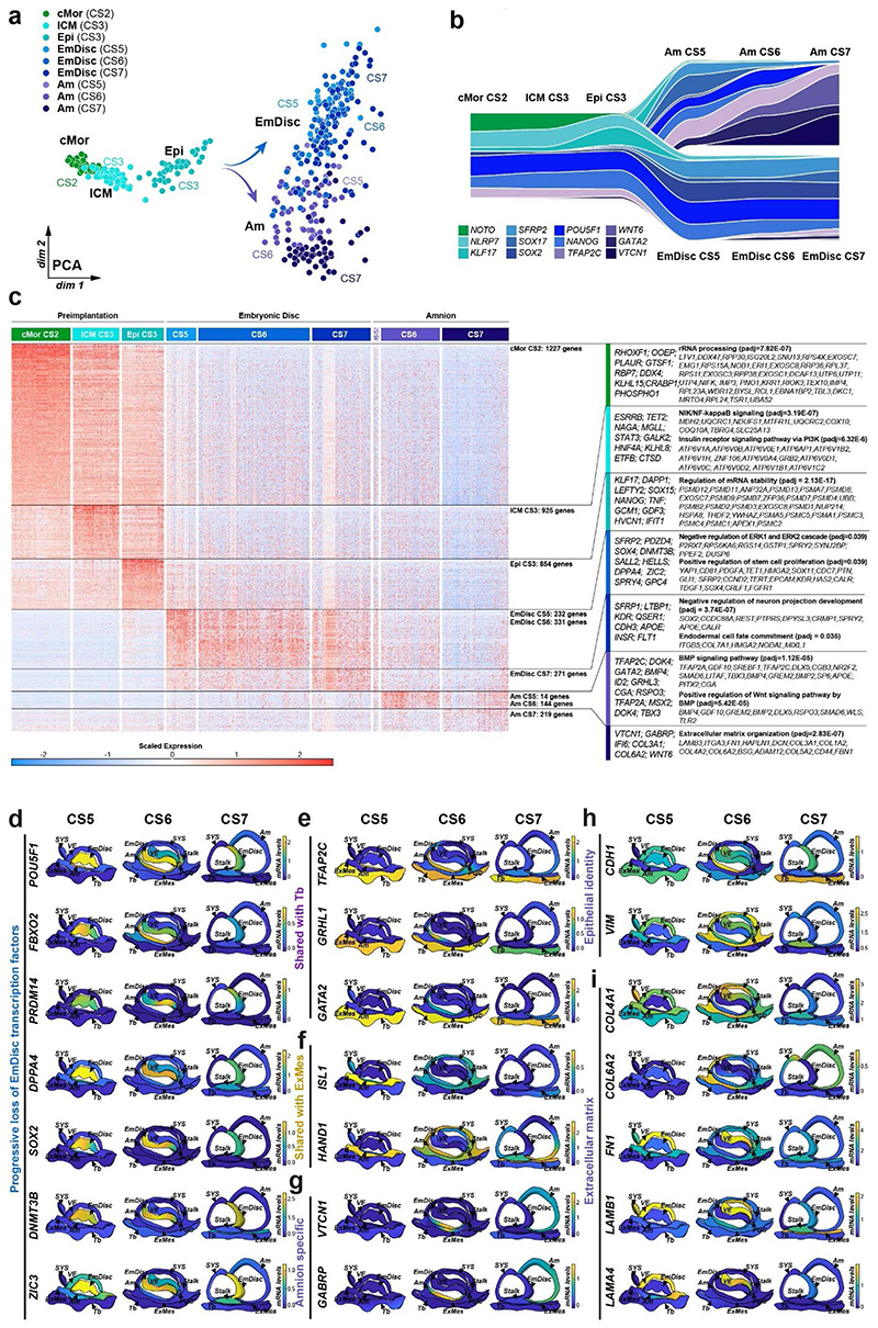
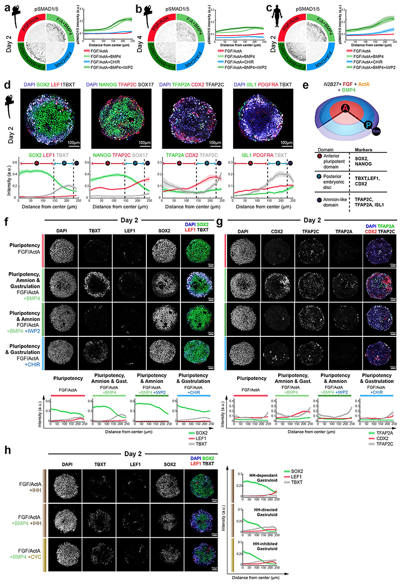
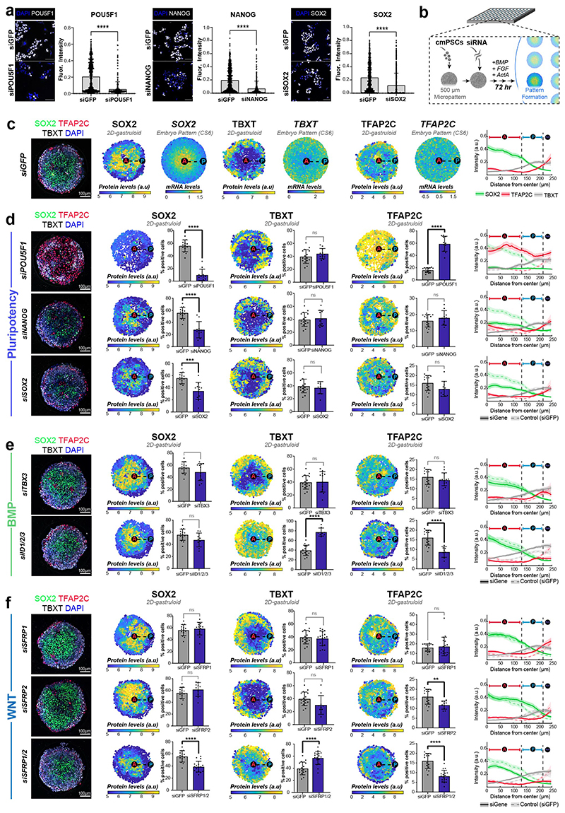
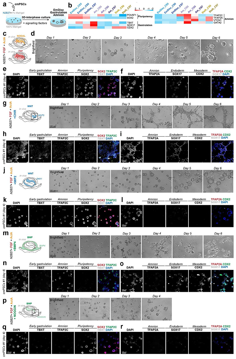
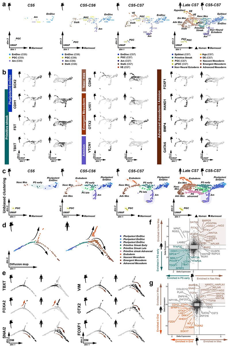
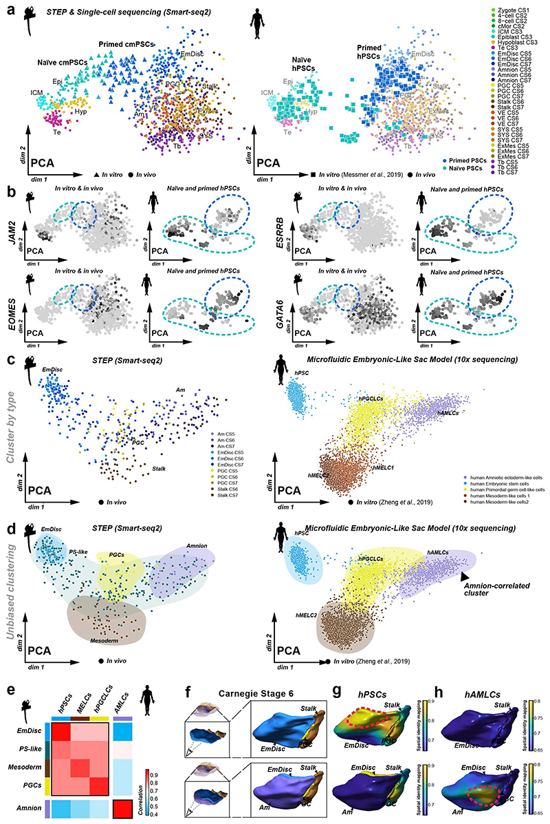
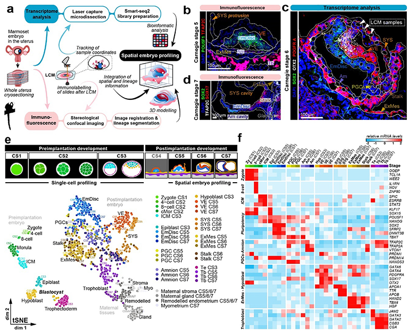
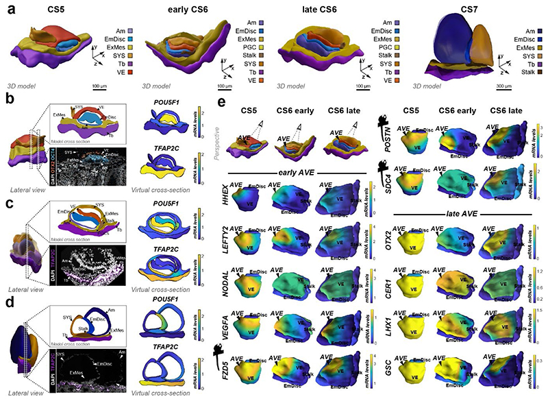
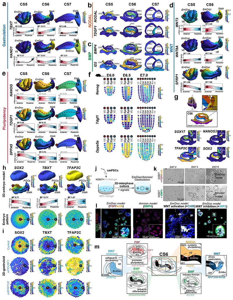
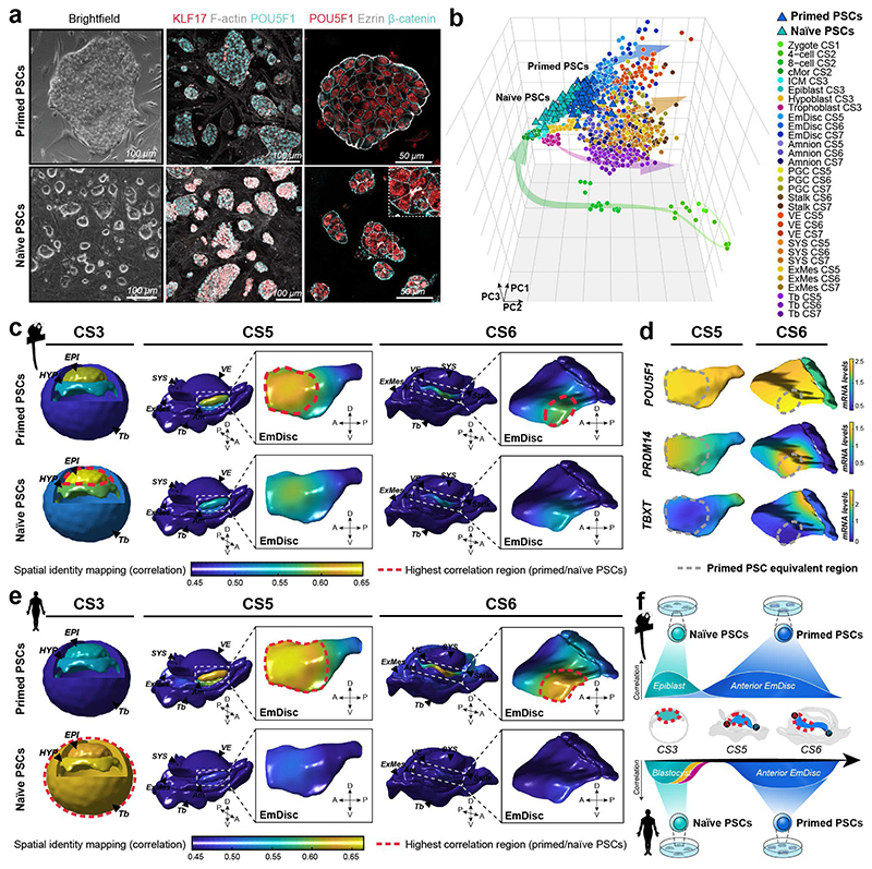
Gastrulation controls the emergence of cellular diversity and axis patterning in the early embryo. In mammals, this transformation is orchestrated by dynamic signalling centres at the interface of embryonic and extraembryonic tissues1-3. Elucidating the molecular framework of axis formation in vivo is fundamental for our understanding of human development4-6 and to advance stem-cell-based regenerative approaches7. Here we illuminate early gastrulation of marmoset embryos in utero using spatial transcriptomics and stem-cell-based embryo models. Gaussian process regression-based 3D transcriptomes delineate the emergence of the anterior visceral endoderm, which is hallmarked by conserved (HHEX, LEFTY2, LHX1) and primate-specific (POSTN, SDC4, FZD5) factors. WNT signalling spatially coordinates the formation of the primitive streak in the embryonic disc and is counteracted by SFRP1 and SFRP2 to sustain pluripotency in the anterior domain. Amnion specification occurs at the boundaries of the embryonic disc through ID1, ID2 and ID3 in response to BMP signalling, providing a developmental rationale for amnion differentiation of primate pluripotent stem cells (PSCs). Spatial identity mapping demonstrates that primed marmoset PSCs exhibit the highest similarity to the anterior embryonic disc, whereas naive PSCs resemble the preimplantation epiblast. Our 3D transcriptome models reveal the molecular code of lineage specification in the primate embryo and provide an in vivo reference to decipher human development.


Conflict of interest statement

Competing interests

The authors declare no competing interests.

Figures

Extended Data Figure 1
Extended Data Figure 1. Overview of the STEP method
1. Cryosectioning of the pregnant uterus: Pregnant marmoset uteri were extracted, embedded in O.C.T. and snap-frozen. To provide the best possible RNA quality, the tissue was processed unfixed for laser capture microdissection (LCM)-mediated Smart-Seq2 sequencing. 2. Stereological immunofluorescence imaging: Immunostainings for established lineage markers were performed for every other to every third section containing the implanted embryo. Tile-scan images were generated tile-scan with a confocal or apotome microscope. 12μm-thick cryosections were catalogued in ascending order, which was used to determine the Z-coordinate of LCM-transcriptomes. 3. LCM assisted sample collection: Every other to every third section was processed for LCM sample collection and all LCM-processed sections were subjected to immunofluorescence (IF) with lineage markers afterwards. For transcriptome sample acquisition, a photo was taken of the section. Then, one to three cells in a region of interest were selected using the LCM software Zeiss PALM and cut out by the laser. In a second step, a pulse laser catapulted the sample into a collection tube with lysis buffer. Then, a second picture was taken of the section at the matching position with the sample removed and matched to the collection tube and image file. Each STEP-transcriptome was assigned an individual ID and lineage identity based on the original location within the embryo cryosection, i.e. the cut-out location of the LCM-sample. 4. Sample annotation: Collected LCM-samples were subjected to the Smart-Seq2 protocol and sequenced to an average depth of > 2 million 150bp paired reads. LCM-sample lineage identity was assigned based on the position within the embryo. Sample annotation was performed manually, side-by-side with phase contrast images acquired during sample collection and the confocal image with lineage markers (e.g. PDGFRA, OTX2, SOX2) of the same section. In addition, annotations were guided by the density and orientation of DAPI-labelled nuclei, which allowed us to discriminate between neighbouring tissues. We refined annotations by integration of lineage marker expression from immunofluorescence stainings or STEP-transcriptome data. Samples with more than one lineage signature were annotated as mixed and removed from downstream analysis. 5. Image registration and lineage segmentation: Images were aligned by image registration in Fiji, whereby each image was registered to the DAPI channel of the previous image. Next, nuclei were segmented into individual objects using Cell Profiler. For lineage segmentation, the segmented nuclei were assigned lineages based on lineage marker immunostaining (e.g. POU5F1 to demarcate the EmDisc and Amnion, PDGFRA for ExMes, VE and SYS, TFAP2C for trophoblast, Amnion and PGCs), and taking into account known anatomical features of the embryo (e.g. EmDisc resides in-between VE and Amnion). 6. Transcriptome coordinate integration: The X and Y coordinates of the annotated transcriptomes were compiled into MATLAB matrices. 7.Virtual 3D embryo reconstruction: We generated primary surfaces in MATLAB by triangulation (see Methods). In a second step, embryonic and extraembryonic surfaces were smoothened in Blender, an open-source 3D modelling and animation software. 8. Gaussian Process Regression (GPR) over LCM samples: LCM spatial transcriptome sample coordinates were integrated into the 3D embryo models and continuous expression patterns between discrete LCM samples were inferred using Gaussian Process Regression (see Methods). Since expression patterns may be discontinuous across tissue types, we inferred an independent GPR model for each tissue at each stage. 9. GPR spatial transcriptome models and virtual cross sections: Final smooth GPR gene expression patterns could be displayed in 3D on embryo models. Defined coordinates were used to extract expression patterns in each lineage for visualisation of virtual cross sections. Scale bars represent 100 μm. cs, Carnegie stage; O.C.T, Optimal cutting temperature compound (used to mount uteri); EmDisc, Embryonic disc; Am, Amnion; SYS, Secondary Yolk Sac; VE, Visceral Endoderm; Tb, Trophoblast; ExMes, Extraembryonic mesoderm; PGCs, Primordial Germ Cells.
Extended Data Figure 2
Extended Data Figure 2. Staging of marmoset postimplantation embryos.
a, Staging of marmoset embryos based on listed hallmarks allowed us to stage blastocysts (Carnegie Stage (CS) 3), CS5, CS6 early and late, and CS7 embryos. Middle panel: illustrative cross section of embryo models for each stage with stage-specific differences in (1) secondary yolk sac, (2) Am, (3) stalk, and (4) ExMes formation indicated. Bottom panel: Representative images from human embryos at corresponding Carnegie Stages. CS3 reprinted from, CS5, CS6 late, and CS7 from, and CS6 early from. b, Principal component analysis of marmoset development of CS5-7 integrated by stage based on whole transcriptome (>20,000 genes). c, Immunofluorescence stainings of marmoset embryo sections. Scale bars represent 100 μm. EmDisc, Embryonic disc; Am, Amnion; SYS, Secondary Yolk Sac; VE, Visceral Endoderm; Tb, trophoblast; ExMes, Extraembryonic mesoderm; PGCs, Primordial Germ Cells.
Extended Data Figure 3
Extended Data Figure 3. Gaussian Process Regression models of spatial transcriptomics in mouse and marmoset embryos.
a, Spatial modelling of mouse postimplantation embryos based on published mouse embryo expression data shown for representative genes markers of anterior/posterior (A/P) epiblast (Epi), primitive streak (PS) and anterior primitive streak (APS). Each kernel on corn plots represents a micro-dissected and sequenced section of the mouse embryo from ref,. Corn plots were transformed into spatial models to match anterior-posterior and left-right axis. Gaussian process regression allowed visualisation of gene expression gradients, which were compared to in-situ hybridisation of postimplantation mouse embryos for validation of gaussian process regression approach. Source publication indicated next to individual images. b, Projection of shots on 3D virtual reconstructions. LCM-samples projected as black dots on 3D virtual models of each developmental stage used for downstream gene expression analysis. EmDisc, Embryonic Disc; VE, Visceral Endoderm; ExMes, Extraembryonic Mesoderm; A, Anterior; P, Posterior. c-d, Spatial embryo profiling LCM-sample lineage assignment examples. LCM-samples that were spatially close were assigned to amnion or ExMes based on PDGFRA expression. PDGFRA immunostaining of the CS6 early embryo demonstrates that amnion (nuclei adjacent to the amniotic cavity) is PDGFRA-negative (highlighted in inset below). Raw PDGFRA feature counts of individual LCM-samples overlaid on DAPI recapitulate immunostaining pattern, showing high expression in ExMes samples and no expression in amnion samples. LCM-samples from other lineages, that showed mixed lineage identity, or did not pass QC are not displayed. Arrowheads indicate PDGFRA-negative amnion. Cross-section of 3D-model (right) represents gaussian process regression-based modelling of all lineages in CS6 early embryo, recapitulating specific PDGFRA expression pattern observed by immunofluorescence and raw transcriptome data. e-h, Spatial embryo profiling processing example. Representative example of a CS6 section processed by spatial embryo profiling and immunofluorescence staining for SOX2 (e) and OTX2 (g). Gaussian process regression-based modelling of EmDisc and VE for SOX2 recapitulates the anterior EmDisc expression pattern observed by immunofluorescence and raw transcriptome data (f), and recapitulated anterior expression of OTX2 recapitulates the anterior VE expression pattern observed by immunofluorescence and raw transcriptome data (h). Upper panel: Relative mRNA levels for gene expression across the model. Lower panel: mRNA expression changes along anterior-posterior axis (dashed line, anterior EmDisc (red, A) to posterior EmDisc (blue, P), anterior VE (yellow, A) to posterior VE (green, P)) change along A-P axis quantified by Bayesian factor (BF).
Extended Data Figure 4
Extended Data Figure 4. Expression gradients and signalling environment in marmoset gastrulation
a, Posterior markers depicted in Gaussian process regression-based 3D models of CS5-7 EmDisc and VE. Upper panels: Relative mRNA levels across the model. Lower panels: mRNA expression change along EmDisc anterior-posterior axis (indicated by dashed line; anterior (red, A) to posterior (blue, P)), quantified by Bayes Factor (BF) (relates to Figure 3a). b, BMP signalling-related gene expression depicted in CS5-7 model cross sections. Schematic (bottom) summarises BMP signalling pathways in the context of amnion differentiation from EmDisc boundaries in CS5 and 6. c, WNT signalling genes shown in CS5-7 in model cross sections. Schematic summarises WNT signalling patterning in the CS6 EmDisc during gastrulation. d, RTK-related gene expression depicted in CS5-7 model cross sections displays VE is the primary source of IGF1, low expression of FGFs involved in mouse gastrulation (FGF8, FGF5), and presence of FGF4 and intracellular FGFs (FGF12, FGF13). e, RTK-related gene expression depicted in EmDisc and VE in CS5 and 6 3D models. Schematic summarizes PDGFA and VEGFA in the CS6 embryo. f, IHH signalling-related gene expression shown in CS5-7 in model cross sections. Schematic summarises proposed paracrine IHH signalling pathways. g, Representative anterior pluripotency genes depicted in CS5-7 EmDisc and VE 3D models (relates to Fig 3e) h, Corn plots of matched pluripotency genes in the gastrulating mouse embryo at E6.0, E6.5, and E7.0. Each kernel represents the average transcriptome of micro-dissected, spatially-defined sections of mouse embryos (relates to Figure 3c). i, Early (NANOG, PRDM1, POU5F1, KLF4) and late (DAZL, MAEL, PRAME) PGC marker and enriched signalling components (FGF4, WNT8A) expression in PGCs, depicted in CS6 model cross sections. EmDisc, Embryonic Disc; SYS, Secondary Yolk Sac; VE, Visceral Endoderm; ExMes, Extraembryonic Mesoderm; Am, Amnion; Tb, Trophoblast.
Extended Data Figure 5
Extended Data Figure 5. Amnion segregation from the Embryonic disc
a, PCA of EmDisc and Am based on the top 5000 most variable genes, PC1=20.7%, PC2=13.6%. b, Marker expression delineates the divergence of EmDisc and Am. Genes enriched in EmDisc and Am are marked in blue and purple, respectively; preimplantation genes are depicted in green. Stream plot track width is scaled to relative expression normalized to the mean across all stages displayed. c, Heatmap of expression of differentially expressed genes (DEG) in embryonic and extraembryonic lineages displayed in (a, b). Representative genes (left panel) and key gene ontology (GO) enrichment analysis (right panel) are shown. Genes shown in heatmap from Seurat function FindAllMarkers (minimum percent 50%, minimum log fold change 0.25) and filtered by adjusted p-value <0.05. d-i, Virtual cross-sections of 3D-transcriptomes at CS5, 6 and 7 depicting mRNA levels of representative genes for (d) pluripotency factor expression in the nascent Am, (e) Am-Tb shared genes, (f) Am-ExMes shared genes, (g) Am-specific genes, (h) epithelial genes, (i) ECM-related genes. Categories indicated on the left of each panel. EmDisc, embryonic disc; Am, amnion.
Extended Data Figure 6
Extended Data Figure 6. Endogenous WNT required for posterior patterning in marmoset 2D gastruloids models
a-c, pSMAD1/5 (phosphorylated SMAD 1/5) activity in marmoset 2D-gastruloids detected by immunostaining at day 2 (a) or day 4 (b) compared to human 2D-gastruloids under conventional conditions at day 2 (c). Micropatterned colonies were treated with self-renewal conditions (10 ng/mL FGF + 20 ng/mL Activin A), conventional gastruloid conditions (10 ng/mL FGF + 20 ng/mL Activin A + 50 ng/mL BMP4) or WNT modulatory conditions (10 ng/mL FGF + 20 ng/mL Activin A + 3 μM IWP-2 or 10 ng/mL FGF + 20 ng/mL Activin A + 3 μM CHIR99021). Representative maximum projection of immunofluorescence images (left). Quantification plots (right) mean +/- SEM across a minimum of 10 gastruloids across 2 wells. F/A = FGF/Activin A. pSMAD1/5 gradient indicates that in the marmoset system, similar to the human, FGF/ActivinA/BMP4 induce a graded response to BMP signalling at day 2, with the highest signalling in the outermost ring of the colony. d, Molecular characterisation of 2D-gastruloids. Representative immunofluorescence images of gastruloids differentiated in conventional gastruloid conditions (10 ng/mL FGF + 20 ng/mL Activin A + 50 ng/mL BMP4) for 2 days. Quantification plots (bottom) display mean +/- SEM across a minimum of 10 gastruloids across 2 experiments. Anterior domain (A, SOX2+, TBXT-, TFAP2C-), posterior domain (P, TBXT high, CDX2 heterogeneous, SOX17 sparse, LEF1 sparse), and amnion domain (Am, TFAP2C high, SOX2-) demarcated. ISL1 and TFAP2A are observed heterogeneously predominantly in the amnion region. PDGFRA expression is low, indicating lack of mature mesoderm. e, Schematic of lineage identities present in 2D-gastruloids and marker patterns that define each region. f, WNT-associated anterior/posterior patterning phenotypes of 2D-gastruloids. Representative immunofluorescence images of gastruloids differentiated in conditions listed at left for 2 days. Quantification plots (bottom) mean +/- SEM across a minimum of 10 gastruloids across 2 wells. g, WNT-associated amniogenesis phenotypes of 2D-gastruloids. Representative immunofluorescence images of gastruloids differentiated in conditions listed at left for 2 days. CDX2 expression is lost upon 3 μM IWP2 treatment, but TFAP2C and TFAP2A remain expressed. 3 μM CHIR treatment (WNT agonist) leads to low CDX2 expression but does not support upregulation of TFAP2C or TFAP2A. Quantification plots (bottom) mean +/- SEM across a minimum of 10 gastruloids across 2 wells. h, No evident change in expression profiles associated with Hedgehog signalling manipulation in 2D-gastruloids. Representative immunofluorescence images of gastruloids differentiated in conditions listed at left for 2 days. Exogenous Indian Hedgehog (IHH, 200 ng/mL) did not lead to loss of pluripotency under FGF/Activin self-renewal conditions, or change expression patterns under conventional gastruloids conditions. Inhibition of hedgehog signalling with Cyclopamine (5 μM) also did not lead to evident changes in expression patterns. Quantification plots (bottom) mean +/- SEM across a minimum of 10 gastruloids across 2 wells.
Extended Data Figure 7
Extended Data Figure 7. siRNA in 2D marmoset gastruloids
a, siRNA knockdown efficiency. Representative immunofluorescence images (left) and quantification of mean fluorescence intensity (right) 24 hours following transfection with siRNA against POU5F1, SOX2, or NANOG. Comparisons to siGFP (green fluorescent protein) control conducted with two-tailed Mann-Whitney test (****, p<0.0001). siRNA: small interfering RNA b, Schematic of siRNA screening approach. cmPSCs (common marmoset pluripotent stem cells) were seeded in micropatterned 96-well plates on day -1 and transfected overnight with siRNA. On day 0, media was changed to gastruloid induction media (10 ng/mL FGF + 20 ng/mL Activin A + 50 ng/mL BMP4). 2D-gastruloids were fixed after 72 hours and stained to assess pattern formation. c, Comparison of siGFP phenotype to in vivo EmDisc patterns. Representative maximum projection immunofluorescence image of siGFP-treated gastruloids differentiated in conventional gastruloid conditions (10 ng/mL FGF + 20 ng/mL Activin A + 50 ng/mL BMP4) for 3 days (left). 2D-gastruloid log expression patterns normalized to maximum intensity plotted for individual channels (SOX2, TBXT, TFAP2C) side by side with virtual embryo pattern of CS6 EmDisc expression patterns generated by Gaussian process regression of anterior-posterior axis expression. Quantification plot (right) shows mean +/- SEM across a minimum of 10 gastruloids with anterior domain (A, SOX2+, TBXT-, TFAP2C-), posterior domain (P, TBXT high), and amnion domain (Am, TFAP2C high, SOX2-) demarcated. d-f. siRNA knockdown phenotypes of pluripotency factors (siPOU5F1, siNANOG, siSOX2), BMP-related genes (siID1/2/3, siTBX3) and WNT-related genes (siSFRP1, siSFRP2, siSFRP1/2). For each siRNA, a representative maximum projection immunofluorescence is shown (left). Representative expression patterns are plotted for individual channels (SOX2, TBXT, TFAP2C) adjacent to quantification of the percent of nuclei positive for each marker per gastruloid (center). Comparison conducted with two-tailed Mann-Whitney test (ns: not significant; p<0.0332 (*); p <0.0021 (**); p<0.0002,(***); p<0.1; p < 0.0001 (****)). Quantification plot (right) shows mean +/- SEM across a minimum of 10 gastruloids across 2 wells. SOX2 = green, TFAP2C = red, TBXT = grey. siGFP patterns plotted for comparison in reduced opacity and dashed line with control anterior domain (A, SOX2+, TBXT-, TFAP2C-), posterior domain (P, TBXT high), and amnion domain (Am, TFAP2C high, SOX2-) demarcated.
Extended Data Figure 8
Extended Data Figure 8. 3D in vitro modelling of the marmoset EmDisc
a, Schematic overview of interphase culture. To model self-organisation of conventional cmPSCs (common marmoset pluripotent stem cells) in 3D culture, cmPSCs were seeded on a 100% Matrigel base overlaid with 1% Matrigel dissolved in N2B27 media supplemented with signalling factors. Interphase culture was amenable to probing signalling requirements that promote anterior embryonic disc-like pluripotency or differentiation into the germ layers of the gastrulating embryo. All experiments were performed in two different cell lines (cell line #1 (New4f), N=2 and cell line #2 (New2f), N=2) showing consistent results. b, Heatmap of marker genes used for molecular characterisation of interphase culture structures. Relative mRNA levels were centred and scaled across all marmoset in vivo samples. c, Summary schematic of NODAL and FGF signalling in the marmoset embryo. The visceral endoderm is the primary source of NODAL and IGF1 in the marmoset embryo, while the EmDisc expresses low levels of FGF4. Relative mRNA expression gradients summarised in CS6 cross section. d, Time series brightfield images of interphase culture with FGF (100ng/ml) and Activin A (20ng/ml). FGF/Activin A culture provides a signalling environment that mimics high NODAL and IGF1 from the VE. Structures formed columnar epithelial cysts, reminiscent of the embryonic disc. Structures first open a lumen at day 3 and expand up to day 6. e-f, Molecular characterisation of EmDisc-like structures at day 4. Representative maximum projection images from immunostaining at day 4 for pluripotency (SOX2), early gastrulation (TBXT), amnion (TFAP2C, TFAP2A), endoderm (SOX17) or mesoderm (CDX2) markers. EmDisc-like structures homogenously expressed SOX2, with heterogeneous low expression of TBXT, SOX17 and TFAP2C indicative of priming toward gastrulation and rare emergence of endoderm. Pluripotent EmDisc-like structures support a role for FGF and Activin/NODAL signalling in promoting pluripotency in the EmDisc. g, Summary schematic of canonical WNT signalling in the marmoset embryo. The posterior EmDisc, stalk and PGCs express WNT3. mRNA expression gradients summarised in CS6 cross section (left). Time series brightfield images of interphase culture with FGF and Activin A + CHIR (CHIR99021, WNT agonist) (right). The emergence of differentiated populations was evident at day 4. h-i, Molecular characterisation of WNT-treated EmDisc-like structures at day 4. Representative maximum projection images from immunostaining at day 4 from staining for pluripotency (SOX2), early gastrulation (TBXT), amnion (TFAP2C, TFAP2A), endoderm (SOX17) or mesoderm (CDX2) markers. Structures exhibited loss of SOX2 expression and upregulation of TBXT and SOX17 in comparison to FGF/Activin A culture, consistent with differentiation into amnion, endoderm, and mesoderm populations. j, Summary schematic of WNT inhibition in the marmoset embryo. The VE and Amnion express canonical WNT inhibitor DKK1. mRNA expression gradients summarised in CS6 cross section (left). Time series brightfield images of interphase culture with FGF and Activin A + IWP-2 (right). Similar to FGF/Activin A culture, structures first open a lumen at day 3 and expand up to day 6. k-l, Molecular characterisation of WNT-inhibited EmDisc-like structures at day 4. Representative maximum projection images from immunostaining at day 4 from staining for pluripotency (SOX2), early gastrulation (TBXT), amnion (TFAP2C, TFAP2A), endoderm (SOX17) or mesoderm (CDX2) markers. EmDisc-like structures homogenously expressed SOX2 and downregulated TBXT and SOX17 in comparison to FGF/Activin A culture, consistent with a role for WNT inhibition in preserving pluripotency in the EmDisc. m, Summary schematic of BMP signalling in the marmoset embryo.The ExMes, amnion, and PGCs are sources of BMP4 in the embryo. mRNA expression gradients summarised in CS6 cross section (left). Time series brightfield images of interphase culture with FGF and Activin A + BMP4 (right).The emergence of disorganized, differentiated populations was evident at day 4. n-o, Molecular characterisation of BMP-treated EmDisc-like structures at day 4.Representative maximum projection images from immunostaining at day 4 from staining for pluripotency (SOX2), early gastrulation (TBXT), amnion (TFAP2C, TFAP2A), endoderm (SOX17) or mesoderm (CDX2) markers. Structures exhibited loss of SOX2 expression and upregulation of TFAP2C, TFAP2A, CDX2 and SOX17 in comparison to FGF/Activin A culture, consistent with a mixed amnion and posteriorized primitive streak-like fate. p, Summary schematic of BMP inhibition in the marmoset embryo. The VE expresses BMP inhibitor NOGGIN in the embryo. mRNA expression gradients summarised in CS6 cross section (left). Time series brightfield images of interphase culture with FGF and Activin A + BMP4 (right).Similar to FGF/Activin A culture, structures first open a lumen at day 3 and form homogenous spheroids. q-r, Molecular characterisation of BMP-inhibited EmDisc-like structures at day 4.Representative maximum projection images from immunostaining at day 4 from staining for pluripotency (SOX2), early gastrulation (TBXT), amnion (TFAP2C, TFAP2A), endoderm (SOX17) or mesoderm (CDX2) markers. Scale bars represent 100 μm. EmDisc-like structures homogenously expressed SOX2 and downregulated TBXT, SOX17 and TFAP2C in comparison to FGF/Activin A culture. This is consistent with a role for BMP inhibition in preserving pluripotency in the EmDisc and inhibiting amnion differentiation.
Extended Data Figure 9
Extended Data Figure 9. Cross-species analysis of primate mesoderm differentiation in vivo
a, Alignment of EmDisc-derived postimplantation lineages in marmoset and human. Visualization based on alignment of embryo in vivo and in vitro datasets of pre- to postimplantation cynomolgus monkey, in vitro-cultured human, in vivo human CS7, preimplantation marmoset (ref and this study), and postimplantation marmoset embryo data (this study) were aligned. UMAP: Uniform Manifold Approximation and Projection for Dimension Reduction. b, UMAP plots of marmoset CS5-7 or human late CS7 lineages showing normalized log expression of marker genes from . Pluripotent EmDisc (Embryonic Disc): SOX2, CDH1; Primitive Streak: CDH1, FST, TBXT; Nasc Mes (Nascent mesoderm): TBXT, CDH2, Emergent Mesoderm: OTX2, LHX1; Advanced Mesoderm: FOXF1, HAND1, BMP4, GATA6; Amnion: VTCN1, HAND1, BMP4; Endoderm subcluster also indicated by co-expression of CDH2, OTX2, GATA6. c, Unbiased clustering of gastrulation stage lineages represented in UMAP in (a) resolves 9 clusters by shared nearest neighbour clustering: Pluripotent EmDisc (Embryonic Disc), PS early (Primitive Streak early), PS late (Primitive Streak late), PS advanced (Primitive Streak advanced), Endoderm, Nasc Mes (Nascent Mesoderm), Em Mes (Emergent Mesoderm), Adv Mes (Advanced Mesoderm), PGCs (Primordial Germ Cells). d., Bifurcation of endoderm and mesoderm from the primitive streak represented in a diffusion map. Human CS7 shows an additional route from primitive streak to advanced mesoderm through emergent mesoderm. Alignment includes data plotted in (c) with amnion and PGC clusters excluded where cluster identity is given by colour. The first 2 diffusion components are shown (dim 1, dim 2). e, Diffusion maps of marmoset CS5-7 or human late CS7 lineages showing normalized log expression of marker genes for primitive streak (TBXT), endoderm (FOXA2), pan-mesoderm (SNAI2, CDH2), emergent mesoderm (OTX2) or nascent mesoderm (FOXF1). f-g, Human vs. marmoset scatterplots of primitive streak vs. nascent mesoderm (f) or nascent mesoderm vs. endoderm (g). Highlighted quadrants show human-marmoset conserved markers for each lineage, whereas white quadrants show species-specific expression patterns. Gene names for transcription factors, ligands, and extracellular matrix molecules are labelled.
Extended Data Figure 10
Extended Data Figure 10. Spatial identity mapping of human in vitro models
a, Alignment of naïve and primed human and marmoset PSCs with in vivo marmoset data. Marmoset in vivo datasets of pre- to postimplantation development and naïve/primed PSCs and human naïve/primed PSCs were aligned. Visualisation of aligned datasets by principal component analysis shows separation of preimplantation (on the left) and postimplantation (on the right) samples, with marmoset PSCs sitting between pre- and post-implantation. Human PSCs are plotted overlayed with transparent marmoset embryo data. Naïve human cells mapped earlier on PC1 than naïve marmoset cells and showed greater heterogeneity in mapping to ICM, Hypoblast, and Tb. Primed human cells mapped closely to the marmoset EmDisc. PCA, Principal component analysis; PSCs, pluripotent stem cells. ICM, inner cell mass; Hyp, hypoblast; SYS, Secondary Yolk Sac; VE, Visceral Endoderm; ExMes, Extraembryonic Mesoderm; EmDisc, Embryonic Disc; Am, Amnion; Tb, Trophoblast. b, Expression of extraembryonic markers in marmoset in vivo data (left) and human naïve and primed PSCs (right) represented in aligned PCAs from (a) showing integrated log expression of trophoblast markers (JAM2), endoderm markers (EOMES, GATA6) and ICM/preimplantation blastocyst marker (ESRRB). Turquoise lines indicate location of naïve cells, blue lines indicate primed cells on the PCA. c, Alignment of human microfluidic embryonic-like sac model to marmoset STEP data. Smart-seq2 profiling of marmoset in vivo lineages (EmDisc, PGC, Amnion, and Stalk populations) was aligned to 10x sequencing profiling of microfluidic embryonic-like sac model. Visualisation of aligned datasets by principal component analysis shows separation hPSC, mesoderm, PGCLCs, and AMLCs. PCA, Principal component analysis; PSC, human pluripotent stem cell; hPGCLC, human primordial germ cell-like cell, hAMLC; human amnion-like cell; hMELC1/2, human mesoderm-like cell population 1 or 2. d. Unbiased Clustering represented in UMAP in (c) resolves 5 clusters by shared nearest neighbour clustering in marmoset data. Four clusters aligned to subpopulation of the human microfluidic embryonic-like sac model and were annotated EmDisc, PGC, Amnion, and Mesoderm. A fifth subcluster in marmoset data contained cells from the gastrulating EmDisc, PGCs, and amnion and was therefore annotated PS-like (primitive streak-like). e, Pearson’s correlation of marmoset and human microfluidic embryonic-like sac model of clusters identified in (d). f-h. Spatial identity mapping of human microfluidic embryonic-like sac model. Subpopulations of the human microfluidic embryonic-like sac model were mapped to the marmoset EmDisc, PGC, Stalk, and Amnion. Spatial identity displayed in the orientations described in (f) for hPSCs (g), which mapped to the anterior EmDisc and hAMLCs (h) which mapped most strongly to the posterior amnion.
Figure 1
Figure 1. Spatial profiling of marmoset embryogenesis
a, SpaTial Embryo Profiling (STEP) to delineate early marmoset postimplantation development. Transcriptome samples are collected by laser capture microdissection (LCM) and processed by Smart-Seq2. Sample coordinates are determined from consecutive cryosections and embedded in virtually reconstructed embryo models based on stereological confocal immunofluorescence images. b-d, Confocal immunofluorescence stainings of marmoset implantation stages at Carnegie stages (CS) 5,6,7. b, Pluripotency factor POU5F1 and Tb / early Am marker TFAP2C at CS5. c, Lineage marker analysis after LCM-processing using pluripotency marker SOX2, VE marker OTX2 and hypoblast/VE/mesoderm marker PDGFRA of a CS6 embryo cryosection. The locations of harvested LCM-samples are indicated with white dashed circles. d, Immunofluorescence of CS7 for SOX2, SOX17 and TFAP2C. e, Schematic overview of marmoset embryonic stages (top panel) from zygote (CS1) to gastrula (CS7); CS4 was not included in transcriptome analysis. The tSNE plot shows the combined Smart-seq2 embryo atlas consisting of 279 preimplantation single-cell samples as well as 866 postimplantation embryo and 193 maternal tissue STEP samples. Lineage colour code was used for all following figures (preimplantation stages=green, embryonic lineage and derivatives=blue, amnion=lavender, hypoblast-derived lineages=yellow, trophoblast-derived lineages=purple, maternal tissue=grey). f, Heatmap of embryonic and extraembryonic lineage markers. Relative mRNA levels were centred and scaled across samples. Zy, Zygote; cMor, Compacted Morula; ICM, Inner cell mass; Epi, Epiblast; Hyp, Hypoblast; Te, Trophectoderm; Am, Amnion; EmDisc, Embryonic disc; VE, Visceral endoderm; ExMes, Extraembryonic Mesoderm; SYS, Secondary Yolk Sac; Tb, Trophoblast; PGCs, Primordial Germ Cells; Myo, Myometrium.
Figure 2
Figure 2. Virtual reconstruction of gastrulating marmoset embryos
a,Virtual 3D-reconstructions of postimplantation implantation stages at CS5, early CS6, late CS6, and CS7 based on stereological confocal imaging and lineage segmentation. ExMes of CS6 early and late embryos is vertically sectioned to expose underlying structures. ExMes of CS7 that overlays amnion and SYS are not displayed, and both amnion and yolk sac are partially transparent. b-d, GPR-based 3D-transcriptome expression of pluripotency marker POU5F1 and Tb/Am marker TFAP2C in CS5 (b), late CS6 (c), and CS7 (d) embryos. Cross-sections are indicated in lateral views of virtual 3D-reconstructions with immunofluorescence staining of corresponding embryo sections. (e) Anterior visceral endoderm (AVE) genes depicted in EmDisc/VE for the stages indicated. Marmoset symbols indicate primate-specificity. GPR, Gaussian process regression; CS, Carnegie stage; Am, Amnion; EmDisc, Embryonic disc; ExMes, Extraembryonic Mesoderm; SYS, Secondary Yolk Sac; Tb, Trophoblast; VE, Visceral endoderm; PGC, Primordial Germ Cells.
Figure 3
Figure 3. 3D-transcriptomes and stem cell-based embryo models delineate body axis formation
a, GPR-based 3D-transcriptomes in EmDisc/VE showing gastrulation marker expression in the posterior EmDisc. Upper panels: Relative mRNA levels in EmDisc, VE, and stalk. Lower panels: mRNA expression change along anterior-posterior axis (dashed line; anterior (red, A) to posterior (blue, P)) in EmDisc, quantified by Bayesian factor (BF). b-c, Relative mRNA levels of (b) NODAL and (c) BMP in virtual embryo cross sections. d, WNT signalling pathway components shown in EmDisc/VE model. e, GPR-models for EmDisc/VE displaying regionalised pluripotency factor transcription in the anterior EmDisc. f, Spatial expression of pluripotency factors in gastrulating mouse embryos at E6.0, E6.5, and E7.0 according to Geo-seq. g, Virtual cross-sections of the PGCs at CS6. GPR, Gaussian process regression; CS, Carnegie stage; EmDisc, Embryonic disc; VE, Visceral endoderm; ExMes, Extraembryonic Mesoderm. Am, Amnion; SYS, Secondary Yolk Sac; Tb, Trophoblast; PGC, Primordial Germ Cells. h, GPR-models for CS6 embryo of anterior marker (SOX2), posterior marker (TBXT) and amnion/PGC maker (TFAP2C). Upper panels: Relative mRNA levels for gene expression in EmDisc, VE, PGCs, Stalk, and Amnion. Am is displayed separately for visualisation. Middle panels: mRNA expression change along anterior-posterior axis (dashed line, anterior (red, A) to posterior (blue, P)) in EmDisc, quantified by Bayesian factor (BF). Lower panel: Virtual embryo pattern of EmDisc expression patterns generated by axis expression in middle panel. i, Expression patterns of in vitro 2D gastruloids segmented nuclei stained for anterior marker (SOX2), posterior marker (TBXT) and amnion/PGC maker (TFAP2C) after differentiation in BMP4 (50 ng/mL), FGF (10ng/mL), and Activin A (20ng/mL) under control conditions (top panel) and following siRNA transfection (bottom panels). Each intensity profile is normalized log expression levels are standardized so that they vary within [0,1]. j, Schematic representation of 3D-interphase culture system. cmPSCs are seeded on a bed of 100% Matrigel and overlaid with 1% Matrigel supplemented N2B27-based culture medium with and without signalling molecules. k, Brightfield images of EmDisc-like structures (N2B27 + 100 ng/mL FGF + 20 ng/mL Activin A) or Amnion-like structures (N2B27 + 50 ng/mL BMP4). l, Immunofluorescence images of structures generated after 4 days in EmDisc- or Amnion-promoting conditions or in EmDisc conditions with WNT modulation through 3 μM CHIR99021 (WNT activator) or 3 μM IWP-2 (WNT production inhibitor). m, Schematic summary diagram of BMP, WNT, FGF and NODAL signalling pathway activities in the marmoset embryo at CS6. PSCs, Pluripotent Stem Cells.
Figure 4
Figure 4. Spatial identity mapping of in vitro cultured cells
a, Brightfield and immunofluorescence images of primed and naïve marmoset pluripotent stem cells (PSCs) cultured in PLAXA medium. b, Principal component analysis (PCA) of embryonic and extraembryonic cells in vivo and naïve and primed PSCs in vitro. PCA based on the top 2000 most variable genes, PC1=22.0%, PC2 =12.4%, PC3=7.5%. c-e, Spatial identity mapping of marmoset (c) and human (e) naïve and primed PSCs. Colour scale represents projection of correlation values onto embryo model surfaces followed by Gaussian process regression mapping. Blastocyst model is a schematic representation with bulk correlation plotted for each lineage. Gene expression in regions of highest correlations for primed PSCs in the pluripotent anterior indicated by dotted line (d). f, Summary of PSC mapping of marmoset and human PSCs to the marmoset in vivo atlas. A, anterior; P, posterior; D, dorsal; V, ventral; EPI, Epiblast; HYP, Hypoblast; Tb, Trophoblast; EmDisc, Embryonic disc; Am, Amnion; SYS, Secondary Yolk Sac; VE, Visceral Endoderm; ExMes, Extraembryonic mesoderm; PGCs, Primordial Germ Cells.

References

    1. Tam PP, Loebel DA. Gene function in mouse embryogenesis: get set for gastrulation. Nat Rev Genet. 2007;8:368–381. - PubMed
    1. Rossant J, Tam PPLL. Blastocyst lineage formation, early embryonic asymmetries and axis patterning in the mouse. Development. 2009;136:701–713. - PubMed
    1. Arnold SJ, Robertson EJ. Making a commitment: cell lineage allocation and axis patterning in the early mouse embryo. Nat Rev Mol Cell Biol. 2009;10:91–103. - PubMed
    1. Rossant J, Tam PPL. Exploring early human embryo development. Science (80-.) 2018;360:1075–1076. - PubMed
    1. Boroviak T, Nichols J. Primate embryogenesis predicts the hallmarks of human naïve pluripotency. Development. 2017;144:175–186. - PMC - PubMed