. 2022 Aug;298(8):102134.
doi: 10.1016/j.jbc.2022.102134. Epub 2022 Jun 13.

A naturally occurring membrane-anchored Gαs variant, XLαs, activates phospholipase Cβ4

Affiliations

A naturally occurring membrane-anchored Gαs variant, XLαs, activates phospholipase Cβ4

Hoa T N Phan et al. J Biol Chem. 2022 Aug.

Abstract

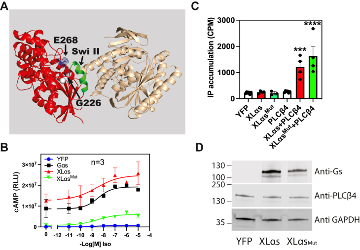
Extra-large stimulatory Gα (XLαs) is a large variant of G protein αs subunit (Gαs) that uses an alternative promoter and thus differs from Gαs at the first exon. XLαs activation by G protein-coupled receptors mediates cAMP generation, similarly to Gαs; however, Gαs and XLαs have been shown to have distinct cellular and physiological functions. For example, previous work suggests that XLαs can stimulate inositol phosphate production in renal proximal tubules and thereby regulate serum phosphate levels. In this study, we show that XLαs directly and specifically stimulates a specific isoform of phospholipase Cβ (PLCβ), PLCβ4, both in transfected cells and with purified protein components. We demonstrate that neither the ability of XLαs to activate cAMP generation nor the canonical G protein switch II regions are required for PLCβ stimulation. Furthermore, this activation is nucleotide independent but is inhibited by Gβγ, suggesting a mechanism of activation that relies on Gβγ subunit dissociation. Surprisingly, our results indicate that enhanced membrane targeting of XLαs relative to Gαs confers the ability to activate PLCβ4. We also show that PLCβ4 is required for isoproterenol-induced inositol phosphate accumulation in osteocyte-like Ocy454 cells. Taken together, we demonstrate a novel mechanism for activation of phosphoinositide turnover downstream of Gs-coupled receptors that may have a critical role in endocrine physiology.

Keywords: G protein; G protein–coupled receptor; Gαs; adenylate cyclase; inositol phosphate; phospholipase C; plasma membrane targeting; signal transduction.


Conflict of interest statement

Conflict of interest The authors declare that they have no conflicts of interest with the contents of this article.

Figures

Figure 1
Figure 1
XLαsactivates PLCβ4.A, splicing of the XL exon to exons 2 to 13 at the GNAS locus results in XLαs. The XL amino-terminal domain contains a proline-rich region (PRR) followed by a highly charged domain (HCD). Red asterisks denote two cysteine residues (C287 and C318). B, COS-7 cells were transfected with indicated plasmid constructs. About 24 h post-transfection, cells were incubated with F-10 media containing 1.5 mCi/well myo[2-3H(N)] inositol and assayed the next day for total [3H]inositol phosphate (IP) accumulation, using Dowex AGX8 anion exchange columns as detailed in the Experimental procedures section. ∗∗∗∗ One-way ANOVA test, Bonferroni post hoc test, p < 0.0001. C, concentration-dependent activation of PLCβ4 by XLαs in COS-7 cells. COS-7 cells were transfected with indicated plasmid constructs. Coexpression of an increasing amount of XLαs (0–200 ng) and a fixed amount of PLCβ4 (200 ng) results in increasing IP accumulation. ∗, ∗∗, ∗∗∗, and ∗∗∗∗ one-way ANOVA, Bonferroni post hoc test, p < 0.05, p < 0.01, p < 0.001, p < 0.0001, respectively. D, Western blot of XLαs (HA tagged) shows increased XLαs protein expression in corresponding to the amount of XLαs plasmid transfected, whereas PLCβ4 expression is unchanged. E, IP accumulation in COS-7 cells transiently transfected with different Gαs variants and PLCβ4. ∗, ∗∗, and ∗∗∗∗ One-way ANOVA, compared with PLCβ4, Bonferroni post hoc test, p < 0.05, p < 0.01, p < 0.0001, respectively. F, Western blot of XLαs, Gαslong, Gαsshort, and GαsQL shows similar protein expression, whereas PLCβ4 expression is unchanged. Data combined from at least three independent experiments are shown as mean ± SEM. Alpha Stimulating activity polypeptide; GNAS, Guanine Nucelotide binding protein, HA, hemagglutinin; PLCβ4, phospholipase Cβ4; XLαs, extra-large stimulatory Gα.
Figure 2
Figure 2
Direct and specific activation of PLCβ4 by XLαsin a reconstituted enzyme assay.A, PLCβ4 and XLαs were purified to ∼95% and ∼30%, respectively. B, titration of PLCβ4 activity with XLαs and (C) Gαq. D, recombinant XLαs stimulates PLCβ4 in a concentration-dependent manner, whereas recombinant Gαs does not increase PLCβ4 enzymatic activity. E, recombinant Gαq stimulates PLCβ3 in a concentration manner, whereas XLαs does not. Data combined from at least three independent experiments are shown as mean ± SEM. PLCβ4, phospholipase Cβ4; XLαs, extra-large stimulatory Gα.
Figure 3
Figure 3
Activation of PLCβ4 by XLαsis not nucleotide state dependent but is inhibited by Gβγ.A, specific activity of PLCβ4 in the presence of different concentrations of XLαs with or without 30 μM AlCl3 and 10 mM NaF (AlF4). B, COS-7 cells cotransfection with PLCβ4 and XLαs or GTPase-deficient XLαs (XLαs R543H) results in higher IP accumulation. XLαs R543H does not lead to a higher IP accumulation. ∗∗ and ∗∗∗∗ One-way ANOVA, Dunnett post test, p < 0.01, p < 0.0001, respectively. C, Western blot of XLαs, XLαsR543H shows similar protein expression, whereas PLCβ4 expression is unchanged. D, effect of addition of purified Gβ1γ2 on XLαs-activated PLCβ4 in reconstituted assay. ∗∗∗ and ∗∗∗∗ Two-way ANOVA, Dunnett post test, p < 0.001, p < 0.0001, respectively, or in E, cellular assay, ∗∗∗∗ one-way ANOVA, Dunnett post test, p < 0.0001. F, Western blot from COS-7 cells coexpressing PLCβ4 with or without Gβ1 and Gγ2 as indicated in E. Data combined from three to four independent experiments are shown as mean ± SEM. IP, inositol phosphate; PLCβ4, phospholipase Cβ4; XLαs, extra-large stimulatory Gα.
Figure 4
Figure 4
Loss-of-function mutation in XLαsdoes not affect its ability to activate PLCβ4.A, mutations at glutamate-268 and glycine-226 (blue) in Gαs (red) render Gαs protein that has impaired agonist-induced cAMP generation. Green helix denotes the switch II region that engages adenylyl cyclase (tan). B, GαsKO HEK293 cells transfected with β2 adrenergic receptor, cAMP Glo, and indicated plasmid constructs (YFP, Gαs, XLαs, and XLαsMut) and cAMP content following isoproterenol addition was assayed as described in the Experimental procedures section. C, COS-7 cells were transfected with indicated plasmid constructs, and total IP accumulation was assayed as described for Figure 1A. ∗∗∗∗ and ∗∗∗ One-way ANOVA test, Dunnett post hoc test, p < 0.0001, p < 0.001, respectively. D, Western blots show similar protein expression of XLαs and XLαsMut, whereas PLCβ4 expression is unchanged. Data combined from three to four independent experiments are shown as mean ± SEM. HEK293, human embryonic kidney 293 cell line; IP, inositol phosphate; PLCβ4, phospholipase Cβ4; XLαs, extra-large stimulatory Gα.
Figure 5
Figure 5
Plasma membrane localization of XLαsis important for PLCβ4 activation.A, schematic diagram of XLαs domain structure, WT XLαs consists of an XL domain, which contains a highly charged domain (HCD), and a proline-rich region (PRRP). Asterisks depict the two conserved cysteines in the XL domain. Immunocytochemical analysis of subcellular distribution for WT and Cys-to-Ser mutants of XLαs in HEK293 cells by using an anti-HA antibody. HEK293 cells were transiently transfected with expression constructs encoding HA-tagged WT or Cys-to-Ser mutants of XLαs (Cys-287 and Cys-318). Twenty-four hours after transfection, subcellular localizations of these XLαs mutants were investigated. The scale bar represents 5 μM. B, total IP accumulation in COS-7 cells expressing WT XLαs or Cys-to-Ser mutants of XLαs and PLCβ4. ∗∗∗∗p < 0.0001 compared with PLCβ4, one-way ANOVA, Tukey post test, ### and #### p < 0.001, p < 0.0001, respectively, compared with XLαs + PLCβ4, one-way ANOVA, Tukey post test. Western blots show expression of PLCβ4 and different XLαs constructs tested in the IP accumulation assays. Data combined from three to four independent experiments are shown as mean ± SEM. HA, hemagglutinin; HEK293, human embryonic kidney 293 cell line; IP, inositol phosphate; PLCβ4, phospholipase Cβ4; XLαs, extra-large stimulatory Gα.
Figure 6
Figure 6
Identifying the region in XLαsthat activates PLCβ4. Membrane-targeting intact Gαs activates PLCβ4. A, sequence alignment of XLαs and the amino terminus of Gαs long (through Gαs amino acid 138. The remainder of Gαs is identical to XLαs). Red asterisks denote XLαs cysteine 287 and 318. Arrow marks the beginning of the post PRR-XLαs and the QMR-XLαs sequence that follow immediately after Lyn membrane targeting motif (Lyn-QMR-XLαs). Red box denotes the N-terminal α-helical Gβγ interaction domains in XLαs and Gαs. BD, COS-7 cells were transfected with indicated plasmid constructs, and total IP accumulation was assayed as described for Figure 1A; protein expression was examined by SDS-PAGE and immunoblotting. For D, above the graph is a schematic depicting the sequence at Lyn-N-terminal Gαs junction in Lyn-Gαs. ∗∗ and ∗∗∗∗ One-way ANOVA test, Dunnett post hoc test, p < 0.01, p < 0.0001, respectively. Data combined from three to four independent experiments are shown as mean ± SEM. IP, inositol phosphate; PLCβ4, phospholipase Cβ4; PRR, prOline-rich region; XLαs, extra-large stimulatory Gα.
Figure 7
Figure 7
Isoproterenol-induced IP accumulation in Ocy454 cells is mediated by PLCβ4.A, isoproterenol induces a concentration-dependent increase in IP accumulation in osteocyte-like Ocy454 cells. ∗p < 0.05 and ∗∗p < 0.01, one-way ANOVA, Dunnett post test. B, representative immunoblot showing reduced PLCβ4 expression in Ocy454 cells transiently transfected with PLCβ4 siRNA or control scramble siRNA (SmartPool) (Scrm, scrambled). C, IP1 concentrations were significantly diminished at baseline and after isoproterenol stimulation in PLCβ4 knockdown-Ocy454 cells. ∗ and ∗∗∗ Two-way ANOVA test, Sidak post hoc test, p < 0.05, p < 0.001, respectively. Data combined from five independent experiments are shown as mean ± SEM. IP, inositol phosphate; PLCβ4, phospholipase Cβ4.

References

    1. Turnham R.E., Scott J.D. Protein kinase A catalytic subunit isoform PRKACA; History, function and physiology. Gene. 2016;577:101–108. - PMC - PubMed
    1. Bos J.L. Epac: a new cAMP target and new avenues in cAMP research. Nat. Rev. Mol. Cell Biol. 2003;4:733–738. - PubMed
    1. Dhallan R.S., Yau K.W., Schrader K.A., Reed R.R. Primary structure and functional expression of a cyclic nucleotide-activated channel from olfactory neurons. Nature. 1990;347:184–187. - PubMed
    1. Pasolli H.A., Klemke M., Kehlenbach R.H., Wang Y., Huttner W.B. Characterization of the extra-large G protein alpha subunit XLalphas: i. Tissue distribution and subcellular localization. J. Biol. Chem. 2000;275:33622–33632. - PubMed
    1. Plagge A., Gordon E., Dean W., Boiani R., Cinti S., Peters J., et al. The imprinted signaling protein XL alpha s is required for postnatal adaptation to feeding. Nat. Genet. 2004;36:818–826. - PubMed

Publication types

MeSH terms

LinkOut - more resources