. 2022 Jun 16;9(1):320.
doi: 10.1038/s41597-022-01401-7.

A large, curated, open-source stroke neuroimaging dataset to improve lesion segmentation algorithms

Sook-Lei Liew #  1   2 Bethany P Lo #  3 Miranda R Donnelly  3 Artemis Zavaliangos-Petropulu  4 Jessica N Jeong  3 Giuseppe Barisano  5   6 Alexandre Hutton  3 Julia P Simon  4 Julia M Juliano  6 Anisha Suri  7 Zhizhuo Wang  3 Aisha Abdullah  3 Jun Kim  4 Tyler Ard  4 Nerisa Banaj  8 Michael R Borich  9 Lara A Boyd  10 Amy Brodtmann  11 Cathrin M Buetefisch  9   12 Lei Cao  13 Jessica M Cassidy  14 Valentina Ciullo  8 Adriana B Conforto  15   16 Steven C Cramer  17 Rosalia Dacosta-Aguayo  18 Ezequiel de la Rosa  19   20 Martin Domin  21 Adrienne N Dula  22 Wuwei Feng  23 Alexandre R Franco  13   24   25 Fatemeh Geranmayeh  26 Alexandre Gramfort  27 Chris M Gregory  28 Colleen A Hanlon  29 Brenton G Hordacre  30 Steven A Kautz  28   31 Mohamed Salah Khlif  32 Hosung Kim  4 Jan S Kirschke  33 Jingchun Liu  34 Martin Lotze  21 Bradley J MacIntosh  35   36 Maria Mataró  37   38 Feroze B Mohamed  39 Jan E Nordvik  40   41 Gilsoon Park  5 Amy Pienta  42 Fabrizio Piras  8 Shane M Redman  42 Kate P Revill  43 Mauricio Reyes  44 Andrew D Robertson  45   46 Na Jin Seo  28   31   47 Surjo R Soekadar  48 Gianfranco Spalletta  8   49 Alison Sweet  42 Maria Telenczuk  27 Gregory Thielman  50 Lars T Westlye  51   52 Carolee J Winstein  53   54 George F Wittenberg  55   56 Kristin A Wong  57 Chunshui Yu  34   58
Affiliations

A large, curated, open-source stroke neuroimaging dataset to improve lesion segmentation algorithms

Sook-Lei Liew et al. Sci Data. .

Abstract

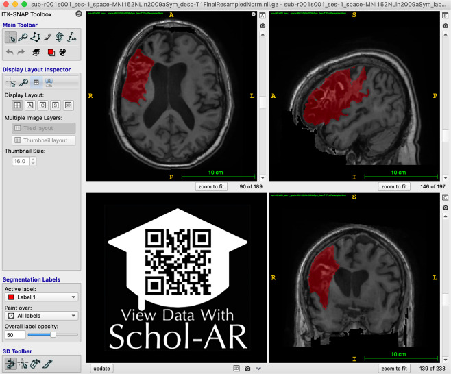
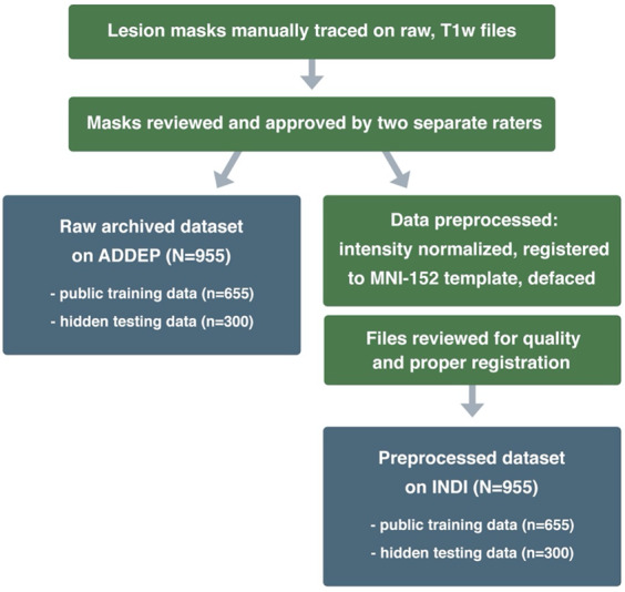
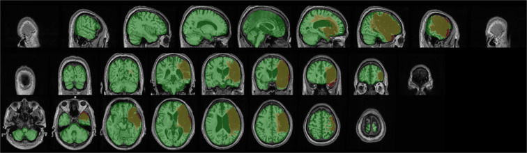
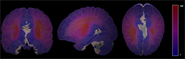
Accurate lesion segmentation is critical in stroke rehabilitation research for the quantification of lesion burden and accurate image processing. Current automated lesion segmentation methods for T1-weighted (T1w) MRIs, commonly used in stroke research, lack accuracy and reliability. Manual segmentation remains the gold standard, but it is time-consuming, subjective, and requires neuroanatomical expertise. We previously released an open-source dataset of stroke T1w MRIs and manually-segmented lesion masks (ATLAS v1.2, N = 304) to encourage the development of better algorithms. However, many methods developed with ATLAS v1.2 report low accuracy, are not publicly accessible or are improperly validated, limiting their utility to the field. Here we present ATLAS v2.0 (N = 1271), a larger dataset of T1w MRIs and manually segmented lesion masks that includes training (n = 655), test (hidden masks, n = 300), and generalizability (hidden MRIs and masks, n = 316) datasets. Algorithm development using this larger sample should lead to more robust solutions; the hidden datasets allow for unbiased performance evaluation via segmentation challenges. We anticipate that ATLAS v2.0 will lead to improved algorithms, facilitating large-scale stroke research.


Conflict of interest statement

A.G.B. serves on the Biogen Australia Dementia Scientific Advisory Committee and editorial boards of Neurology and International Journal of Stroke. S.C.C. serves as a consultant for Abbvie, Constant Therapeutics, MicroTransponder, Neurolutions, SanBio, Panaxium, NeuExcell, Elevian, Medtronic, and TRCare. E.d.L.R. is employed by icometrix. C.A.H. serves on the Advisory Board for Welcony Magstim and as a Consultant for Brainsway. B.G.H. has a clinical partnership with Fourier Intelligence. J.S.K. is cofounder of BoneScreen GmbH. G.F.W. serves on the Scientific Advisory Board for Myomo, Inc.

Figures

Fig. 1
Fig. 1
Example of Lesion Segmentation in ITK-SNAP. An example of the ITK-SNAP interface displaying a lesion segmentation mask (red) in radiological convention (the left hemisphere is shown on the right side of the screen). Axial (top left), sagittal (top right), and coronal (bottom right) planes are shown. A video of the example lesion mask in ITK-SNAP can be viewed through Schol-AR by scanning the QR code in the bottom left with a mobile device, or by opening this PDF with a non-mobile web browser at www.Schol-AR.io/reader.
Fig. 2
Fig. 2
Lesion Tracing and Preprocessing Pipeline. A flowchart diagram demonstrating the process for creating the two archived datasets: a raw dataset in native space archived with the Archive of Data on Disability to Enable Policy and research (ADDEP) (left blue box) and a preprocessed dataset in MNI-152 space archived with the International Neuroimaging Data-Sharing Initiative (INDI) (right blue box).
Fig. 3
Fig. 3
Example of Visual Quality Control. Example of an image used to ensure proper registration of each subject’s brain (gray) and lesion segmentation mask (reddish brown) to the MNI template (green).
Fig. 4
Fig. 4
Probabilistic Lesion Overlap Map on the MNI_icbm152 Template. Visualization of the lesion overlap across all subjects (N = 955) overlaid on the MNI template, with hotter colors representing more subjects with lesions at that voxel. An interactive volumetric 3D display of this data may be viewed through Schol-AR by scanning the QR code from Fig. 1 with a mobile device, or by opening this PDF with a non-mobile web browser at www.Schol-AR.io/reader.

References

    1. Liew S-L, et al. The ENIGMA Stroke Recovery Working Group: Big data neuroimaging to study brain-behavior relationships after stroke. Human brain mapping. 2022;43:129–148. doi: 10.1002/hbm.25015. - DOI - PMC - PubMed
    1. Liew, S.-L. et al. Smaller spared subcortical nuclei are associated with worse post-stroke sensorimotor outcomes in 28 cohorts worldwide. Brain Communications, 10.1093/braincomms/fcab254 (2021). - PMC - PubMed
    1. Boyd LA, et al. Biomarkers of stroke recovery: Consensus-based core recommendations from the Stroke Recovery and Rehabilitation Roundtable. Neurorehabilitation and neural repair. 2017;31:864–876. doi: 10.1177/1545968317732680. - DOI - PubMed
    1. Feng W, et al. Corticospinal tract lesion load: An imaging biomarker for stroke motor outcomes. Ann Neurol. 2015;78:860–870. doi: 10.1002/ana.24510. - DOI - PMC - PubMed
    1. Kim B, Winstein C. Can neurological biomarkers of brain impairment be used to predict poststroke motor recovery? A systematic review. Neurorehabilitation and neural repair. 2017;31:3–24. doi: 10.1177/1545968316662708. - DOI - PubMed