. 2022 Jun 16;12(1):10036.
doi: 10.1038/s41598-022-13689-x.

CRISPR/Cas9 editing of the MYO7A gene in rhesus macaque embryos to generate a primate model of Usher syndrome type 1B

Affiliations

CRISPR/Cas9 editing of the MYO7A gene in rhesus macaque embryos to generate a primate model of Usher syndrome type 1B

Junghyun Ryu et al. Sci Rep. .

Abstract

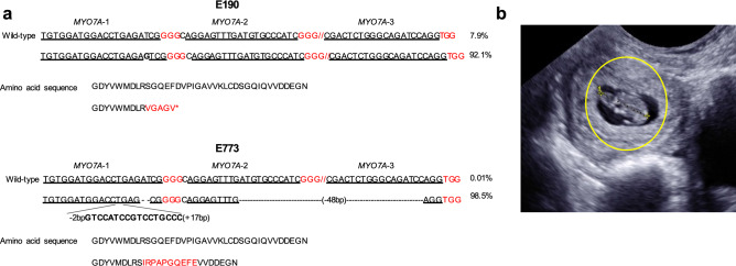
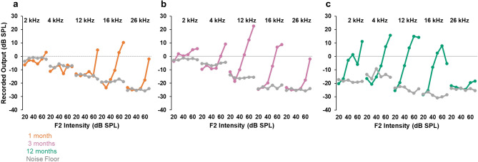
Mutations in the MYO7A gene lead to Usher syndrome type 1B (USH1B), a disease characterized by congenital deafness, vision loss, and balance impairment. To create a nonhuman primate (NHP) USH1B model, CRISPR/Cas9 was used to disrupt MYO7A in rhesus macaque zygotes. The targeting efficiency of Cas9 mRNA and hybridized crRNA-tracrRNA (hyb-gRNA) was compared to Cas9 nuclease (Nuc) protein and synthetic single guide (sg)RNAs. Nuc/sgRNA injection led to higher editing efficiencies relative to mRNA/hyb-gRNAs. Mutations were assessed by preimplantation genetic testing (PGT) and those with the desired mutations were transferred into surrogates. A pregnancy was established from an embryo where 92.1% of the PGT sequencing reads possessed a single G insertion that leads to a premature stop codon. Analysis of single peripheral blood leukocytes from the infant revealed that half the cells possessed the homozygous single base insertion and the remaining cells had the wild-type MYO7A sequence. The infant showed sensitive auditory thresholds beginning at 3 months. Although further optimization is needed, our studies demonstrate that it is feasible to use CRISPR technologies for creating NHP models of human diseases.


Conflict of interest statement

The authors declare no competing interests.

Figures

Figure 1
Figure 1
MYO7A exon 3 targeting strategy and assessment of gRNA targeting efficiency. (a) Using gRNA design programs described in the “Methods” section, four gRNAs were identified that correspond to exon 3 of the rhesus macaque MYO7A gene. Two gRNAs (#2 and #4) had overlapping targeting sites and gRNA #4 was not used further. Black letters represent the gRNA protospacer/target sequence and red letters indicate the PAM sequence. Gray arrows represent the primers used to amplify the flanking region of the target site. Black arrows were primers used to generate the PCR amplicons needed to perform the T7E1 editing assay. The distances between sgRNA indicated PAM to PAM distances. (b) Targeting efficiency was assessed using the rhesus macaque CMMT cell line, where each gRNA was tested separately and as a mixture (MYO7A-mix). Yellow arrowheads indicate the digested PCR amplicons by CRISPR/Cas9. MYO7A-mix + contained 50 ng of wild-type amplicon mixed with sample amplicon before performing the T7E1 assay. The negative control (–) contains Cas9 protein without gRNA, and the positive control is provided as part of the Integrated DNA Technologies Surveyor kit used to perform the T7E1 assay. PCR product size was 888 bp, and expected cleavage products were 500 bp and 388 bp for MYO7A-1, 524 bp and 364 bp for MYO7A-2, and 564 bp and 324 bp for MYO7A-3.
Figure 2
Figure 2
MYO7A genotype of TE biopsies from two embryos transferred into a recipient animal and resulted in a singleton pregnancy. (a) TE cell genotyping results of two expanded blastocysts selected for transfer to surrogate females. Double slashes denote omitted DNA sequence. Genotyping by NGS revealed E190 possessed a single G insertion in exon 3 of the MYO7A gene in 92.1% of sequencing reads, whereas 98.5% of the sequencing reads from E773 possessed an insertion and deletion in the target region. The single G insertion observed in E190 generates a premature stop codon (*). The mutation observed in E773 included a 50 bp deletion and a 17 bp insertion, with the net result being a change of 10 amino acids. (b) Ultrasound confirmed a singleton pregnancy at 28 days after transfer of E190 and E773. Yellow circle indicated implanted embryo.
Figure 3
Figure 3
MYO7A mutant rhesus macaque infant and single cell genotyping results. (a) A healthy female rhesus macaque infant was delivered on day 150 of gestation following the transfer of two MYO7A edited embryos. (b) Genomic DNA was isolated from duplicate blood, buccal, placenta, and skin samples. Exon 3 of MYO7A was PCR amplified and the PCR amplicons were sequenced by NGS. In all tissues, 40–50% of the sequencing reads possessed a single G insertion, confirming the pregnancy was the result of implantation of E190. (c) Genotyping results from individual FACs-sorted leukocytes demonstrated that Mya is homozygous mosaic for a G insertion. The top panel shows wild-type MYO7A sequence was present in 9 out of 22 cells, whereas the bottom panel indicates a homozygous G insertion (red arrow) was detected in 13 out of 22 individual cells (59%).
Figure 4
Figure 4
ABR thresholds at 1, 3, 6 and 12 months. (a) ABR thresholds in left (circle) and right (X) ears at one (gold line) and three (magenta line) months. Arrows indicate that the threshold, if present, is above 85 dB SPL, the highest stimulus intensity tested. The control range was defined using an in-ear acoustic probe (see “Methods”). (b) ABR thresholds in left and right ears at 6 (blue trace) and 12 (green trace) months. The control range was defined using an open field paradigm (see “Methods”).
Figure 5
Figure 5
Distortion product otoacoustic emissions at 1, 3, and 12 months. DPOAE responses (2f1–f2; colored traces) at 2, 4, 12, 16 and 26 kHz at one month (gold trace), 3 months (magenta trace), and 12 months (green trace). DPOAE responses were recorded at 6 intensities from 20 to 70 dB SPL incremented in 10 dB steps. The noise floor is in grey for each test age. An authentic DPOAE response profile is characterized by an increase in the magnitude of the distortion product with increasing stimulus intensity.
Figure 6
Figure 6
Retinal imaging and retinal layer thickness at 2, 4, and 6 months. (a) Color fundus photograph and (b) macular sdOCT scan of Mya at 6 months of age, showing normal retinal morphology. (c) The thickness of inner + outer segment layers (in µm) in right and left eye of Mya at 2, 4, 6 and 12 months of age, in comparison to data for 8 normal rhesus monkey infants (box plots show mean, interquartile range and range of values for the normal infants). Values were slightly below the normal range in the central 1 mm (fovea) and in each concentric annulus (1–2, 2–3, and 3–6 mm diameter annuli, centered on the fovea). Thickness of the total retina, inner retinal layers and outer nuclear layer were within the normal range except within the fovea, where total retinal thickness was just below the normal range (not shown).

References

    1. Boughman JA, Vernon M, Shaver KA. Usher syndrome: Definition and estimate of prevalence from two high-risk populations. J. Chronic Dis. 1983;36:595–603. doi: 10.1016/0021-9681(83)90147-9. - DOI - PubMed
    1. Kimberling WJ, et al. Frequency of Usher syndrome in two pediatric populations: Implications for genetic screening of deaf and hard of hearing children. Genet. Med. 2010;12:512–516. doi: 10.1097/GIM.0b013e3181e5afb8. - DOI - PMC - PubMed
    1. Keats BJ, Corey DP. The usher syndromes. Am. J. Med. Genet. 1999;89:158–166. doi: 10.1002/(SICI)1096-8628(19990924)89:3<158::AID-AJMG6>3.0.CO;2-#. - DOI - PubMed
    1. Mathur P, Yang J. Usher syndrome: Hearing loss, retinal degeneration and associated abnormalities. Biochim. Biophys. Acta. 2015;1852:406–420. doi: 10.1016/j.bbadis.2014.11.020. - DOI - PMC - PubMed
    1. Kremer H, van Wijk E, Marker T, Wolfrum U, Roepman R. Usher syndrome: Molecular links of pathogenesis, proteins and pathways. Hum. Mol. Genet. 2006;15 Spec No 2:262–270. doi: 10.1093/hmg/ddl205. - DOI - PubMed

Publication types

Supplementary concepts